Review
. 2013 Apr 5;7(2):21501.
doi: 10.1063/1.4799787.

Hydrodynamic mechanisms of cell and particle trapping in microfluidics

Affiliations
Review

Hydrodynamic mechanisms of cell and particle trapping in microfluidics

A Karimi et al. Biomicrofluidics. .

Abstract

Focusing and sorting cells and particles utilizing microfluidic phenomena have been flourishing areas of development in recent years. These processes are largely beneficial in biomedical applications and fundamental studies of cell biology as they provide cost-effective and point-of-care miniaturized diagnostic devices and rare cell enrichment techniques. Due to inherent problems of isolation methods based on the biomarkers and antigens, separation approaches exploiting physical characteristics of cells of interest, such as size, deformability, and electric and magnetic properties, have gained currency in many medical assays. Here, we present an overview of the cell/particle sorting techniques by harnessing intrinsic hydrodynamic effects in microchannels. Our emphasis is on the underlying fluid dynamical mechanisms causing cross stream migration of objects in shear and vortical flows. We also highlight the advantages and drawbacks of each method in terms of throughput, separation efficiency, and cell viability. Finally, we discuss the future research areas for extending the scope of hydrodynamic mechanisms and exploring new physical directions for microfluidic applications.
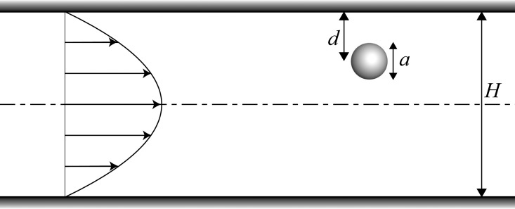
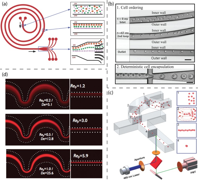
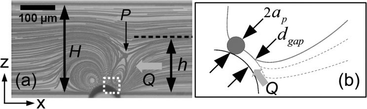
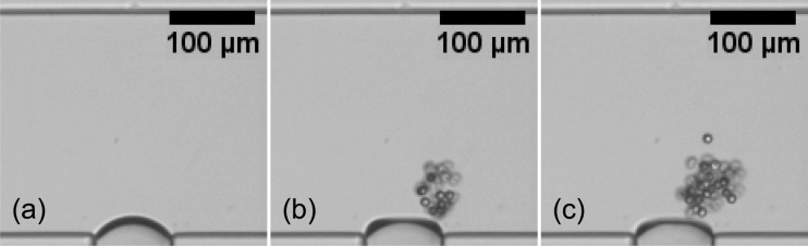

Figures

Figure 1
Figure 1
Schematic illustration showing channel and particle dimensions and the fully developed velocity profile.
Figure 2
Figure 2
Some examples of applications of inertial migration in microfluidics. (a) Schematics showing the dimensions and the working principle of a microdevice designed to separate and enrich larger cells by exploiting the difference in the inertial lift forces. Reprinted with permission from Hur et al., Biomicrofluidics 5, 022206 (2011). Copyright 2011 American Institute of Physics. (b) Experimental images of the particles flowing through a multiorifice microchannel at Re63. Due to lateral migration induced by the contraction/expansion cavities, two separate focused streams of particles are formed at the outlet. Reprinted with permission from Park et al., Lab Chip 9, 939–948 (2009). Copyright 2009 Royal Society of Chemistry. (c) Schematics illustrating the design and mechanism of a rare cell isolating microdevice. It consists of focusing, pinching, and extraction regions to trap the larger cells of interest in the vicinity of channel axis. Reprinted with permission from Bhagat et al., Lab Chip 11, 1870–1878 (2011). Copyright 2011 Royal Society of Chemistry.
Figure 3
Figure 3
(a) The top image illustrates the particle focusing along the centerline in a viscoelastic fluid with almost constant viscosity, while the bottom image shows the bistability of particle distribution in a shear-thinning fluid. The images are the results of experimental observations of particulate flows in a cylindrical microchannel. Reprinted with permission from G. D'Avino et al., Lab Chip 12, 1638–1645 (2012). Copyright 2012 Royal Society of Chemistry. (b) Experimental images of the elasto-inertial particle focusing in a straight rectangular channel. By increasing the flow rate and hence the elasticity number, focusing along the centerline is achieved due to synergetic effects of the forces induced by inertia and viscoelasticity. Reprinted with permission from Yang et al., Lab Chip 11, 266–273 (2011). Copyright 2011 Royal Society of Chemistry. (c) The top figure shows a schematic demonstrating the design of a microdevice used to separate cells and particles flowing in a viscoelastic fluid. The design includes sheath and sample flows. The particles are segregated based on their size due to difference in their lateral migration. Reprinted with permission from Nam et al., Lab Chip 12, 1347–1354 (2012). Copyright 2012 Royal Society of Chemistry.
Figure 4
Figure 4
Some applications of the deformability-induced migration in the microfluidics. (a) Schematics showing the dimensions and mechanism of operation of a microdevice designed to separate RBCs from the platelets. Reprinted with permission from Geislinger et al., Appl. Phys. Lett. 100, 183701 (2012). Copyright 2012 American Institute of Physics. (b) The top figure shows a schematic illustrating the mechanism of deformability-selective cell separation in a viscoelastic medium. The bottom figure is an experimental snapshot demonstrating the separation of WBCs from RBCs due to difference in their deformability characteristics. Reprinted with permission from Yang et al., Soft Matter 8, 5011–5019 (2012). Copyright 2012 Royal Society of Chemistry. (c) Deformability-selective separation of blood and cancer cells in an inertial flow. Reprinted with permission from Hur et al., Lab Chip 11, 912–920 (2011). Copyright 2011 Royal Society of Chemistry.
Figure 5
Figure 5
Some applications of Dean flow in microfluidic systems. (a) Schematic of a microdevice to separate different particles based on their size using combined inertial and secondary flow effects. In this spiral microchannel, larger particles equilibrate along the inner wall, while smaller ones aggregate around the centerline. Reprinted with permission from Kuntaegowdanahalli et al., Lab Chip 9, 2973–2980 (2009). Copyright 2009 Royal Society of Chemistry. (b) Experimental pictures showing the cell ordering and encapsulation using a spiral microchannel. Reprinted with permission from Kemna et al., Lab Chip 12, 2881–2887 (2012). Copyright 2012 Royal Society of Chemistry. (c) Schematic of a microfluidic system to focus the particles using a curved microchannel as well as horizontal and vertical sheath flows. The lateral position of the particles at different locations are depicted in panels 1-4. Reprinted with permission from Mao et al., Lab Chip 9, 1583–1589 (2009). Copyright 2009 Royal Society of Chemistry. (d) Fluorescent images of the particulate flow in a serpentine microchannel show that by increasing flow rate and thus Dean number, particle focusing along a single stream is achieved. Reprinted with permission from Oakey et al., Anal. Chem. 82, 3862–3867 (2010). Copyright 2010 American Chemical Society.
Figure 6
Figure 6
Mechanism of trapping: (a) streak image highlighting the hyperbolic point P and critical streamline and (b) schematic detail of the boxed region of panel (a). Reprinted with permission from Wang et al., Appl. Phys. Lett. 99, 034101 (2011). Copyright 2011 American Institute of Physics.
Figure 7
Figure 7
Accumulation of microparticles around an oscillating bubble. Snapshots (b) and (c) are taken 2 s and 4 s after (a), respectively. Reprinted with permission from Wang et al., Biomicrofluidics 6, 012801 (2012). Copyright 2012 American Institute of Physics.
Figure 8
Figure 8
Schematic illustration of characteristic loops of large and small particles. Reprinted with permission from Wang et al., Biomicrofluidics 6, 012801 (2012). Copyright 2012 American Institute of Physics.
Figure 9
Figure 9
Bacterial collection in a vortical flow of an oscillating bubble trapped in a horse-shoe structure for (a) low motile bacteria (E. coli DH5α) and (b) high motile (E. coli RP437). (c) Rapid biofilm formation in the vicinity of the bubble. Reprinted with permission from S. H. Yazdi and A. M. Ardekani, Biomicrofluidics 6, 044114 (2012). Copyright 2012 American Institute of Physics.
Figure 10
Figure 10
(a) Schematic representation of the flow channel and the symmetry planes as observed in experiments. (b) The image of particle pathlines at plane A for particle size ap=500μm. (c) Schematic streamlines of streaming flow at planes B and C. Reprinted with permission from Lutz et al., Fluids 17, 023601 (2005). Copyright 2012 American Institute of Physics.

References

    1. Danova M., Torchio M., and Mazzini G., “Isolation of rare circulating tumor cells in cancer patients: Technical aspects and clinical implications,” Expert Rev. Mol. Diagn. 11, 473–485 (2011). 10.1586/erm.11.33 - DOI - PubMed
    1. Colter D. C., Sekiya I., and Prockop D. J., “Identification of a subpopulation of rapidly self-renewing and multipotential adult stem cells in colonies of human marrow stromal cells,” Proc. Natl. Acad. Sci. 98, 7841–7845 (2001). 10.1073/pnas.141221698 - DOI - PMC - PubMed
    1. Jo K., Chen Y.-L., de Pablo J. J., and Schwartz D. C., “Elongation and migration of single DNA molecules in microchannels using oscillatory shear flows,” Lab Chip 9, 2348–2355 (2009). 10.1039/b902292a - DOI - PMC - PubMed
    1. Gascoyne P., Satayavivad J., and Ruchirawat M., “Microfluidic approaches to malaria detection,” Acta Tropica 89, 357–369 (2004). 10.1016/j.actatropica.2003.11.009 - DOI - PMC - PubMed
    1. van de Stolpe A., Pantel K., Sleijfer S., Terstappen L. W., and den Toonder J. M. J., “Circulating tumor cell isolation and diagnostics: Toward routine clinical use,” Cancer Res. 71, 5955–5960 (2011). 10.1158/0008-5472.CAN-11-1254 - DOI - PubMed