. 2014 Jan 3;9(1):e83955.
doi: 10.1371/journal.pone.0083955. eCollection 2014.

Human mammospheres secrete hormone-regulated active extracellular vesicles

Affiliations

Human mammospheres secrete hormone-regulated active extracellular vesicles

Esperanza Gonzalez et al. PLoS One. .

Abstract

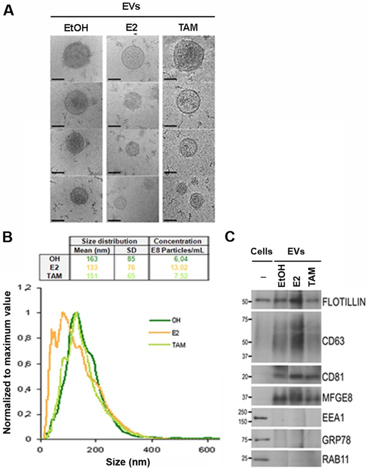
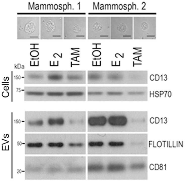
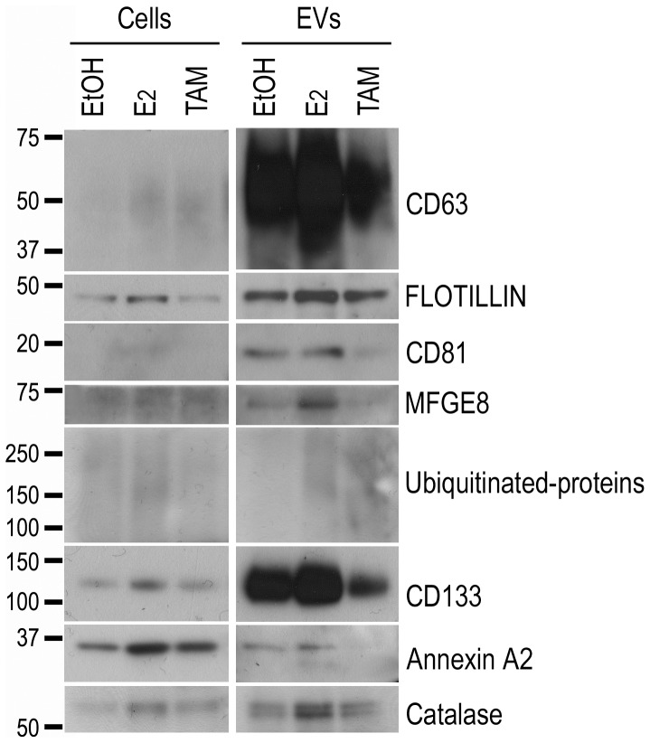
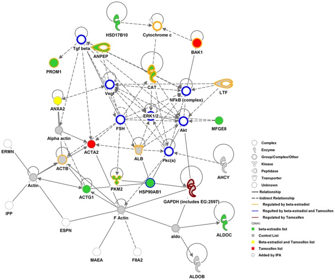
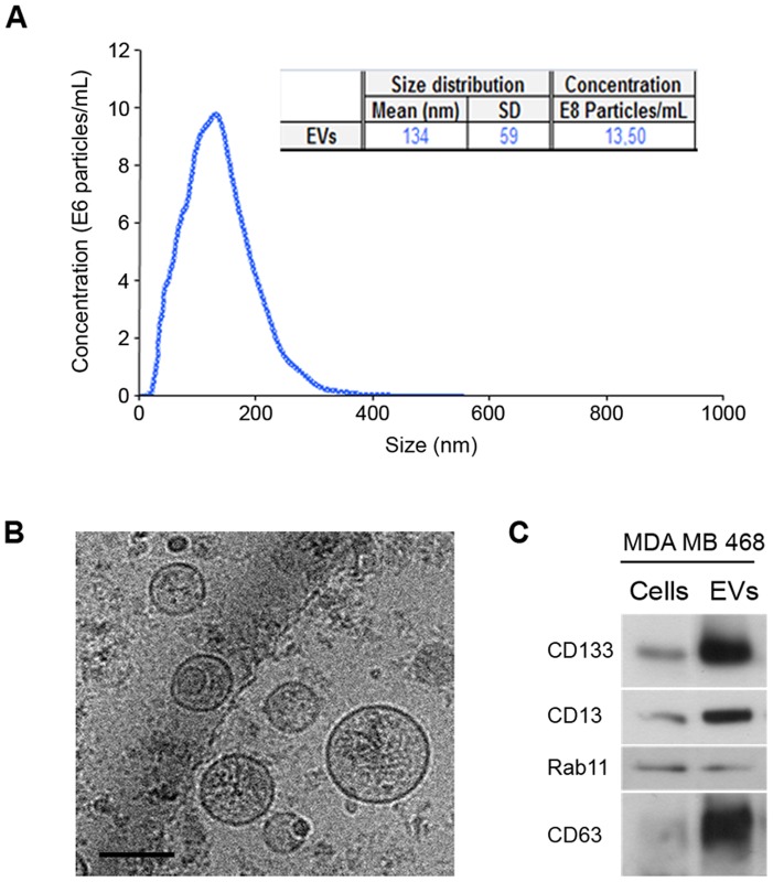
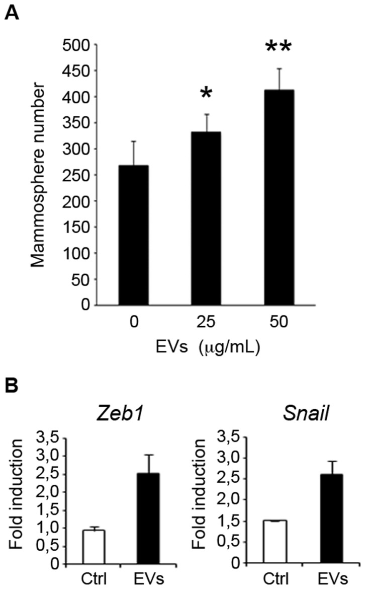
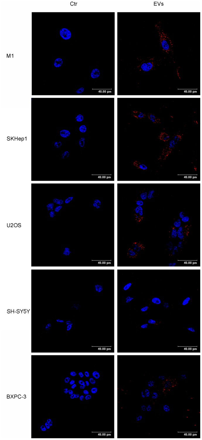
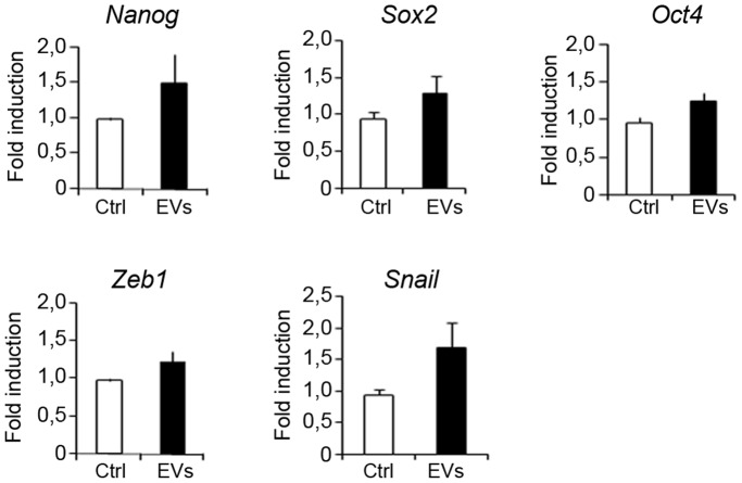
Breast cancer is a leading cause of cancer-associated death worldwide. One of the most important prognostic factors for survival is the early detection of the disease. Recent studies indicate that extracellular vesicles may provide diagnostic information for cancer management. We demonstrate the secretion of extracellular vesicles by primary breast epithelial cells enriched for stem/progenitor cells cultured as mammospheres, in non-adherent conditions. Using a proteomic approach we identified proteins contained in these vesicles whose expression is affected by hormonal changes in the cellular environment. In addition, we showed that these vesicles are capable of promoting changes in expression levels of genes involved in epithelial-mesenchymal transition and stem cell markers. Our findings suggest that secreted extracellular vesicles could represent potential diagnostic and/or prognostic markers for breast cancer and support a role for extracellular vesicles in cancer progression.


Conflict of interest statement

Competing Interests: The authors have declared that no competing interests exist.

Figures

Figure 1
Figure 1. Characterization and comparison of primary mammosphere-derived EVs cultured with or without hormone treatments.
(A) Representative cryo-electron micrographs (Bar, 100 nm). (B) Normalized representation of size distribution by NTA analysis of EVs in one primary breast epithelial cell preparation. Mean, SD and particle concentration values are indicated in the table (EtOH in green, E2 in orange and TAM in light green). (C) Western blot analysis of cell extracts and EVs derived from mammospheres treated with ethanol (EtOH), estrogen (E2) or tamoxifen (TAM). Antibodies against exosomes (Flotillin-1, CD63, CD81 and MFGE8), early (EEA1) and recycling (Rab11) endosomes, or endoplasmic reticulum (Grp78) protein markers were assayed. The molecular mass (kDa) for each protein is indicated.
Figure 2
Figure 2. EV content is modulated by hormone treatment.
Western blot analysis of total cell extracts and EVs of two independent human primary mammosphere preparations cultured in the presence of ethanol (EtOH), estrogen (E2) or tamoxifen (TAM). Protein expression was analyzed by immunoblotting with antibodies against CD13, Flotillin-1 and CD81 proteins. The molecular mass (kDa) for each protein is indicated. Representative micrographs of mammospheres formed under the different conditions are also shown. Bar, 50 µm.
Figure 3
Figure 3. Validation of proteomics profiling of EVs from primary human mammospheres by immunoblotting.
Western blot analysis of protein extracts obtained from cells (left) or EVs (right) secreted by mammospheres cultured in the presence of ethanol (EtOH), estrogen (E2) or tamoxifen (TAM). In addition to EV protein markers (Flotillin-1, CD63, CD81 and MFGE8) used as positive controls, the presence of proteins detected in the proteomic analysis, including prominin1/CD133, annexin A2 and catalase, were evaluated. Three different tissue samples were examined and one representative example is shown. The molecular mass (kDa) for each protein is indicated.
Figure 4
Figure 4. Cancer-related networks detected by the data mining IPA software.
A list of 37 proteins was recognized by ingenuity pathway analysis software to build a connectivity network. This network was highly associated to cancer (p-value = 5.9E-12) and integrates the data from the list of proteins identified in control (grey-filled circles), in estrogen (green-filled circles), in tamoxifen (red-filled circles) or in both estrogen and tamoxifen (yellow-filled circles) treatments. Some molecules (unfilled) were added by IPA software to complete the pathway. The proteins known to be regulated by estrogen (surrounded by an orange outline), by tamoxifen (surrounded by violet outline) or both (surrounded by blue outline) are also indicated.
Figure 5
Figure 5. Characterization of EVs secreted by MDA-MB-468 mammospheres.
(A) NTA analysis indicating mean, standard deviation and concentration of the particles present in the preparation. (B) Representative cryo-electron micrographs (Bar, 100 nm). (C) Western blot analysis of protein extracts prepared from cells or from EVs using antibodies against indicated proteins.
Figure 6
Figure 6. Effect of EVs secreted by MDA-MB-468 mammospheres on MCF7 cells.
(A) MCF-7 cells were incubated with 0, 25 and 50 µg/mL of MDA-MB-468 EVs and the number of mammospheres formed after 7-days was counted [data are mean ±SD; n = 3, *p<0.05, **p<0.01, relative to the values in the control]. (B) Quantitative polymerase chain reaction analysis was conducted to examine the expression of the factors Zeb1 and Snaill [data are mean ±SD; n = 3, p<0.05 and 0.05 respectively, relative to the values in the control].
Figure 7
Figure 7. Uptake of EVs by cell lines of different tissue origin as putative metastatic destinations.
Left panels represent images of M1 (fibroblast), SK-Hep1 (endothelial, liver adenocarcinoma), U2SO (osteosarcoma), SH-SY5Y (neuroblastoma) and BXPC3 (pancreatic adenocarcinoma) human acceptor cells under control conditions. Right panels are individual frames showing the internalization of CD133-positive EVs from MDA-MB-468 into the acceptor cells (Bar, 40 µm).
Figure 8
Figure 8. Effect of EVs from MDA-MB-468 on stem cell and EMT markers in U2OS cells.
U2OS cells were incubated with 0 or 50 µg/mL of MDA-MB-468-derived EVs and quantitative polymerase chain reaction analysis was conducted to examine the expression of the transcription factors Nanog, Oct-4, Sox2, Zeb1 and Snaill involved in development and maintenance of stem cells [data are mean ±SD; n = 3, p<0.05, 0.05, 0.01, 0.05 and 0.01, respectively, relative to the values in the control].

References

    1. Bocchinfuso WP, Lindzey JK, Hewitt SC, Clark JA, Myers PH, et al. (2000) Induction of mammary gland development in estrogen receptor-alpha knockout mice. Endocrinology 141: 2982–2994. - PubMed
    1. Ali S, Coombes RC (2002) Endocrine-responsive breast cancer and strategies for combating resistance. Nat Rev Cancer 2: 101–112. - PubMed
    1. Reya T, Morrison SJ, Clarke MF, Weissman IL (2001) Stem cells, cancer, and cancer stem cells. Nature 414: 105–111. - PubMed
    1. Dontu G, Abdallah WM, Foley JM, Jackson KW, Clarke MF, et al. (2003) In vitro propagation and transcriptional profiling of human mammary stem/progenitor cells. Genes Dev 17: 1253–1270. - PMC - PubMed
    1. Vivanco M (2010) Function follows form: defining mammary stem cells. Sci Transl Med 2: 31ps22. - PubMed

Publication types