. 2014 Jan 3;9(1):e84704.
doi: 10.1371/journal.pone.0084704. eCollection 2014.

Rab27a is essential for the formation of neutrophil extracellular traps (NETs) in neutrophil-like differentiated HL60 cells

Affiliations

Rab27a is essential for the formation of neutrophil extracellular traps (NETs) in neutrophil-like differentiated HL60 cells

Tatsumi Kawakami et al. PLoS One. .

Abstract

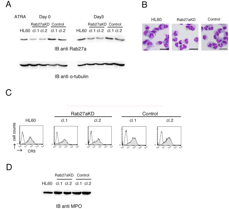
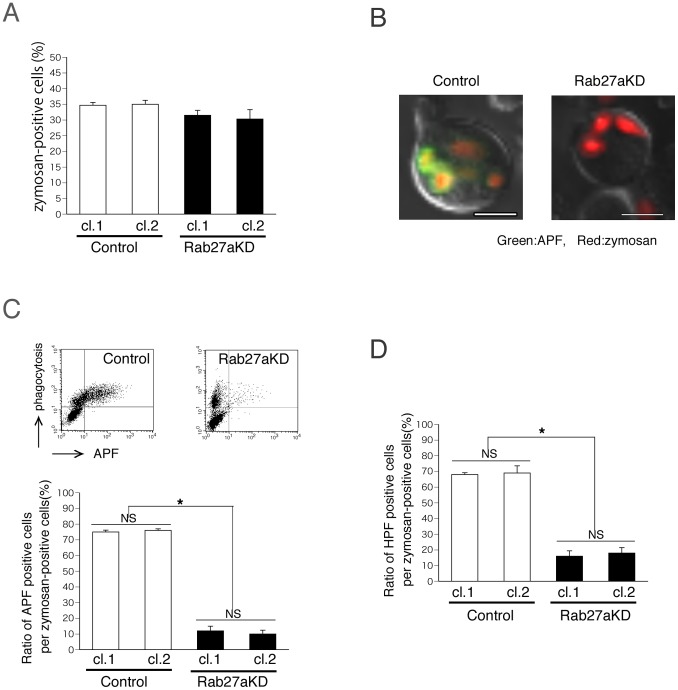
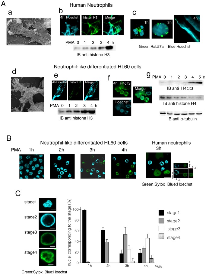
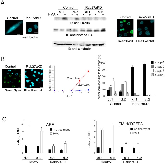
Neutrophils play a crucial role in host defence. In response to a variety of inflammatory stimulation, they form neutrophil extracellular traps (NETs). NETs are extracellular structures composed of chromatin fibers decorated with antimicrobial proteins and developing studies indicate that NETs contribute to extracellular microbial killing. While the intracellular signaling pathways that regulate NET formation remain largely unknown, there is growing evidence that generation of reactive oxygen species (ROS) is a key event for NET formation. The Rab family small GTPase Rab27a is an important component of the secretory machinery of azurophilic granules in neutrophils. However, the precise mechanism of NET formation and whether or not Rab27a contributes to this process are unknown. Using neutrophil-like differentiated HL60 cells, we show here that Rab27a plays an essential role in both phorbol myristate acetate (PMA)- and Candida albicans-induced NET formation by regulating ROS production. Rab27a-knockdown inhibited ROS-positive phagosome formation during complement-mediated phagocytosis. To investigate the role of Rab27a in neutrophil function in detail, both primary human neutrophils and neutrophil-like differentiated HL60 cells were treated with PMA, and NET formation process was assessed by measurement of release of histone H3 into the medium, citrullination of the arginine in position 3 of histone H4 and chase of the nuclear change of the living cells in the co-existence of both cell-permeable and -impermeable nuclear indicators. PMA-induced NET formation occured sequentially in both neutrophil-like differentiated HL60 cells and primary neutrophils, and Rab27a-knockdown clearly inhibited NET formation in association with reduced ROS production. We also found that serum-treated Candida albicans triggers NET formation in a ROS-dependent manner, and that Rab27a-knockdown inhibits this process as well. Our findings demonstrate that Rab27a plays an important role in NET formation induced by both Candida albicans infection and PMA treatment by regulating ROS production.


Conflict of interest statement

Competing Interests: The authors have declared that no competing interests exist.

Figures

Figure 1
Figure 1. Effects of Rab27a-knockdown on neutrophil-like differentiaiton of HL60 cells.
(A–C) HL60 cells, Rab27a-knockdown cells transfected with shRNA-Rab27a using a lentiviral system (Rab27aKD; clone 1 and clone 2) and control-shRNA transferred HL60 cells (Control; clone 1 and clone 2) were treated with ATRA for 3 days. (A) The expression of Rab27a in similarly treated cells were analyzed by immunoblotting analysis. (B) Morphology and (C) cell surface expression of CR3 were analyzed by May-Giemsa staining and flow cytometry, respectively. In (B), Rab27aKD clone 1 and Control clone 1 are shown as representative images. (D) Expression of myeloperoxidase (MPO) in whole cell lysates were analyzed by immunoblotting analysis. Scale bars indicate 10 µm.
Figure 2
Figure 2. Decreased production of highly reactive oxygen species production induced by complement-mediated phagocytosis in Rab27a-knockdown cells.
(A–D) Both Rab27a-knockdown HL60 cells (Rab27aKD; clone 1 and clone 2) and control-shRNA transferred HL60 cells (Control; clone 1 and clone 2) were treated with ATRA for 3 days and complement-mediated phagocytosis assay was performed using serum-opsonized Texas Red-labeled zymosan. (A) At 30 min after the onset of phagocytosis, uptake of zymosan particles in Rab27a-knockdown cells was analyzed by flow cytometry. In the presence of APF, (B) microscopic analysis and (C) flow cytometric analysis were performed at 30 min after the onset of phagocytosis. In (B) and the cytograms of (C), the results of Rab27aKD clone 1 and Control clone 1 are shown as representative data. In (B), hypochlorite appears green and Texas Red-labeled zymosan is red. Scale bars indicate 5 µm. (D) In the presence of HPF, flow cytometric analysis was performed at 30 min after the onset of phagocytosis. In (A), (C), (D), the data are the mean with SD from three independent experiments. Asterisks (*) mean that the difference is statistically significant (p values<0.01). NS means no statistical significance.
Figure 3
Figure 3. Process of NET formation after PMA treatment both in primary neutrophils and neutrophil-like differentiated HL60 cells.
(A–C) Primary human neutrophils and neutrophil-like differentiated HL60 cells were stimulated with PMA. (A) NET formation was observed both in human neutrophils (a–c) and neutrophil-like differentiated HL60 cells (d–g). (a and d) Scanning electron microscopy (SEM) images of both cells indicating NET structures. Scale bars indicate 10 µm. (b and e) PFA-fixed cells were stained with antibody against histone H3 in the presence of Hoechst 33342. Scale bars indicate 20 µm. Extracellular histone H3 content in DNase I-treated supernatants were assessed by immunoblotting analysis in the time course study after PMA treatment. (c) PFA-fixed cells were stained with antibody against Rab27a in the presence of Hoechst 33342. (f) PFA-fixed cells were stained with antibody against H4cit3 in the presence of Hoechst 33342. Scale bars indicate 10 µm. (g) The amounts of histone H4cit3 and histone H4 in the cells in the time course study after PMA treatment were assesed by immunoblotting analysis using specific antibodies.(B) Both primary neutrophils and neutrophil-like differentiated HL60 cells were stimulated with PMA with a mixture of Hoechst 33342 and Sytox Green. Representative fluorescence images at indicated time points are shown. In the images of human neutrophils (right), x-y-z section images are also shown to indicate the spherical forms of the nuclei. Scale bars indicate 10 µm. Blue and green colors show Hoechst 33342-positive and Sytox Green-positive, respectively. (C) Cell stages in NET formation were classified into four groups by the degree of nuclei expansion and types of reagent staining (stage1–4). Nuclei are stained with Hoechst 33342 and showed lobulated shape and decondensation does not occur judging from z-section scanning (stage1), nuclei are stained with Hoechst 33342 and chromatin decondensation occurs like a spherical form (stage2), chromatin stained with Sytox Green like a spherical form (stage3), chromatin stained with Sytox Green shows a cloud-like spread form (stage4). The histogram shows the percentage of the cells in the individual stage at indicated time points. The data are the mean with SD from three independent experiments. Scale bars indicate 5 µm.
Figure 4
Figure 4. The effects of Rab27a-knockdown on PMA-induced NET formation.
(A–C) Neutrophil-like differentiated control HL60 cells (Control; clone 1 and clone 2) and Rab27a-knockdown cells (Rab27aKD; clone 1 and clone 2) were stimulated with PMA. (A) Both cells fixed with PFA were stained with Hoechst 33342 (left). Cell lysates were assesed by immunoblotting analysis using specific antibody against H4cit3 and histone H4 (middle). PFA-fixed cells were stained with antibody against H4cit3 in the presence of Hoechst 33342 (right). Scale bars indicate 10 µm. (B) Representative fluorescence images at 4 hours after PMA treatment in the presence of both Hoechst 33342 and Sytox Green. Scale bars indicate 10 µm. Blue and green colors show Hoechst 33342-positive and Sytox Green-positive, respectively (left). The results of Rab27aKD clone 1 and Control clone 1 are shown as representative data. Frequency of Sytox Green-positive cells at indicated time points after PMA treatment (middle). The percentage of the cells in the individual stages at 4 h after PMA treatment (right). (C) The mean fluorescence intensity (MFI) after PMA treatment in the presence of APF at 30 min (left) or CM-H2DCFDA at 20min (right). The mean value of MFI of control cells was adjusted to 1. In (B) and (C), the data are the mean with SD from three independent experiments.
Figure 5
Figure 5. NET formation induced by complement-mediated phagocytosis against the invasion of C. albicans.
(A) Serum-treated or non-treated C. albicans were added to neutrophil-like differentiated HL60 cells (the ratio of C. albicans to cell; 5∶1) in the presence of Sytox Green for 3 h at 37°C. Left panel shows representative fluorescence images and right panel shows frequency of Sytox green-positeve cells. Green shows Sytox Green staining. (B) Serum-treated C. albicans were added to neutrophil-like differentiated control cells and Rab27a-knockdown cells in the presence of both Hoechst 33342 and Sytox Green for 3 h (The ratio of C. albicans to cell; 5∶1). Left panel shows representative fluorescence images. Blue shows Hoechst 33342 staining and Green Sytox Green. Right panel shows the percentage of the cells in the individual stage (black: Hoechst 33342–positive; white: Sytox Green-positive and decondensed; shaded area: Sytox Green-positive and cloud-like shape). (C) Serum-treated C. albicans were added to neutrophil-like differentiated control cells and Rab27a-knockdown cells in the presence or absence of DPI for 3 h. Frequency of Sytox green-positive cells are shown. In (A) and (B), Scale bars indicate 10 µm. In (A), (B) and (C), the results of Rab27aKD clone 1 and Control clone 1 are shown and the data are the mean with SD from three independent experiments. Asterisks (*) mean that the difference is statistically significant (p values <0.01).

References

    1. Stenmark H (2009) Rab GTPases as coordinators of vesicle traffic. Nat Rev Mol Cell Biol 10: 513–525 10.1038/nrm2728.PubMed:19603039 - DOI - PubMed
    1. Ostrowski M, Carmo NB, Krumeich S, Fanget I, Raposo G, et al. (2010) Rab27a and Rab27b control different steps of the exosome secretion pathway. Nat Cell Biol 12: 19–30 10.1038/ncb2000.PubMed:19966785 - DOI - PubMed
    1. Kuroda TS, Fukuda M (2004) Rab27A-binding protein Slp2-a is required for peripheral melanosome distribution and elongated cell shape in melanocytes. Nat Cell Biol 6: 1195–1203. PubMed: 15543135. - PubMed
    1. Ménasché G, Pastural E, Feldmann J, Certain S, Ersoy F, et al... (2000) Mutations in RAB27A Cause Griscelli Syndrome Associated with Haemophagocytic Syndrome. Nat Genet 25: 173–176. PubMed: 10835631. - PubMed
    1. Ménager MM, Ménasché G, Romao M, Knapnougel P, Ho CH, et al... (2007) Secretory cytotoxic granule maturation and exocytosis require the effector protein hMunc13–4. Nat Immunol 8: 257–267. PubMed: 17237785. - PubMed

Publication types

LinkOut - more resources