

Proliferation and survival signaling from both Jak2-V617F and Lyn involving GSK3 and mTOR/p70S6K/4EBP1 in PVTL-1 cell line newly established from acute myeloid leukemia transformed from polycythemia vera
- PMID: 24404189
- PMCID: PMC3880321
- DOI: 10.1371/journal.pone.0084746
Proliferation and survival signaling from both Jak2-V617F and Lyn involving GSK3 and mTOR/p70S6K/4EBP1 in PVTL-1 cell line newly established from acute myeloid leukemia transformed from polycythemia vera
Abstract
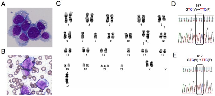
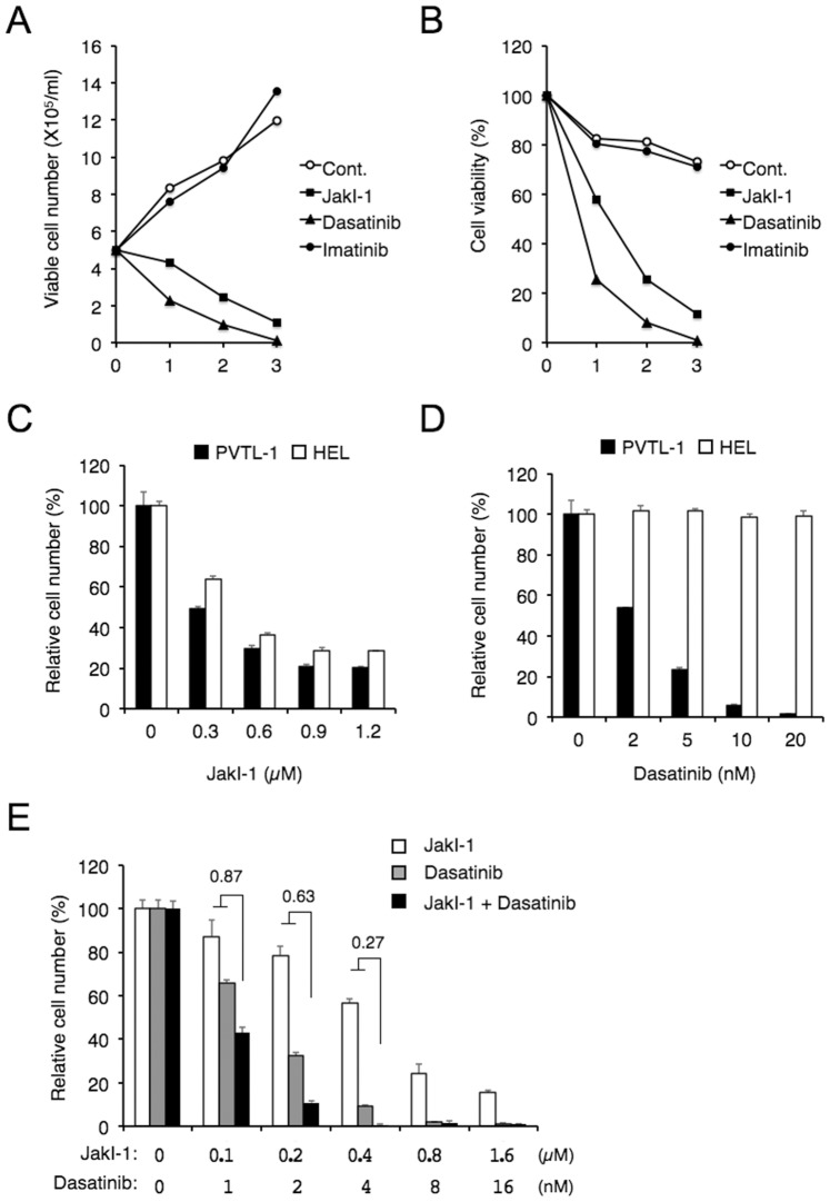
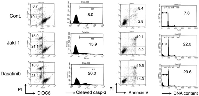
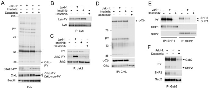
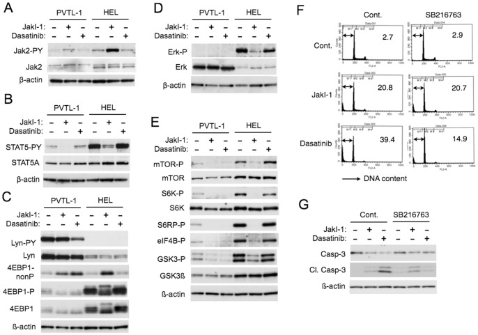
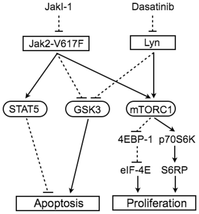
The gain of function mutation JAK2-V617F is very frequently found in myeloproliferative neoplasms (MPNs) and is strongly implicated in pathogenesis of these and other hematological malignancies. Here we report establishment of a new leukemia cell line, PVTL-1, homozygous for JAK2-V617F from a 73-year-old female patient with acute myeloid leukemia (AML) transformed from MPN. PVTL-1 is positive for CD7, CD13, CD33, CD34, CD117, HLA-DR, and MPO, and has complex karyotypic abnormalities, 44,XX,-5q,-7,-8,add(11)(p11.2),add(11)(q23),-16,+21,-22,+mar1. Sequence analysis of JAK2 revealed only the mutated allele coding for Jak2-V617F. Proliferation of PVTL-1 was inhibited and apoptosis was induced by the pan-Jak inhibitor Jak inhibitor-1 (JakI-1) or dasatinib, which inhibits the Src family kinases as well as BCR/ABL. Consistently, the Src family kinase Lyn was constitutively activated with phosphorylation of Y396 in the activation loop, which was inhibited by dasatinib but not by JakI-1. Further analyses with JakI-1 and dasatinib indicated that Jak2-V617F phosphorylated STAT5 and SHP2 while Lyn phosphorylated SHP1, SHP2, Gab-2, c-Cbl, and CrkL to induce the SHP2/Gab2 and c-Cbl/CrkL complex formation. In addition, JakI-1 and dasatinib inactivated the mTOR/p70S6K/4EBP1 pathway and reduced the inhibitory phosphorylation of GSK3 in PVTL-1 cells, which correlated with their effects on proliferation and survival of these cells. Furthermore, inhibition of GSK3 by its inhibitor SB216763 mitigated apoptosis induced by dasatinib but not by JakI-1. Together, these data suggest that apoptosis may be suppressed in PVTL-1 cells through inactivation of GSK3 by Lyn as well as Jak2-V617F and additionally through activation of STAT5 by Jak2-V617F. It is also speculated that activation of the mTOR/p70S6K/4EBP1 pathway may mediate proliferation signaling from Jak2-V617F and Lyn. PVTL-1 cells may provide a valuable model system to elucidate the molecular mechanisms involved in evolution of Jak2-V617F-expressing MPN to AML and to develop novel therapies against this intractable condition.
Conflict of interest statement
Figures
Similar articles
-
The PIM inhibitor AZD1208 synergizes with ruxolitinib to induce apoptosis of ruxolitinib sensitive and resistant JAK2-V617F-driven cells and inhibit colony formation of primary MPN cells.Oncotarget. 2015 Nov 24;6(37):40141-57. doi: 10.18632/oncotarget.5653. Oncotarget. 2015. PMID: 26472029 Free PMC article.
-
Inhibition of the PI3K/Akt/GSK3 pathway downstream of BCR/ABL, Jak2-V617F, or FLT3-ITD downregulates DNA damage-induced Chk1 activation as well as G2/M arrest and prominently enhances induction of apoptosis.PLoS One. 2013 Nov 18;8(11):e79478. doi: 10.1371/journal.pone.0079478. eCollection 2013. PLoS One. 2013. PMID: 24260231 Free PMC article.
-
Loss of p53 induces leukemic transformation in a murine model of Jak2 V617F-driven polycythemia vera.Oncogene. 2017 Jun 8;36(23):3300-3311. doi: 10.1038/onc.2016.478. Epub 2017 Jan 9. Oncogene. 2017. PMID: 28068330
-
Changing concepts of diagnostic criteria of myeloproliferative disorders and the molecular etiology and classification of myeloproliferative neoplasms: from Dameshek 1950 to Vainchenker 2005 and beyond.Acta Haematol. 2015;133(1):36-51. doi: 10.1159/000358580. Epub 2014 Aug 7. Acta Haematol. 2015. PMID: 25116092 Review.
-
The role of JAK2 V617F mutation, spontaneous erythropoiesis and megakaryocytopoiesis, hypersensitive platelets, activated leukocytes, and endothelial cells in the etiology of thrombotic manifestations in polycythemia vera and essential thrombocythemia.Semin Thromb Hemost. 2006 Jun;32(4 Pt 2):381-98. doi: 10.1055/s-2006-942759. Semin Thromb Hemost. 2006. PMID: 16810614 Review.
Cited by
-
Activation of signaling pathways in models of t(6;9)-acute myeloid leukemia.Ann Hematol. 2022 Oct;101(10):2179-2193. doi: 10.1007/s00277-022-04905-9. Epub 2022 Aug 8. Ann Hematol. 2022. PMID: 35941390 Free PMC article.
-
Gab2 mediates hepatocellular carcinogenesis by integrating multiple signaling pathways.FASEB J. 2017 Dec;31(12):5530-5542. doi: 10.1096/fj.201700120RR. Epub 2017 Aug 21. FASEB J. 2017. PMID: 28842424 Free PMC article.
-
The pan-PIM inhibitor INCB053914 displays potent synergy in combination with ruxolitinib in models of MPN.Blood Adv. 2019 Nov 26;3(22):3503-3514. doi: 10.1182/bloodadvances.2019000260. Blood Adv. 2019. PMID: 31725895 Free PMC article.
-
Src family kinase inhibitor bosutinib enhances retinoic acid-induced differentiation of HL-60 leukemia cells.Leuk Lymphoma. 2018 Dec;59(12):2941-2951. doi: 10.1080/10428194.2018.1452213. Epub 2018 Mar 23. Leuk Lymphoma. 2018. PMID: 29569971 Free PMC article.
-
Molecular Pathways Involved in the Development of Congenital Erythrocytosis.Genes (Basel). 2021 Jul 28;12(8):1150. doi: 10.3390/genes12081150. Genes (Basel). 2021. PMID: 34440324 Free PMC article. Review.
References
-
- Ihle JN (1995) Cytokine receptor signalling. Nature 377: 591–594. - PubMed
-
- Ihle JN, Gilliland DG (2007) Jak2: normal function and role in hematopoietic disorders. Curr Opin Genet Dev 17: 8–14. - PubMed
-
- Tefferi A (2012) Polycythemia vera and essential thrombocythemia: 2012 update on diagnosis, risk stratification, and management. Am J Hematol 87: 285–293. - PubMed
-
- LaFave LM, Levine RL (2012) JAK2 the future: therapeutic strategies for JAK-dependent malignancies. Trends Pharmacol Sci 33: 574–582. - PubMed
-
- Quintas-Cardama A (2013) The role of Janus kinase 2 (JAK2) in myeloproliferative neoplasms: therapeutic implications. Leuk Res 37: 465–472. - PubMed
Publication types
MeSH terms
Substances
LinkOut - more resources
Full Text Sources
Other Literature Sources
Medical
Molecular Biology Databases
Research Materials
Miscellaneous
