. 2014 Jan 3;9(1):e85343.
doi: 10.1371/journal.pone.0085343. eCollection 2014.

The potential role of As-sumo-1 in the embryonic diapause process and early embryo development of Artemia sinica

Affiliations

The potential role of As-sumo-1 in the embryonic diapause process and early embryo development of Artemia sinica

Bing Chu et al. PLoS One. .

Abstract

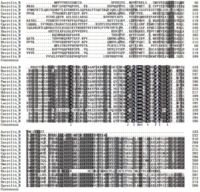
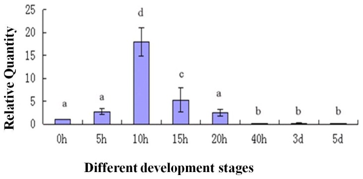
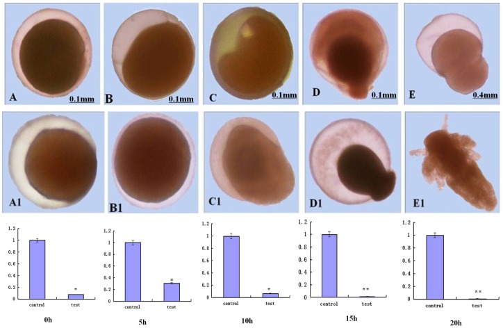
During embryonic development of Artemia sinica, environmental stresses induce the embryo diapause phenomenon, required to resist apoptosis and regulate cell cycle activity. The small ubiquitin-related modifier-1 (SUMO), a reversible post-translational protein modifier, plays an important role in embryo development. SUMO regulates multiple cellular processes, including development and other biological processes. The molecular mechanism of diapause, diapause termination and the role of As-sumo-1 in this processes and in early embryo development of Artemia sinica still remains unknown. In this study, the complete cDNA sequences of the sumo-1 homolog, sumo ligase homolog, caspase-1 homolog and cyclin B homolog from Artemia sinica were cloned. The mRNA expression patterns of As-sumo-1, sumo ligase, caspase-1, cyclin B and the location of As-sumo-1 were investigated. SUMO-1, p53, Mdm2, Caspase-1, Cyclin B and Cyclin E proteins were analyzed during different developmental stages of the embryo of A. sinica. Small interfering RNA (siRNA) was used to verify the function of sumo-1 in A. sinica. The full-length cDNA of As-sumo-1 was 476 bp, encoding a 92 amino acid protein. The As-caspases-1 cDNA was 966 bp, encoding a 245 amino-acid protein. The As-sumo ligase cDNA was 1556 bp encoding, a 343 amino acid protein, and the cyclin B cDNA was 739 bp, encoding a 133 amino acid protein. The expressions of As-sumo-1, As-caspase-1 and As-cyclin B were highest at the 10 h stage of embryonic development, and As-sumo ligase showed its highest expression at 0 h. The expression of As-SUMO-1 showed no tissue or organ specificity. Western blotting showed high expression of As-SUMO-1, p53, Mdm2, Caspase-1, Cyclin B and Cyclin E at the 10 h stage. The siRNA caused abnormal development of the embryo, with increased malformation and mortality. As-SUMO-1 is a crucial regulation and modification protein resumption of embryonic diapause and early embryo development of A. sinica.


Conflict of interest statement

Competing Interests: The authors have declared that no competing interests exist.

Figures

Figure 1
Figure 1. Nucleotide and deduced amino acid sequences of As-sumo-1 and putative protein domain.
(A) Sequence analysis of the cDNA and predicted peptide sequences of As-sumo-1. The start codon is indicated in yellow; the stop codon is indicated in green. The region defined by a straight purple line is the UBQ domain. (B) Result of domain analysis of putative As-SUMO-1 protein. The mature putative protein includes a UBQ domain.
Figure 2
Figure 2. Multiple sequence alignment of the As-SUMO-1 protein.
Sequence alignment of known sumo-1 isoforms from 20 species. Black shading indicates identical amino-acid residues. Gray shading indicates less conserved residues. Pale gray shading indicates somewhat similar residues. The sequences and the accession numbers of sumo-1 are as follows: Assumo-1, Artemia sinica, AFH36133.1; Afsumo-1, Artemia franciscana, ABQ41279.1; Cfsumo-1, Coptotermes formosanus AGM32544.1; Amsumo-1, Apis mellifera XP_392826.1; Btsumo-1, Bombus terrestris sumo-1, XP_003394722.1; Nvsumo-1, Nasonia vitripennis XP_001607301.1; Lvsumo-1, Litopenaeus vannamei, ACR56783.1; Pmsumo-1, Penaeus monodon sumo-1, ACD13593.1; Ccsumo-1, Caligus clemensi, ACO15528.1; Lssumo-1, Lepeophtheirus salmonis, ACO12186.1; Cesumo-1, Caenorhabditis elegans, NP_490842.1; Llsumo-1, Loa loa, XP_003137066.1; Bmsumo-1, Brugia malayi, XP_001900504.1; Onsumo-1, Oreochromis niloticus, XP_003446582.1; Trsumo-1, Takifugu rubripes sumo-1, XP_003966620.1; Ggsumo-1, Gallus gallus,NP_989466.1; Acsumo-1, Anolis carolinensis XP_003223593.1; Lasumo-1, Loxodonta africana, XP_003406178.1; Xlsumo-1, Xenopus laevis, NP_001083717.1; Clsumo-1, Canis lupus familiaris, NP_001239192.1; Hssumo-1, Homo sapiens sumo-1, NP_003343.1. The ubiquitin domain is boxed in red and the C-terminal double Gly-Gly residues for conjugation are boxed in blue. The sumoylation consensus ψ KXE/D motif is boxed in green. In ψ KXE/D,‘ ψ’ represents a hydrophobic amino acid residue and ‘ X ’ any residue.
Figure 3
Figure 3. The neighbor-joining phylogenetic analysis of SUMO-1 protein.
Phylogenetic tree of aligned amino-acid sequences of sumo-1 from Artemia sinica and 20 other species. A neighbor-joining phylogenetic tree was constructed using sumo-1 sequences from A. sinica and 20 other sequences from GenBank, using the sequence analysis tool MEGA 4.1. The sequences and their accession numbers are indicated in the legend of Fig. 2. A circle (•) indicates sumo-1 from A. sinica.
Figure 4
Figure 4. Nucleotide and deduced amino acid sequences of As-caspase-1 and putative protein domain.
A. Sequence analysis of the cDNA and predicted peptide sequences of As-caspase-1. The start codon is indicated in purple; the stop codon is indicated in green. The region defined by a wavy red line is the CASc domain. B. Result of domain analysis of putative As-Caspase-1 protein. The mature putative protein includes a CASc domain.
Figure 5
Figure 5. Multiple sequence alignment of the As-Caspase-1 protein.
Sequence alignment of known caspase-1 isoforms from 14 species. Black shading indicates identical amino-acid residues. Gray shading indicates less conserved residues. Pale gray shading indicates somewhat similar residues. The sequences and the accession numbers of caspase-1 are as follows: Ascaspase-1,Artemia sinica,AGB84766.1; Mdcaspase-1, Musca domestica, ACF71490.1; Cqcaspase-1, Culex quinquefasciatus, XP_001842236.1; Hmcaspase-1, Heliconius melpomene, ACU11588.1; Dpcaspase-1, Danaus plexippus, EHJ68333.1; Gmcaspase-1, Galleria mellonella, AEH76885.1; Slcaspase-1, Spodoptera litura, BAM62940.1; Bicaspase-1, Bombus impatiens, XP_003487262.1; Cgcaspase-1, Crassostrea gigas, AEB54801.1; Dlcaspase-1, Dicentrarchus labrax, ABB05054.1; Rncaspase-1, Rattus norvegicus, NP_036894.2; Ggcaspase-1, Gallus gallus, AAC69917.1; Sscaspase-1, Sus scrofa, NP_999327.1; Sacaspase-1, Sparus aurata, CAM32183.1;Hacaspase-1, Helicoverpa armigera, ABO93468.1; Cicaspase-1, Ciona intestinalis, XP_002129655.1; Bmcaspase-1, Bombyx mori, NP_001037050.1.
Figure 6
Figure 6. The neighbor-joining phylogenetic analysis of Caspase-1 protein.
Phylogenetic tree of aligned amino acid sequences of caspase-1 from A. sinica and 14 other species. A neighbor-joining phylogenetic tree was constructed using MEGA4.0, based on the sequences from A. sinica (this study) and 14 other species from GenBank. The sequences and their caspase-1 accession numbers are shown in Fig. 5. A red circle (•) indicates As-caspase-1 from A. sinica.
Figure 7
Figure 7. Nucleotide and deduced amino acid sequences of As-sumo ligase and putative protein domain.
Sequence analysis of the cDNA and predicted peptide sequences of As-sumo ligase. The start codon is indicated in blue; the stop codon is indicated in green. The region defined by a straight red line shows the zf-MIZ domain. The zf-Nse domain is indicated in yellow. B. Result of domain analysis of putative As-sumo ligase protein. The mature putative protein includes a zf-MIZ domain and a zf-Nse.
Figure 8
Figure 8. Multiple sequence alignment of the As-Sumo ligase protein.
Sequence alignment of known sumo ligase isoforms from 15 species. Black shading indicates identical amino-acid residues. Gray shading indicates less conserved residues. Pale gray shading indicates somewhat similar residues. The sequences and the accession numbers of sumo ligase are as follows: Assumo ligase, Artemia sinica, AGO51518.1; Tcsumo ligase, Tribolium castaneum, XP_974023.2; Hssumo ligase, Harpegnathos saltator, EFN82639.1; Nvsumo ligase, Nasonia vitripennis, XP_003428228.1; Bmsumo ligase, Bombyx mori, XP_004922442.1; Bisumo ligase, Bombus impatiens, XP_003485893.1; Mrsumo ligase, Megachile rotundata, XP_003706512.1; Aasumo ligase, Aedes aegypti, XP_003706512.1; Drsumo ligase, Danio rerio, XP_692921.2; Clsumo ligase, Columba livia, EMC81308.1; Spsumo ligase, Strongylocentrotus purpuratus, XP_783836.3; Cgsumo ligase, Crassostrea gigas, EKC30788.1; Olsumo ligase, Oryzias latipes, XP_004067009.1; Hgsumo ligase, Heterocephalus glaber, XP_004855644.1; Opsumo ligase, Ochotona princeps, XP_004578008.1; Oosumo ligase, Orcinus orca, XP_004276281.1.
Figure 9
Figure 9. The neighbor-joining phylogenetic analysis of Sumo ligase protein.
Neighbor-joining phylogenetic tree based on the amino acid sequences of As-sumo ligase and 15 other species from GenBank, using the sequence analysis tool MEGA 4.0. The sequences and their accession numbers are indicated in the legend of Fig. 8. A red dot indicates As-sumo ligase from A. sinica.
Figure 10
Figure 10. Nucleotide and deduced amino acid sequences of As-cyclin B and putative protein domain.
Sequence analysis of the cDNA and predicted peptide sequences of As-cyclin B. The start codon is indicated in yellow; the stop codon is indicated in red; The region indicated in green shows the cyclin_N domain. B. Result of domain analysis of putative As-cyclin B protein. The mature putative protein includes a cyclin_N domain.
Figure 11
Figure 11. Multiple sequence alignment of the As-Cyclin B protein.
Sequence alignment of known cyclin B isoforms from 15 species. Black shading indicates identical amino-acid residues. Gray shading indicates less conserved residues. Pale gray shading indicates somewhat similar residues The sequences and the accession numbers of cyclin B are as follows: Ascyclin B, Artemia sinica, KF149987.1; Bocyclin B, Bombina orientalis, ACJ12072.1; Cicyclin B, Ciona intestinalis, XP_002126215.2;Sscyclin B, Sus scrofa, NP_001107754.1; Pmcyclin B, Penaeus monodon, ACH72068.1; Ggcyclin B, Gallus gallus, NP_001004369.1; CgcyclinB, Crassostrea gigas, EKC39097.1; Hdcyclin B, Haliotis diversicolor supertexta, ADP06655.1; Skcyclin B, Saccoglossus kowalevskii, NP_001158480.1; Omcyclin B, Oncorhynchus mykiss, NP_001118131.1; Lacyclin B, Loxodonta Africana, XP_003418445.1; Btcyclin B, Bos Taurus, AAX46547.1; Cpcyclin B, Cryptomonas paramecium, XM_003239530.1; Cacyclin B, Carassius auratus, EU333815.1; Drcyclin B, Danio rerio, AF268043.1; Spcyclin B, Scylla paramamosain, FJ595022.1.
Figure 12
Figure 12. The neighbor-joining phylogenetic analysis of Cyclin B protein.
Phylogenetic tree of aligned amino-acid sequences of cyclin B from Artemia sinica and 14 other species. A neighbor-joining phylogenetic tree was constructed using cyclin B sequences from A. sinica and 15 other sequences in GenBank, using the sequence analysis tool MEGA 4.1. The sequences and their accession numbers are indicated in the legend of Fig. 11. Circle (•) indicates cyclin B from A. sinica.
Figure 13
Figure 13. Real-time quantitative PCR analysis of As-sumo-1 expression during different stages of Artemia sinica development.
The expression of sumo-1 was measured at various time points during development. The x-axis indicates the developmental stage (0 h–5 d); the y-axis indicates the expression level relative to expression at 0 h. Data are the means ± SD of triplicate experiments. Significant differences between developmental stages (P<0.05) were analyzed by one-way analysis of variance (ANOVA) and are indicated with letters (a, b, c and d).
Figure 14
Figure 14. Real-time quantitative PCR analysis of As-caspase-1 expression during different stages of Artemia sinica development.
The expression of As-caspase-1 was measured at various time points during development. The x-axis indicates the developmental stage (0 h–5 d); the y-axis indicates the expression level relative to the expression level at 0 h. Data are the means ± SD of triplicate experiments. Significant differences between developmental stages (P<0.05) were analyzed by one-way analysis of variance (ANOVA) and are indicated with letters (a, b, c and d).
Figure 15
Figure 15. Real-time quantitative PCR analysis of As-sumo ligase expression during different stages of Artemia sinica development.
The expression of As-sumo ligase was measured at various time points during development. The x-axis indicates the developmental stage (0 h–5 d); the y-axis indicates the expression level relative to the expression level at 0 h. Data are the means ± SD of triplicate experiments. Significant differences between developmental stages (P<0.05) were analyzed by one-way analysis of variance (ANOVA) and are indicated with letters (a, b, c and d).
Figure 16
Figure 16. Real-time quantitative PCR analysis of As-cyclin B expression during different stages of Artemia sinica development.
The expression of As-cyclin B was measured at various time points during development. The x-axis indicates the developmental stage (0 h–5 d); the y-axis indicates the expression level relative to the expression level at 0 h. Data are the means ± SD of triplicate experiments. Significant differences between developmental stages (P<0.05) were analyzed by one-way analysis of variance (ANOVA) and are indicated with letters (a, b, c and d).
Figure 17
Figure 17. The results of prokaryotic expression of As-SUMO-1 like protein.
(A) Expression of Artemia sinica As-SUMO-1 recombinant protein. M: protein markers from 12–100 kDa. Lanes 1–4 show the expression of As-SUMO-1 recombinant protein from four induction treatments (1 mM IPTG at 37°C, 1 mM IPTG at 30°C, 0.25 mM IPTG at 37°C, and 0.25 mM IPTG at 30°C, respectively). The arrow shows the position of the expressed recombinant protein. Lane 5: total proteins from non-induced cells. Lane 6: total proteins from induced cells harboring pET-30a (control). (B) Detection of soluble Artemia sinica As-SUMO-1 recombinant protein. Lane 1: total proteins from induced cells harboring pET-30a-sumo-1. Lane 2: soluble fraction of the lysate from induced cells harboring pET-30a-sumo-1. Lane 3: insoluble fraction of the lysate from induced cells harboring pET-30a-sumo-1. (C) Purification of recombinant Artemia sinica As-SUMO-1 protein. M: protein markers from 12–100 kDa. Lane 1: total proteins extracted from induced cells harboring pET-30a-sumo-1. Lane 2: flow-through eluate of total proteins. lanes 3–8: column elution with elutant containing 20 mM, 40 mM, 60 mM, 80 mM,100 mM and 300 mM imidazole, respectively. (D) Detection of the His-tag in the purified protein. M: protein markers from 14–100 kDa. Lane 1: total proteins from induced cells harboring pET-30a-SUMO-1. Lane 2: purified recombinant pET-30a-SUMO-1 protein. (E) Western blot showing specific binding of the antibody to the purified protein.
Figure 18
Figure 18. Western blot analysis of As-SUMO-1, As-Caspase-1, As-Mdm2, As-p53, As-Cyclin E, As-Cyclin B.
(A) Western blot showing the expression of As-SUMO-1, As-Caspase-1, As-Mdm2, As-p53, As-Cyclin E, As-Cyclin B protein at different developmental stages in A. sinica. The band intensities for these proteins were normalized against the GAPDH protein. (B) Values are expressed as arbitrary units of relative value. The expression of these proteins at 0 h was used as the reference, and asterisks indicate statistically significant differences.
Figure 19
Figure 19. Immunohistochemical analysis of the expression of As-SUMO-1 at different developmental stages in Artemia sinica.
A–H represent experimental groups and A 1-H 1 represent the control groups. (A) 0 h, gastrula stage of Artemia cysts; (B, C, and D) 5 h, 10 h, 15 h, embryonic stage; (E and F) 20 h and 40 h, nauplius stage; (G) 3 d, metanauplius larval stage; (H) 5 d, pseudoadult stage.
Figure 20
Figure 20. The relative level of sumo-1 mRNA expression in larvae soaked with dsRNAs for different times.
sumo-1-RNAi depleted expression of As-sumo-1 at different developmental stages from 0 h to 20 h in Artemia sinica. A–E represent experimental groups treated with sumo-1-RNAi and A1-E1 represent the control groups.

Similar articles

Cited by

References

    1. Jiang LJ, Hou L, Zou HL, Zhang XY, Wang RF, et al. (2007) Cloning and expression analysis of p26 gene in Artemia sinica. Acta Biochem Biophys Sin 39: 351–358. - PubMed
    1. Conte FP (2007) Structure and function of the crustacean larval salt gland. Int Rev Cytol 91: 46–106.
    1. Capili AD, Lima CD (2007) Taking it step by step: mechanistic insights from structural studies of ubiquitin/ubiquitin-like protein modification pathways. Current opinion in structural biology 17: 726–735. - PMC - PubMed
    1. Bayer P, Arndt A, Metzger S, Mahajan R, et al. (1998) Structure determination of the small ubiquitin-related modifier SUMO-1. J. Mol. Biol 280: 275–286. - PubMed
    1. Rohit M, Christian D, Tinglu G, et al. (2007) A Small Ubiquitin-Related Polypeptide Involved in Targeting RanGAP1 to Nuclear Pore Complex Protein RanBP2. Cell 88: 98–107. - PubMed

Publication types

MeSH terms