. 2014 Jan 10;16(1):R7.
doi: 10.1186/ar4433.

Characterization of T cell phenotype and function in a double transgenic (collagen-specific TCR/HLA-DR1) humanized model of arthritis

Characterization of T cell phenotype and function in a double transgenic (collagen-specific TCR/HLA-DR1) humanized model of arthritis

Bo Tang et al. Arthritis Res Ther. .

Abstract

Introduction: T cells orchestrate joint inflammation in rheumatoid arthritis (RA), yet they are difficult to study due to the small numbers of antigen-specific cells. The goal of this study was to characterize a new humanized model of autoimmune arthritis and to describe the phenotypic and functional changes that occur in autoimmune T cells following the induction of pathological events.

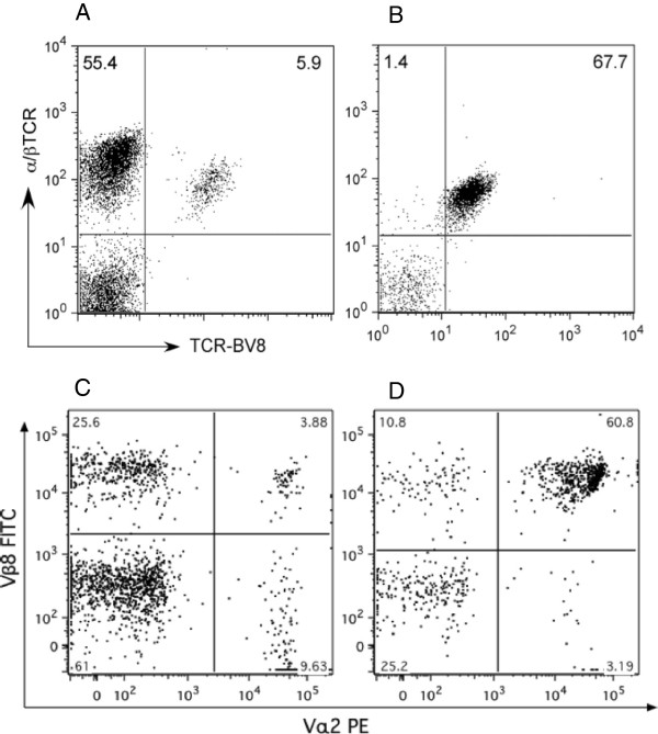
Methods: We developed a double transgenic mouse containing both the HLA-DR1 transgene and an HLA-DR1-restricted collagen-specific TCR in order to obtain large numbers of antigen-specific T cells that can be used for immunologic studies.

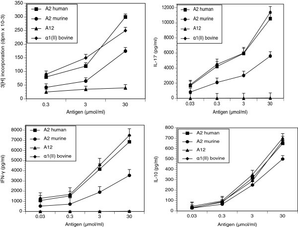
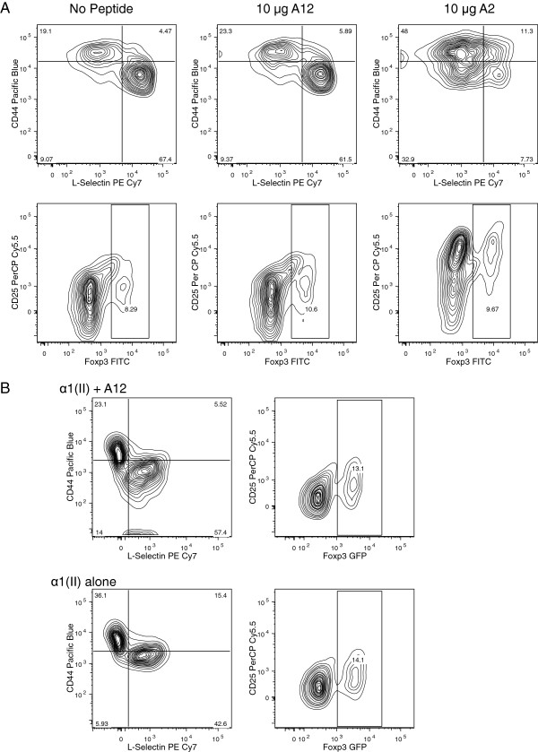
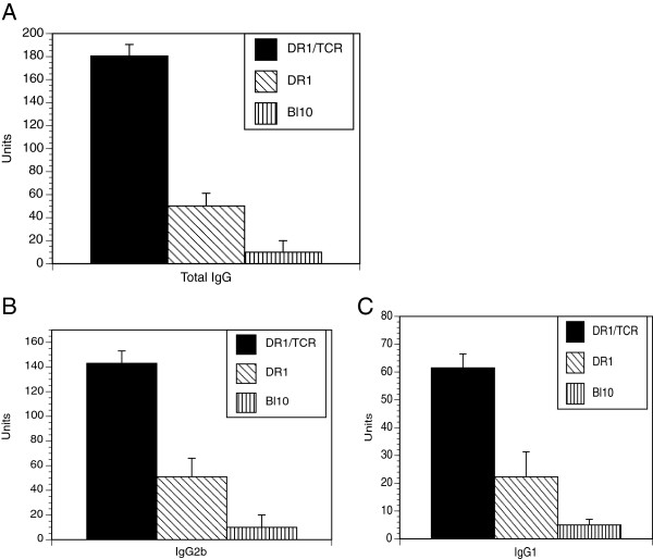
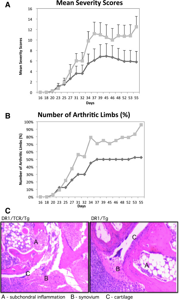
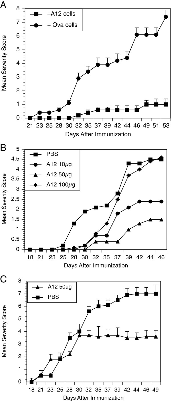
Results: In vitro, CII-specific T cells from this mouse proliferated vigorously in response to the CII immunodominant peptide A2 and the cells altered their phenotype to become predominately CD62Llow and CD44high "activated" T cells. The response was accompanied by the production of Th1, Th2, and Th17-type cytokines. Following immunization with bovine CII/CFA, these mice develop an accelerated arthritis compared to single transgenic HLA-DR1 mice. On the other hand, when the mice were treated orally with the analog peptide A12, (a suppressive analog of collagen we have previously described), arthritis was significantly suppressed, despite the fact that >90% of the CD4+ T cells express the TCR Tg. In GALT tissues taken from the A12-treated mice, IL-2, IFN-γ, and IL-17 production to the autoimmune collagen determinant dropped while high levels of IL-10 and IL-4 were produced.

Conclusions: We have developed a humanized model of autoimmune arthritis that will be useful for the study of T cell directed therapies as well as T cell mediated mechanisms of autoimmune diseases.


Figures

Figure 1
Figure 1
Development of a double-transgenic DR1-T cell receptor (TCR) Tg mouse model of autoimmune arthritis. The double-transgenic DR1-TCR Tg mouse model was developed and backcrossed onto DR1 transgenic mice as described in Methods. To detect the presence of the transgene, spleen cells were stained with a combination of anti-Vβ8- fluorescein isothiocyanate (FITC) and anti- TCR-phycoerythrin (PE). (A) Representative data obtained from the single-transgenic DR1 mouse; (B) representative data obtained from the double-transgenic mouse. In a similar manner, peripheral blood cells were obtained from the single-transgenic DR1 mouse (C) and compared to cells from the double-transgenic mouse (D), staining with antibodies specific for CD4+, Vβ8.1 and Vα2. The majority of the CD4+ cells in the double-transgenic mouse express both Vβ8.1 and Vα2. Data are based on the analysis of 10,000 gated events with the gate set on forward versus side scatter to exclude non-lymphoid cells and dead cells.
Figure 2
Figure 2
Naive spleen cells from DR1-T cell receptor (TCR) Tg mice respond to culture with type II collagen (CII). Spleen cells from naive DR1-TCR Tg mice were cultured with human A2, murine A2, A12 or bovine α1(II) chains with titrated doses. Cytokines are expressed as pg/ml. Proliferation was measured by incorporation of (3H)-thymidine and is expressed as the mean disintegrations per minute (dpm) of triplicate cultures. Data are expressed as means ± standard error of the mean of experiments using three separate mice. Responses to the murine determinant differ significantly from those induced to either human A2 or bovine α1(II) when comparing proliferation, IFN-γ, IL-17, or IL-10 (P ≤0.05 using the Mann–Whitney test). Responses to A12 differ significantly from the responses to human A2, α 1(II) and murine A2 in proliferation, IFN- γ and IL-17 (P ≤0.05 using the Mann–Whitney test) but the A12-induced IL-10 response was not different.
Figure 3
Figure 3
Phenotype of splenocytes from double-transgenic mice cultured with peptides. (A) CD4+ splenocytes from double-transgenic mice cultured with peptide A2, developed a CD62Lhi CD62Llo memory phenotype, whereas CD4+ cultured with A12 peptide or media alone did not change (%CD44hiCD62LloCD4+ T cells = 18 ± 3 for No Ag, 23 ± 5 for A12, and 47 ± 6 for A2; P ≤0.002 for A2 versus No Ag and P ≤0.006 for A2 versus A12). Splenocytes from the double-transgenic mouse cultured with peptide A2 or A12 had no significant differences in numbers of Treg cells (%CD25hiFoxp3hiCD4+ T cells = 8 ± 2 for No Ag, 9 ± 2 for A12, and 9 ± 3 for A2). (B) Phenotype of splenocytes from double-transgenic mice tested directly ex vivo: α1(II) was administered in vivo (100 μg/mouse intravenously) alone, or with 100 μg peptide A12. Three days later, CD4+ T cells treated with α1(II) developed a CD62Lhi CD62Llo memory phenotype, whereas A12 peptide significantly decreased expression of activation markers (%CD44hiCD62LloCD4+ T cells = 22 ± 7 for A12 + α1(II)-treated mice, and 37 ± 6 for α1(II)-treated mice; P ≤0.02 for α1(II)-treated versus α1(II) + A12 treated). Staining for intracellular Foxp3 was not different between groups tested directly ex vivo (15 ± 7 for α1(II)-treated mice versus 13 ± 5 for α1(II) + A12-treated mice. Negative control (naïve double-transgenic T cells) had the pattern: (%CD44hiCD62LloCD4+ T cells = 16 ± 4 and Foxp3 + CD4 T cells = 12 ± 5). Data representative of three separate experiments (A and B).
Figure 4
Figure 4
Production of antibody (Ab) specific for murine type II collagen (mCII) by double DR1-T cell receptor (TCR) Tg and DR1 Tg mice. Double- and single-transgenic littermates were immunized with 100 μg of bCII emulsified in CFA (Complete Freund’s Adjuvant) as described in Methods. Serum samples were collected 27 days after immunization and analyzed for quantity of mCII-specific Ab by ELISA. Units of Ab were calculated using standard reference sera. Data are expressed as mean titers of 10 mice per group ± standard error of the mean (total IgG titers, double-transgenic versus single-transgenic, 180.5 ± 34 versus 50.2 ± 11, P = 0.001; IgG1 titers, double-transgenic versus single-transgenic: 61.5 ± 22.3 versus 12.3 ± 6.1, P = 0.007; IgG2b titers 143.45.9 versus 51.3 ± 16.6, P = 0.0008; IgG3 titers 13.1 ± 5.6 versus 14.2 ± 3.2, P = 0.69).
Figure 5
Figure 5
Disease severity in DR1-T cell receptor (TCR) Tg mice (panels A and B). The severity (panel A) and percentage of arthritic limbs (panel B) of disease in double-transgenic mice (squares, n = 13) compared with DR1 Tg littermate controls (diamonds, n = 15). All animals were challenged with a dose of 100 μg of bovine type II collagen (bCII) emulsified in CFA (Complete Freund’s Adjuvant) for the induction of disease. Mice were scored for arthritis severity as described in Methods. Panel A, P = 0.009 on day 55 when comparing the mean severity scores ± standard error of the mean using the Mann–Whitney test; panel B, P = 0.002 on day 55 when comparing the number of arthritic limbs using Fischer’s exact test. The final incidence was 70% for the single-transgenic mice and 90% for the double-transgenic mice. Histology of joints of arthritic animals (panel C): left, joint from double-transgenic animal showing more eroding pannus and greater amounts of damaged articular cartilage compared to a representative joint (right), from a single-transgenic animal. The animals were sacrificed eight weeks after immunization. Joints were collected, fixed with formaldehyde, and decalcified prior to staining with H&E. The tissues were photographed using an inverted-phase contrast microscope (original magnification: 50×). The data shown are representative of data obtained by analyzing numerous sections from hindpaws taken from 6 animals per group.
Figure 6
Figure 6
Treatment with either A12-immune cells or A12 peptide can suppress arthritis. Panel A: CD4+ T cells from the double transgenic mice treated with A12 can transfer the suppression of arthritis. Single-transgenic DR1 mice (10/group) were infused with 5 × 10⁵ cells of A12-primed CD4+ T cells from double-transgenic mice or Ova-primed CD4+ T cells from single-transgenic DR1 mice. Recipient animals were immunized with bovine type II collagen (bCII)/CFA and observed for arthritis. Only A12-immune cells prevented collagen-induced arthritis (final severity scores 1.0 ± 0.5 versus 7.4 ± 1.2, P ≤0.0001; final incidence 90% versus 10% in the treatment group). (Panel B) Oral treatment with peptide A12 (prevention protocol). Groups of 12 double-transgenic mice were administered PBS or A12 peptide (oral gavage three times/week), beginning the day after immunization with bCII/CFA, continuing for the duration of the experiment. On day 46, mice fed PBS had a severity score of 4.5 ± 0.4, which differed from mice fed 10 μg of A12 (2.4 ± 0.5, P ≤0.05) and mice fed 50 μg of A12 (1.5 ± 0.04, P ≤0.01; final incidence = 80% in the control and 20% in the 50-μg group). (Panel C) Oral treatment with peptide A12 (treatment protocol). Groups of 10 double-transgenic mice were administered PBS or A12 peptide (50 μg/dose, oral gavage three times/week) beginning the day of arthritis onset. On day 46, mice fed PBS had a severity score of 6.8 ± 0.6, which differed from mice fed 50 μg of A12 (3.6 ± 0.4, P ≤0.05) (Mann–Whitney test). (Final incidence = 90% in controls and 60% in the treatment group).

Similar articles

Cited by

References

    1. Meyer AL, Trollmo C, Crawford F, Marrack P, Steere AC, Huber BT, Kappler J, Hafler DA. Direct enumeration of Borrelia-reactive CD4 T cells ex vivo by using MHC class II tetramers. Proc Natl Acad Sci USA. 2000;16:11433–11438. doi: 10.1073/pnas.190335897. - DOI - PMC - PubMed
    1. Oh S, Aitken M, Simons DM, Basehoar A, Garcia V, Kropf E, Caton AJ. Requirement for diverse TCR specificities determines regulatory T cell activity in a mouse model of autoimmune arthritis. J Immunol. 2012;16:4171–4180. doi: 10.4049/jimmunol.1103598. - DOI - PMC - PubMed
    1. Rosloniec EF, Cremer M, Kang AH, Myers LK, Brand DD. Collagen-induced arthritis. Curr Protoc Immunol. 2010;16:11–25. - PubMed
    1. Rosloniec EF, Brand DD, Myers LK, Whittington KB, Gumanovskaya M, Zaller DM, Woods A, Altmann DM, Stuart JM, Kang AH. An HLA-DR1 transgene confers susceptibility to collagen-induced arthritis elicited with human type II collagen. J Exp Med. 1997;16:1113–1122. doi: 10.1084/jem.185.6.1113. - DOI - PMC - PubMed
    1. Kouskoff V, Signorelli K, Benoist C, Mathis D. Cassette vectors directing expression of T cell receptor genes in transgenic mice. J Immunol Methods. 1995;16:273–280. doi: 10.1016/0022-1759(95)00002-R. - DOI - PubMed

Publication types