. 2014 Jan 10:11:6.
doi: 10.1186/1742-2094-11-6.

Docosahexaenoic acid attenuates the early inflammatory response following spinal cord injury in mice: in-vivo and in-vitro studies

Affiliations

Docosahexaenoic acid attenuates the early inflammatory response following spinal cord injury in mice: in-vivo and in-vitro studies

Irene Paterniti et al. J Neuroinflammation. .

Abstract

Background: Two families of polyunsaturated fatty acid (PUFA), omega-3 (ω-3) and omega-6 (ω-6), are required for physiological functions. The long chain ω-3 PUFAs, eicosapentaenoic acid (EPA) and docosahexaenoic acid (DHA), have significant biological effects. In particular, DHA is a major component of cell membranes in the brain. It is also involved in neurotransmission. Spinal cord injury (SCI) is a highly devastating pathology that can lead to catastrophic dysfunction, with a significant reduction in the quality of life. Previous studies have shown that EPA and DHA can exert neuroprotective effects in SCI in mice and rats. The aim of this study was to analyze the mechanism of action of ω-3 PUFAs, such as DHA, in a mouse model of SCI, with a focus on the early pathophysiological processes.

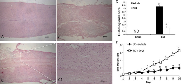
Methods: In this study, SCI was induced in mice by the application of an aneurysm clip onto the dura mater via a four-level T5 to T8 laminectomy. Thirty minutes after compression, animals received a tail vein injection of DHA at a dose of 250 nmol/kg. All animals were killed at 24 h after SCI, to evaluate various parameters implicated in the spread of the injury.

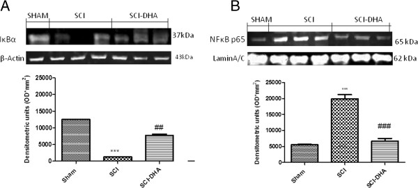
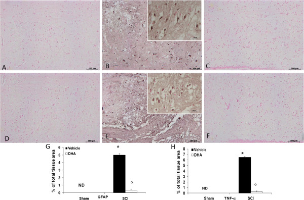
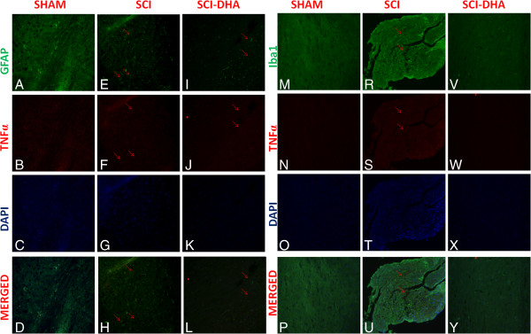
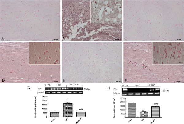
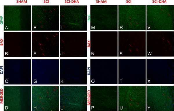
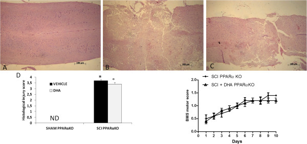
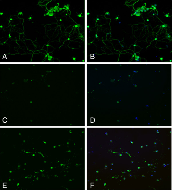
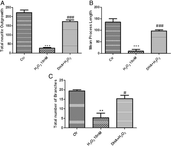
Results: Our results in this in-vivo study clearly demonstrate that DHA treatment reduces key factors associated with spinal cord trauma. Treatment with DHA significantly reduced: (1) the degree of spinal cord inflammation and tissue injury, (2) pro-inflammatory cytokine expression (TNF-α), (3) nitrotyrosine formation, (4) glial fibrillary acidic protein (GFAP) expression, and (5) apoptosis (Fas-L, Bax, and Bcl-2 expression). Moreover, DHA significantly improved the recovery of limb function.Furthermore, in this study we evaluated the effect of oxidative stress on dorsal root ganglion (DRG) cells using a well-characterized in-vitro model. Treatment with DHA ameliorated the effects of oxidative stress on neurite length and branching.

Conclusions: Our results, in vivo and in vitro, clearly demonstrate that DHA treatment reduces the development of inflammation and tissue injury associated with spinal cord trauma.


Figures

Figure 1
Figure 1
Effect of DHA treatment on histological alterations of the spinal cord tissue 24 h after injury and on hind limb motor disturbance. No histological alterations have been found in the spinal cord tissue collected from sham-operated mice (A, see histological score D). A significant damage to the spinal cord was assessed in SCI-operated mice stained with H & E (B see histological score D). The treatment with DHA resulted in a significant decrease in the extent and severity of the histological signs (C, see histological score D). Moreover, the degree of motor disturbance was assessed every day until 10 days after SCI by Basso mouse scale (BMS) open-field score. Treatment with DHA administered 30 min after trauma reduces the motor disturbance after SCI (E). This figure is representative of at least three experiments performed on different experimental days. ND: not detectable. Values shown are mean ± SEM of ten mice for each group; *P < 0.01 vs sham; °P < 0.01 vs SCI.
Figure 2
Figure 2
Effects of DHA treatment on IκBα and nuclear NF-κB p65. By Western blot analysis, a basal level of IκB-α was detected in the spinal cord from sham-operated animals (A), whereas IκB-α levels were substantially reduced in SCI mice (A). DHA treatment reduced the SCI-induced IκB-α degradation (A). In addition, SCI caused a significant increase in nuclear NF-κB p65 compared with sham-operated mice (B). DHA treatment significantly reduced the phosphorylation of p65 on Ser536 and the translocation into the nucleus of NF-κB p65 (B). A representative blot of lysates obtained from each group is shown, and densitometry analysis of all animals is reported. Respective densitometry analysis of protein bands from three separated experiments is reported (***P < 0.001 vs. Sham; ###P < 0.005 vs. SCI; ## P<0.05 vs.SCI).
Figure 3
Figure 3
Effects of DHA on astrocyte activation and TNF-α expression in spinal cord tissue. Spinal cord sections were processed 24 h after SCI to determine the immunohistological staining for GFAP and TNF-α expression. Sham animals never express GFAP (A, see densitometry analysis G), the number of GFAP+ cells was significantly increased upon induction of SCI, due to astrocytes proliferation around the central canal (B, see densitometry analysis G).Treatment with DHA significantly decreased the activation of astrocyte-GFAP+ cells (C, see densitometry analysis G).Moreover, no positive staining for TNF-α was found in the spinal cord tissue from sham-operated mice (D, see densitometry analysis H). A substantial increase in TNF-α expression was found in spinal cord tissues from SCI mice 24 h after SCI (E, see densitometry analysis H). DHA treatment significantly attenuated TNF-α levels in the spinal cord (F, see densitometry analysis H). (G,H) Densitometry analysis of immunohistochemistry photographs (n = 5 photos from each sample collected from all mice in each experimental group) for GFAP and TNF-α from spinal cord tissues. The figure is representative of at least three experiments performed on different experimental days. For each SCI group see the high magnification of the images. Data are expressed as percentage of total tissue area. Data are mean ± SEM of ten mice for each group.*P < 0.05 vs. Sham. °P < 0.01 vs. SCI. ND: not detectable.
Figure 4
Figure 4
Colocalization of GFAP/TNFα and Iba1/TNFα after SCI. Results are shown for (A-D, M-P) sham-operated mice, (E-H, R-U) mice with SCI and (I-L, V-Y) mice with SCI treated with DHA. Spinal cord sections were double stained with antibodies against GFAP (A,E,I, green), Iba1 (M,R,V, green) and TNFα (B,F,J,N,S,W, red). Spinal cord sections revealed increased astrogliosis (GFAP + cells) in SCI mice (E, R). GFAP immunoreactivity was reduced in DHA-treated mice (I, V). Yellow spots indicate co-localizations (H, U) and revealed a high colocalization between GFAP/TNFα and Iba1/TNFα double staining.
Figure 5
Figure 5
Effects of DHA on nitrotyrosine formation and iNOS expression. Immunohistochemical and Western blot analysis in the spinal cord section 24 h after SCI. Sections from sham-operated mice did not stain for iNOS (A, see densitometry analysis G), whereas those from SCI-operated mice exhibited staining for iNOS (B, see densitometry analysis G) mainly in various inflammatory cells in the gray matter. Treatment with DHA reduced the degree of staining for iNOS (C, see densitometry analysis G). Moreover, by Western blot analysis we observed high levels of iNOS in the SCI group (H), whereas the DHA treatment group revealed a low expression of iNOS (H). Thus, the tissue sections obtained from SCI mice demonstrate staining for nitrotyrosine mainly localized in inflammatory cells and in Schwann cell nuclei in the white and gray matter (E, see densitometry analysis I). DHA treatment reduced the degree of staining for nitrotyrosine (F, see densitometry analysis I). Conversely, spinal cord from sham-operated mice did not stain for nitrotyrosine (D, see densitometry analysis I). For each SCI group, see the high magnification of the images. (G,I) Densitometry analysis of immunocytochemistry photographs (n = 5 photos from each sample collected from all mice in each experimental group) for iNOS and nitrotyrosine. Data expressed as a percentage of total tissue area. This figure is representative of at least three experiments performed on different experimental days. Data are mean ± SEM of ten mice for each group.*P < 0.01 vs sham; °P < 0.01 vs SCI; ND: not detectable.
Figure 6
Figure 6
DHA treatment reduced Fas-ligand expression in the perilesional spinal cord tissue. No positive staining for Fas ligand was found in the spinal cord tissue collected from sham-operated mice (A, see densitometry analysis D). A substantial increase in Fas-ligand expression was found in inflammatory cells, and in white and gray matter of spinal cord tissues collected at 24 h after SCI (B, see densitometry analysis D). Spinal cord levels of Fas ligand were significantly attenuated by DHA treatment (C, see densitometry analysis D). (D) Densitometry analysis of immunocytochemistry photographs (n = 5 photos from each sample collected from all mice in each experimental group) for Fas ligand. Moreover Western blot analysis revealed an increased expression of Fas-L in the SCI group (E), whereas DHA treatment significantly reduced the increased levels of Fas-L (E). Figure is representative of at least three experiments performed on different experimental days. Data are mean ± SEM of ten mice for each group. *P < 0.01 vs Sham; °P < 0.01 vs SCI. # P<0.01 vs SCI; ND: not detectable.
Figure 7
Figure 7
Effects of DHA on intrinsic apoptotic pathway. Staining for Bax expression was absent in the sham group (A). Twenty-four hours after SCI, spinal cord tissue from injured animals showed positive staining for Bax (B). DHA treatments significantly reduced the SCI-induced Bax (C). On the contrary, positive staining for Bcl-2 was observed in the spinal cord tissues from sham-operated mice (D) while the staining was significantly reduced in SCI mice (E). DHA treatment attenuated the loss of positive staining for Bcl-2 in the spinal cord from SCI- subjected mice (F). For Bax staining in SCI groups, see the high magnification of the images; for Bcl2 staining, see high magnification for sham and DHA groups. Moreover, an increase in Bax expression was evidenced by Western blot analysis (G). DHA treatment reduced the expression for Bax in the spinal cord ganglia). A basal level of Bcl-2 expression was detected in spinal cord from sham-operated mice (H). Twenty-four hours after SCI, Bcl-2 expression was significantly reduced in spinal cord from SCI mice (H). DHA treatments significantly reduced the SCI-induced inhibition of Bcl-2 expression (H). A representative blot of lysates obtained from each group is shown, and densitometry analysis of all animals is reported. Respective densitometry analysis of protein bands from three separated experiments is reported. Data are mean ± SEM of ten mice for each group; ***P < 0.001 vs. sham; ###P < 0.005 vs. SCI. ND: not detectable.
Figure 8
Figure 8
Colocalization of GFAP/Bax and Iba1/Bax after SCI. Results are shown for (A-D, M-P) sham-operated mice, (E-H, R-U) mice with SCI and (I-L, V-Y) mice with SCI treated with DHA. Spinal cord sections were double stained with antibodies against GFAP (A,E,I, green), Iba1 (M,R,V, green) and Bax (B,F,J,N,S,W, red). Spinal cord sections revealed increased Bax expression in SCI mice (F,S). Bax expression was reduced in DHA-treated mice (J,W). Yellow spots indicate co-localizations (H,U) and revealed a high colocalization between GFAP/Bax and Iba1/Bax double staining.
Figure 9
Figure 9
Involvement of peroxisome proliferator-activated receptors on the protective actions of DHA following spinal cord trauma. Following spinal cord compression, significant damage to the spinal cord from PPARα KO mice (B) at the perilesional zone was observed by H & E staining when compared with spinal cord tissue collected from the sham group (A). The genetic absence of the PPAR-α receptor significantly blocked the effect of the DHA treatment (C). The histological score (D) was made by an independent observer. These figures are representative of at least three experiments performed on different experimental days. Data are mean ± SEM of ten mice for each group. *P < 0.01 vs sham; °P < 0.01 vs SCI.
Figure 10
Figure 10
Representative fluorescence microscopy images of DRG cultures stained with neuronal marker. DRG cells were stained with β-tubulin III to visualize the neurites. Oxidative stress induced by the administration of H2O2 significantly decreased the outgrowth of the DRG neurons in culture (C, D) compared with the control DRG cells group (A, B), whereas the treatment with DHA increased the percentage of cells with complex growth, similar to the control DRG cells (E, F). Cells were counterstained with Hoechst.
Figure 11
Figure 11
Effect of DHA on neurite outgrowth. Neurite process length and number of branches were significantly reduced in H2O2-injured cultures (A,B,C), compared with control DRG cells (A,B,C). Total outgrowth, length of longest neurite and number of branches were all markedly increased in the DRG cells treated with DHA (A,B,C). Data are mean ± SEM of ten mice for each group. ***P < 0.001 vs Ctr; ### P < 0.005 vs H2O2; ** P < 0.01 vs Ctr; # P < 0,05 vs H2O2.

References

    1. Siddall PJ, McClelland JM, Rutkowski SB, Cousins MJ. A longitudinal study of the prevalence and characteristics of pain in the first 5 years following spinal cord injury. Pain. 2003;103:249–257. doi: 10.1016/S0304-3959(02)00452-9. - DOI - PubMed
    1. Cardenas DD, Hoffman JM, Kirshblum S, McKinley W. Etiology and incidence of rehospitalization after traumatic spinal cord injury: a multicenter analysis. Arch Phys Med Rehabil. 2004;85:1757–1763. doi: 10.1016/j.apmr.2004.03.016. - DOI - PubMed
    1. Popovich PG, Wei P, Stokes BT. Cellular inflammatory response after spinal cord injury in Sprague–Dawley and Lewis rats. J Comp Neurol. 1997;377:443–464. doi: 10.1002/(SICI)1096-9861(19970120)377:3<443::AID-CNE10>3.0.CO;2-S. - DOI - PubMed
    1. Streit WJ, Graeber MB, Kreutzberg GW. Functional plasticity of microglia: a review. Glia. 1988;1:301–307. doi: 10.1002/glia.440010502. - DOI - PubMed
    1. Schwartz M, Moalem G, Leibowitz-Amit R, Cohen IR. Innate and adaptive immune responses can be beneficial for CNS repair. Trends Neurosci. 1999;22:295–299. doi: 10.1016/S0166-2236(99)01405-8. - DOI - PubMed

Publication types

MeSH terms