. 2014 Jan 10;9(1):2.
doi: 10.1186/1750-9378-9-2.

Escherichia coli-derived outer membrane vesicles are genotoxic to human enterocyte-like cells

Affiliations

Escherichia coli-derived outer membrane vesicles are genotoxic to human enterocyte-like cells

Peter C Tyrer et al. Infect Agent Cancer. .

Abstract

Background: Colorectal cancers are the third most common type in the world. The causes of the disease are poorly understood, but since the discovery of Helicobacter pylori as a causative agent of gastric cancer, attention has turned to bacteria as a possible trigger for colorectal cancer. Recently H. pylori outer membrane vesicles (OMVs) were revealed as potentially genotoxic which can be important first step in carcinogenesis. We therefore investigated whether OMVs from intestinal Escherichia coli could be genotoxic.

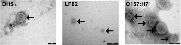
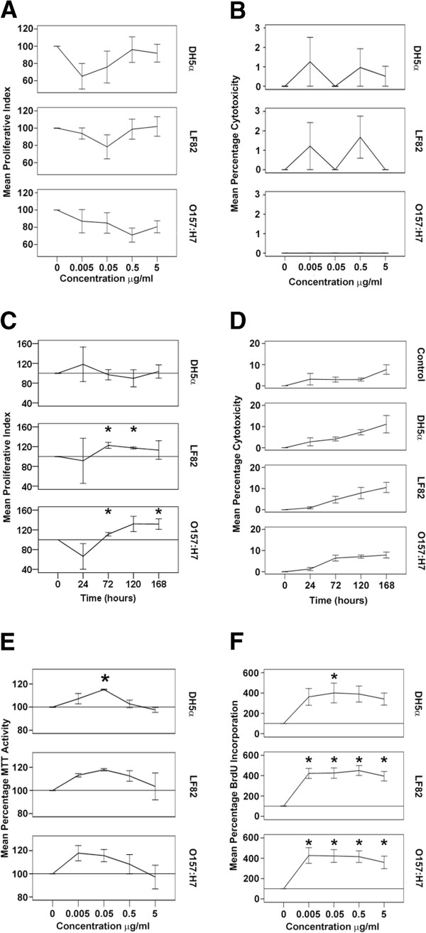
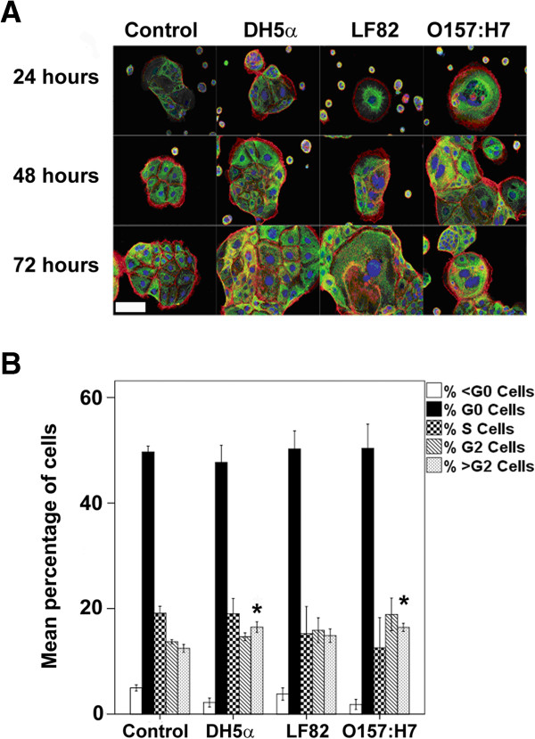
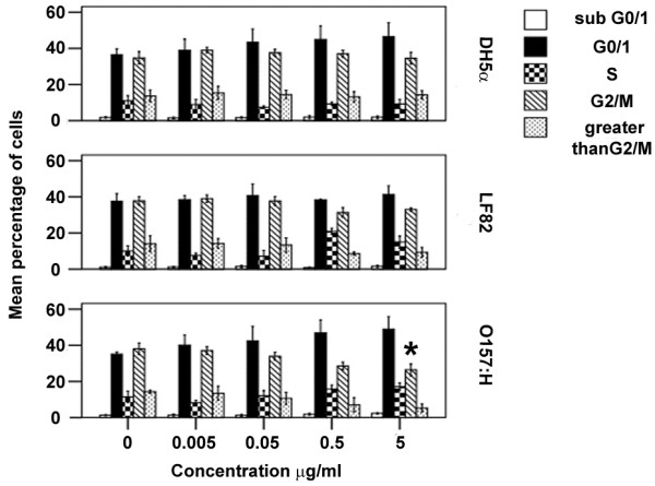
Methods: OMVs from the avirulent DH5α strain, a pathogenic adherent-invasive E. coli (AIEC) and an enterohaemolytic (EHEC) strain of E. coli were enriched by ultracentrifugation. The effect on the growth and viability of human enterocyte-like Caco-2 cells by OMVs was determined by trypan blue exclusion, MTT and BrdU incorporation assays. The ability of OMVs to induce DNA damage was assayed by single-cell gel electrophoresis, and 8-oxo-dG and γH2Ax immunofluorescence staining. Cytopathological changes were assessed by microscopy. The induction of aneuploidy by the OMVs was measured by flow cytometry in Caco-2 and LoVo cells.

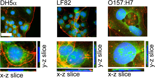
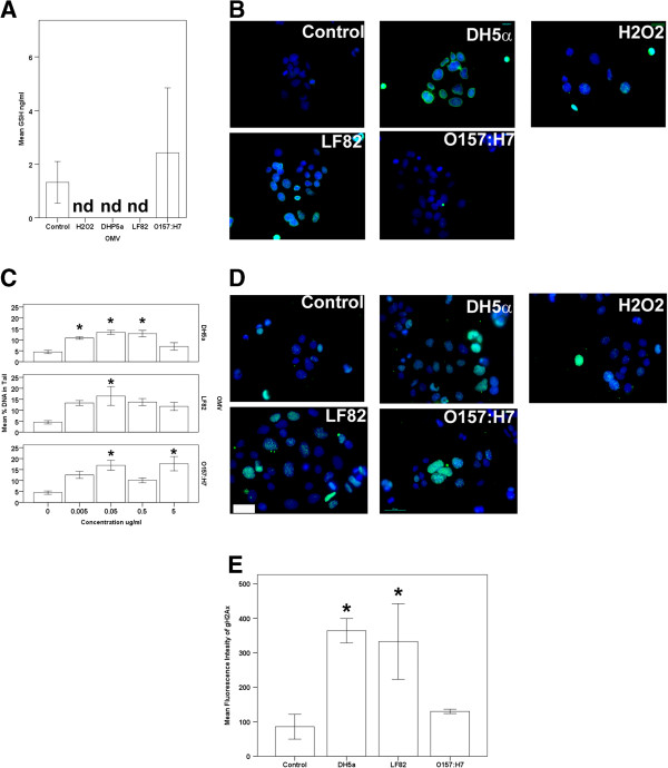
Results: We found that OMVs derived were internalised by Caco-2 cells, increased cell numbers, induced double-stranded DNA breaks, recruited γH2Ax to the nucleus, initiated DNA rereplication, and produced distended multinucleate cells. DH5α and AIEC OMVs caused free radical generation as indicated by the reduction of glutathione in cells, leading to the development of mutagenic 8-oxo-dG adducts in DNA. Flow cytometry revealed that DH5α and EHEC OMVs increased aneuploidy in p53 mutant Caco-2 cells, but not in p53 wild type LoVo cells.

Conclusion: We conclude that E. coli derived OMVs, whether from avirulent or pathogenic strains are potentially genotoxic.


Figures

Figure 1
Figure 1
Electron micrographs of preparations of outer membrane vesicles.E. coli OMVs were recovered following the overnight growth of bacteria in LB broth. OMVs are indicated by black arrows. Magnification 93 000. Size bar indicates 200 nm.
Figure 2
Figure 2
E. coli OMVs are internalised by and localise to the perinuclear regions of Caco-2 cells. Fluorescent DiO–labelled OMVs (green), were added to Caco-2 cells for 4 hours before the cells were stained for actin (red) and nuclei (blue). Image stacks were taken 0.28 μm apart. The top images represent a maximum intensity z-projection constructed in ImageJ whereas the bottom images are insets of those above with their x-z and y-z orthogonal projections to show that OMVs are internalised by Caco-2 cells. Images are from a single representative experiment. Original magnification = 40×, NA1.3 objective used. Note that the figure for O157:H7 OMV treated Caco-2 cells shows an enlarged, intoxicated nucleus.
Figure 3
Figure 3
E. coli OMVs increase Caco-2 cell proliferation. (A) Dose dependent proliferation indices and (B) percentage cytotoxicity of Caco-2 cultures after 7 days exposure to OMVs, determined by trypan blue assay. (C) Time dependent proliferation indices and (D) percentage cytotoxicity of Caco-2 cultures after 7 days exposure to OMVs, determined by trypan blue assay. (E) OMVs increase MTT reduction by Caco-2 cells. (F) OMVs increase the synthesis of DNA in Caco-2 cells as determined by BrdU incorporation assay. *, results are significantly different from untreated controls (p ≤ 0.05). ANOVA, followed by two-sided Dunnet’s post hoc test. Results are ± SEM of three independent experiments. In c, e and f, the line denotes 100% i.e. unexposed control values.
Figure 4
Figure 4
E. coli OMVs induce reactive oxygen species generation and DNA damage. (A) DH5α and LF82 derived OMVs decrease cellular GSH, indicating an increase in ROS. nd denotes not detected. (B) DH5α and LF82 derived OMVs are associated with the formation of mutagenic 8-oxo-dG adducts (green) in Caco-2 nuclei (blue). (C) OMVs induce DNA double strand breaks in Caco-2 cells as measured by Comet assay. (D) OMVs induce the recruitment of γH2Ax histone (green) to sites of damaged nuclear DNA (blue). (E) Flow cytometric analysis of γH2Ax expression during the G1 phase of the cell cycle of Cacco-2 cells. (A), (C) and (E), *, results are significantly different from untreated controls (p ≤ 0.05). ANOVA, followed by two-sided Dunnet’s post hoc test. Results are ± SEM of three independent experiments. (B) and (D), Immunofluorescence microscopy images are from a single representative experiment. Original magnification = 40×, NA1.3 objective used, scale bar indicates 50 μm.
Figure 5
Figure 5
E. coli OMVs produce morphopathogenic changes to Caco-2 cells consistent with endoreplication and can induce aneuploidy. (A) Immunofluorescence labelling of actin (red), cytokeratin 18 (green), and nuclei blue was used to show that OMVs cause endoreplication in Caco-2 cells over time. Images are from a single representative experiment. Original magnification = 20×, NA 0.50 objective used, scale bar indicates 50 μm. (B) OMVs (5 μg/ml) are associated with the generation of aneuploidy in Caco-2 cells exposed for 7 days to DH5α and O157:H7 OMVs, as measured using flow cytometry of propidium iodide stained and RNase 1 treated cells. *, results are significantly different from untreated controls (p ≤ 0.05). ANOVA, followed by two-sided Dunnet’s post hoc test. Results are ± SEM of three independent experiments.
Figure 6
Figure 6
O157:H7 E. coli induce cell cycle changes in LoVo cells. LoVo cells were exposed to OMVs for 4 days, before being subjected to cell cycle analysis. O157:H7 OMV induced a dose dependent G1 arrest. *, results are significantly different from untreated controls (p ≤ 0.05). ANOVA, followed by two-sided Dunnet’s post hoc test. Results are ± SEM of three independent experiments.

Similar articles

Cited by

References

    1. Jemal A, Smith RA, Ward E. Worldwide variations in colorectal cancer. CA Cancer J Clin. 2009;59:336–378.
    1. Frizelle FA. Colorectal cancer in New Zealand. N Z Med J. 2007;120:1–6. - PubMed
    1. Takayama T, Miyanishi K, Hayashi T, Sato Y, Niitsu Y. Colorectal cancer: genetics of development and metastasis. J Gastroenterol. 2006;41:185–192. doi: 10.1007/s00535-006-1801-6. - DOI - PubMed
    1. Buc E, Kwiatkowski F, Alves A, Panis Y, Mantion G, Slim K. Tobacco smoking: a factor of early onset of colorectal cancer. Dis Colon Rectum. 2006;49:1893–1896. doi: 10.1007/s10350-006-0704-1. - DOI - PubMed
    1. Kato I, Boleij A, Kortman GA, Roelofs R, Djuric Z, Severson RK, Tjalsma H. Partial associations of dietary iron, smoking and intestinal bacteria with colorectal cancer risk. Nutr Cancer. 2013;65:169–177. doi: 10.1080/01635581.2013.748922. - DOI - PMC - PubMed