

Giant axon formation in mice lacking Kell, XK, or Kell and XK: animal models of McLeod neuroacanthocytosis syndrome
- PMID: 24405768
- PMCID: PMC3936324
- DOI: 10.1016/j.ajpath.2013.11.013
Giant axon formation in mice lacking Kell, XK, or Kell and XK: animal models of McLeod neuroacanthocytosis syndrome
Abstract
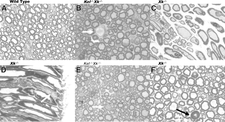
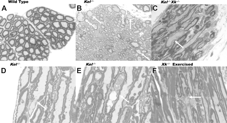
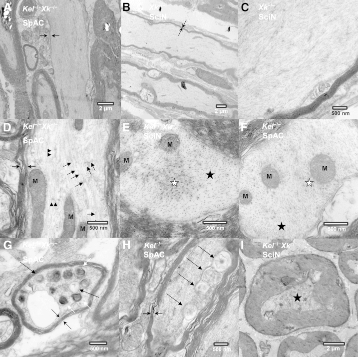
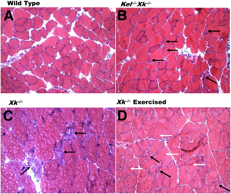
McLeod neuroacanthocytosis syndrome (MLS) is a rare X-linked multisystem disease caused by XK gene mutations and characterized by hematological and neurological abnormalities. XK, a putative membrane transporter, is expressed ubiquitously and is covalently linked to Kell, an endothelin-3-converting enzyme (ECE-3). Absence of XK results in reduction of Kell at sites where both proteins are coexpressed. To elucidate the functional roles of XK, Kell, and the XK-Kell complex associated with pathogenesis in MLS, we studied the pathology of the spinal cord, anterior roots, sciatic nerve, and skeletal muscle from knockout mouse models, using Kel(-/-), Xk(-/-), Kel(-/-)Xk(-/-), and wild-type mice aged 6 to 18 months. A striking finding was that giant axons were frequently associated with paranodal demyelination. The pathology suggests probable anterograde progression from the spinal cord to the sciatic nerve. The neuropathological abnormalities were found in all three genotypes, but were more marked in the double-knockout Kel(-/-)Xk(-/-) mice than in either Kel(-/-) or Xk(-/-) mice. Skeletal muscles from Xk(-/-) and Kel(-/-)Xk(-/-) mice showed mild abnormalities, but those from Kel(-/-) mice were similar to the wild type. The more marked neuropathological abnormalities in Kel(-/-)Xk(-/-) mice suggest a possible functional association between XK and Kell in nonerythroid tissues.
Copyright © 2014 American Society for Investigative Pathology. Published by Elsevier Inc. All rights reserved.
Figures
Similar articles
-
Changes in red cell ion transport, reduced intratumoral neovascularization, and some mild motor function abnormalities accompany targeted disruption of the Mouse Kell gene (Kel).Am J Hematol. 2009 Aug;84(8):492-8. doi: 10.1002/ajh.21453. Am J Hematol. 2009. PMID: 19544475
-
Ablation of the Kell/Xk complex alters erythrocyte divalent cation homeostasis.Blood Cells Mol Dis. 2013 Feb;50(2):80-5. doi: 10.1016/j.bcmd.2012.10.002. Epub 2012 Oct 31. Blood Cells Mol Dis. 2013. PMID: 23122227 Free PMC article.
-
Expression profiles of mouse Kell, XK, and XPLAC mRNA.J Histochem Cytochem. 2007 Apr;55(4):365-74. doi: 10.1369/jhc.6A7126.2006. Epub 2006 Dec 22. J Histochem Cytochem. 2007. PMID: 17189525
-
Kell and Kx blood group systems.Immunohematology. 2015;31(1):14-9. Immunohematology. 2015. PMID: 26308465 Review.
-
The Kell blood group system: Kell and XK membrane proteins.Semin Hematol. 2000 Apr;37(2):113-21. doi: 10.1016/s0037-1963(00)90036-2. Semin Hematol. 2000. PMID: 10791880 Review.
Cited by
-
Treatment with the Glycosphingolipid Modulator THI Rescues Myelin Integrity in the Striatum of R6/2 HD Mice.Int J Mol Sci. 2023 Mar 22;24(6):5956. doi: 10.3390/ijms24065956. Int J Mol Sci. 2023. PMID: 36983032 Free PMC article.
-
Transcriptome-based analysis of blood samples reveals elevation of DNA damage response, neutrophil degranulation, cancer and neurodegenerative pathways in Plasmodium falciparum patients.Malar J. 2021 Sep 26;20(1):383. doi: 10.1186/s12936-021-03918-5. Malar J. 2021. PMID: 34565410 Free PMC article.
-
Genetic Basis of Aerobically Supported Voluntary Exercise: Results from a Selection Experiment with House Mice.Genetics. 2020 Nov;216(3):781-804. doi: 10.1534/genetics.120.303668. Epub 2020 Sep 25. Genetics. 2020. PMID: 32978270 Free PMC article.
-
A patient with McLeod syndrome showing involvement of the central sensorimotor tracts for the legs.BMC Neurol. 2019 Nov 27;19(1):301. doi: 10.1186/s12883-019-1526-9. BMC Neurol. 2019. PMID: 31775676 Free PMC article.
-
Does aberrant membrane transport contribute to poor outcome in adult acute myeloid leukemia?Front Pharmacol. 2015 Jul 2;6:134. doi: 10.3389/fphar.2015.00134. eCollection 2015. Front Pharmacol. 2015. PMID: 26191006 Free PMC article.
References
-
- Danek A., Rubio J.P., Rampoldi L., Ho M., Dobson-Stone C., Tison F., Symmans W.A., Oechsner M., Kalckreuth W., Watt J.M., Corbett A.J., Hamdalla H.H., Marshall A.G., Sutton I., Dotti M.T., Malandrini A., Walker R.H., Daniels G., Monaco A.P. McLeod neuroacanthocytosis: genotype and phenotype. Ann Neurol. 2001;50:755–764. - PubMed
-
- Danek A., Walker R.H. Neuroacanthocytosis. Curr Opin Neurol. 2005;18:386–392. - PubMed
-
- Jung H.H., Danek A., Frey B.M. McLeod syndrome: a neurohaematological disorder. Vox Sang. 2007;93:112–121. - PubMed
-
- Hewer E., Danek A., Schoser B.G., Miranda M., Reichard R., Castiglioni C., Oechsner M., Goebel H.H., Heppner F.L., Jung H.H. McLeod myopathy revisited: more neurogenic and less benign. Brain. 2007;130:3285–3296. - PubMed
-
- Ohno N., Terada N., Yamakawa H., Komada M., Ohara O., Trapp B.D., Ohno S. Expression of protein 4.1G in Schwann cells of the peripheral nervous system. J Neurosci Res. 2006;84:568–577. - PubMed
Publication types
MeSH terms
Substances
Supplementary concepts
Grants and funding
LinkOut - more resources
Full Text Sources
Other Literature Sources
Molecular Biology Databases
