. 2013 Sep 17;4(5):114.
doi: 10.1186/scrt325.

Pleiotropic effects of cancer cells' secreted factors on human stromal (mesenchymal) stem cells

Pleiotropic effects of cancer cells' secreted factors on human stromal (mesenchymal) stem cells

Mashael Al-toub et al. Stem Cell Res Ther. .

Abstract

Introduction: Studying cancer tumors' microenvironment may reveal a novel role in driving cancer progression and metastasis. The biological interaction between stromal (mesenchymal) stem cells (MSCs) and cancer cells remains incompletely understood. Herein, we investigated the effects of tumor cells' secreted factors as represented by a panel of human cancer cell lines (breast (MCF7 and MDA-MB-231); prostate (PC-3); lung (NCI-H522); colon (HT-29) and head & neck (FaDu)) on the biological characteristics of MSCs.

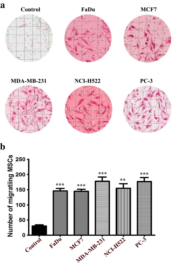
Methods: Morphological changes were assessed using fluorescence microscopy. Changes in gene expression were assessed using Agilent microarray and qRT-PCR. GeneSpring 12.1 and DAVID tools were used for bioinformatic and signaling pathway analyses. Cell migration was assessed using a transwell migration system. SB-431542, PF-573228 and PD98059 were used to inhibit transforming growth factor β (TGFβ), focal adhesion kinase (FAK), and mitogen activated protein kinase kinase (MAPKK) pathways, respectively. Interleukin-1β (IL1β) was measured using ELISA.

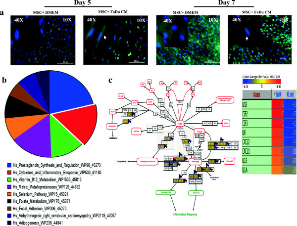
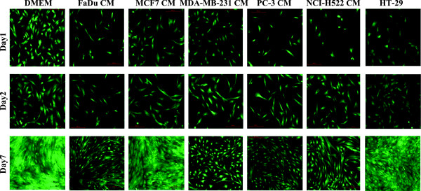
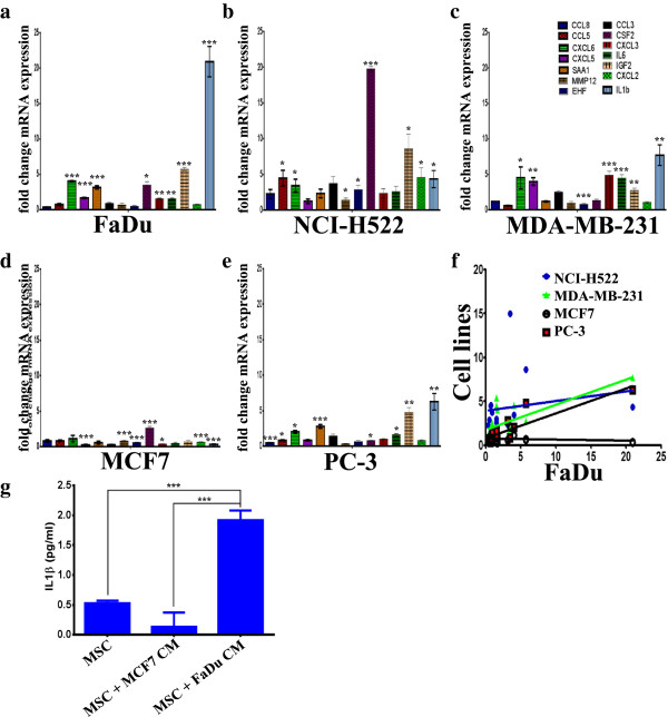
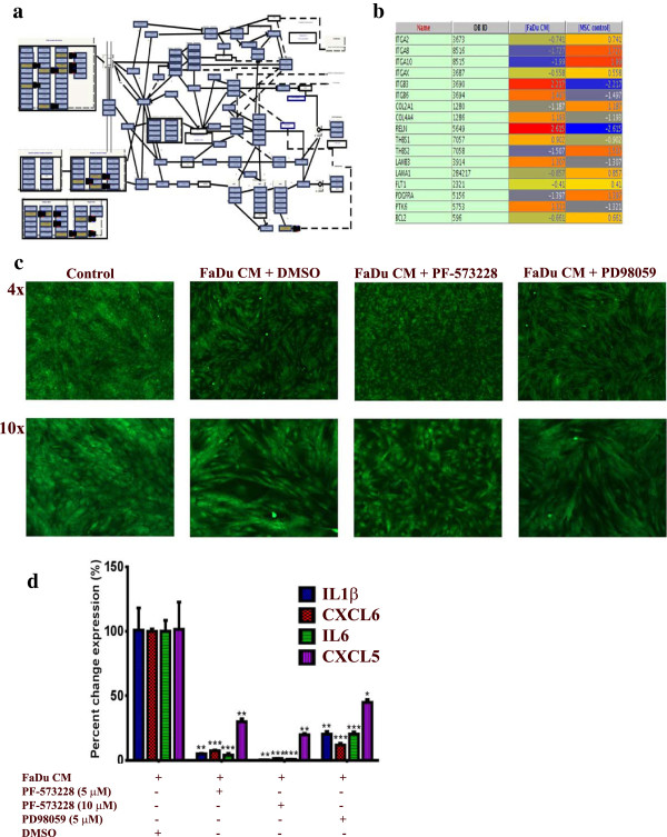
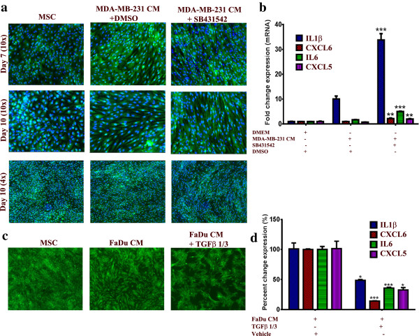
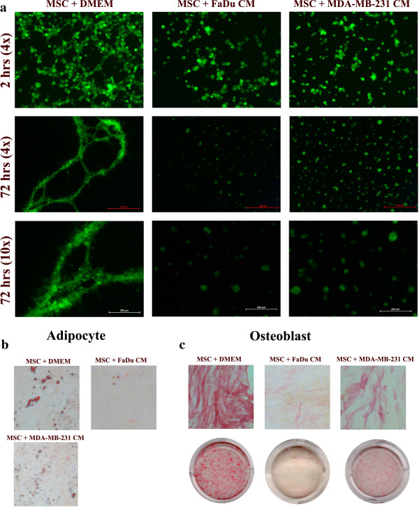
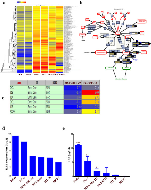
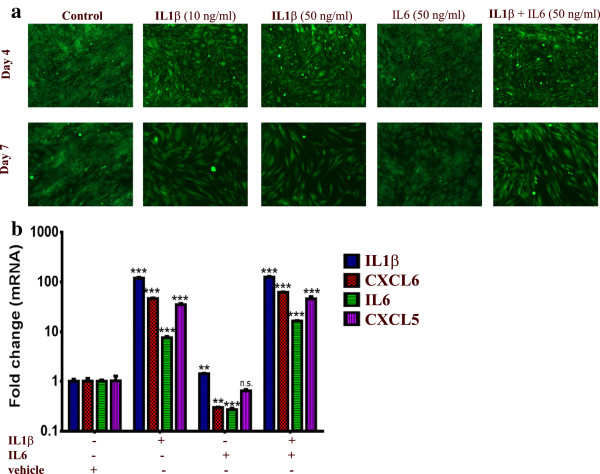
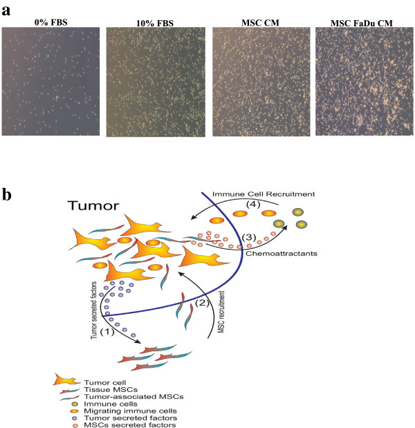
Results: MSCs exposed to secreted factors present in conditioned media (CM) from FaDu, MDA-MB-231, PC-3 and NCI-H522, but not from MCF7 and HT-29, developed an elongated, spindle-shaped morphology with bipolar processes. In association with phenotypic changes, genome-wide gene expression and bioinformatics analysis revealed an enhanced pro-inflammatory response of those MSCs. Pharmacological inhibitions of FAK and MAPKK severely impaired the pro-inflammatory response of MSCs to tumor CM (approximately 80% to 99%, and 55% to 88% inhibition, respectively), while inhibition of the TGFβ pathway was found to promote the pro-inflammatory response (approximately 3-fold increase). In addition, bioinformatics and pathway analysis of gene expression data from tumor cell lines combined with experimental validation revealed tumor-derived IL1β as one mediator of the pro-inflammatory phenotype observed in MSCs exposed to tumor CM.

Conclusions: Our data revealed tumor-derived IL1β as one mediator of the pro-inflammatory response in MSCs exposed to tumor CM, which was found to be positively regulated by FAK and MAPK signaling and negatively regulated by TGFβ signaling. Thus, our data support a model where MSCs could promote cancer progression through becoming pro-inflammatory cells within the cancer stroma.


Figures

Figure 1
Figure 1
Effects of FaDu conditioned medium (CM) on human MSC morphology and gene expression. (a) Representative micrographs of MSC-GFP cells grown under normal conditions (left panel) or exposed to FaDu CM (right panel). Hoechst 33342 was used for nuclear staining and images were obtained at the indicated time points (10× magnification, 200 μm scale bar). Arrow heads point to MSCs with fibroblastic morphology in CM treated cells. (b) MSCs grown under normal conditions or exposed to FaDu CM were subjected to microarray analysis. Differentially upregulated genes in MSCs exposed to FaDu CM were subsequently subjected to pathway analysis as described in Methods. The pie chart represents the top ten pathways where the pie size represents percent enrichment of the pathway. (c) Genes in the cytokine and inflammatory response pathway were among the most highly enriched category in the microarray data. MSCs, mesenchymal stem cells.
Figure 2
Figure 2
Comparative analysis of morphological changes in MSCs exposed to conditioned medium (CM) from a panel of human cancer cell lines. MSCs were grown under normal conditions ((D)MEM) or were exposed to CM from the indicated cancer cell lines (FaDu, MCF7, MDA-MB-231, PC-3, NCI-H522 and HT-29) and, subsequently, images were obtained on days1, 2 and 7. Representative micrographs from at least three independent experiments are shown. All images were taken using 10x magnification. MSCs, mesenchymal stem cells.
Figure 3
Figure 3
Validation of selected genes in the cytokine, inflammation, and metalloproteases pathways in MSCs exposed to conditioned medium (CM) from a panel of human cancer cell lines. MSCs were exposed to CM as described in Figure  2, then qRT-PCR was utilized to validate the expression of selected genes in the cytokine, inflammation, and metalloproteases pathways in MSCs exposed to CM of FaDu (a), NCI-H522 (b), MDA-MB-231 (c), MCF7 (d) and PC-3 (e). Data are presented as mean fold change (relative to control MSCs) ± S.D. from at least two independent experiments, n = 4. P values * <0.05, ** <0.005, *** <0.0005. (f) Correlative analysis of the expression of the aforementioned genes in FaDu versus the other cell lines. (g) Quantification of secreted IL1β by ELISA from MSCs exposed to MCF7 or FaDu CM. Data are presented as mean ± S.D. n = 3. MSCs, mesenchymal stem cells.
Figure 4
Figure 4
Inhibition of FAK and MAPK abrogates the pro-inflammatory response in MSCs exposed to FaDU CM. (a) The focal adhesion kinase (FAK) pathway was among the top upregulated pathways in MSCs exposed to FaDu CM. (b) The list of differentially expressed genes in the FAK pathway in MSCs exposed to FaDu CM. (c) Pharmacological inhibition of FAK (5 μM, PF-573228, Sigma) or MAPKK (5 μM, PD98059, Sigma) led to significant inhibition of the pro-inflammatory response in MSCs exposed to FaDu CM. (d) Quantification of a representative set of genes in the cytokine and inflammatory response pathway in MSCs exposed to FaDu CM in the presence of DMSO, FAK, or MAPKK inhibitors. Data are presented as percent change in gene expression relative to MSCs exposed to FaDu CM + DMSO. Data are presented as mean ± S.D. n = 3. CM, conditioned media; DMSO, dimethyl sulfoxide, FAK, focal adhesion kinase; MAPKK, mitogen activated protein kinase kinase; MSCs, mesenchymal stem cells.
Figure 5
Figure 5
TGFβ signaling negatively regulates the pro-inflammatory response of MSCs exposed to tumor CM. (a) MSCs were cultured as described in Methods and then were exposed to MDA-MB-231 CM in the presence of 10 μM SB-431542 or DMSO. On the indicated days, nuclei were stained with Hoechst 33342 and cells were visualized under a florescent microscope. Data are representative of at least three independent experiments. (b) Quantification of a representative set of genes in the cytokine and inflammatory response pathway in MSCs exposed to MDA-MB-231 CM in the presence of 10 μM SB-431542 or DMSO from a. Data are presented as the fold change in gene expression relative to control MSCs. Data are presented as mean ± S.D. n = 3. (c) MSCs were cultured as described in Methods and then were exposed to FaDu CM in the presence of 10 μg/ml TGFβ1 and TGFβ3. On Day 5, cells were visualized under a florescent microscope (4x). (d) Quantification of a representative set of genes in the cytokine and inflammatory response pathway in MSCs exposed to FaDu CM in the presence of 10 μg/ml TGFβ1 and TGFβ3 from c. Data are presented as percent change in gene expression relative to MSCs exposed to FaDu CM + vehicle (dH2O). Data are presented as mean ± S.D. n = 3. CM, conditioned media; DMSO, dimethyl sulfoxide; MSCs, mesenchymal stem cells.
Figure 6
Figure 6
MSCs exposed to tumor CM lose their multipotent differentiation potential. (a) Control MSCs or MSCs exposed to FaDu or MDA-MB-231 CM (10 days) were harvested and seeded on top of Matrigel® as indicated in Methods sections. Vessel-like tubular formation was assessed at 2 hours and 72 hours using a fluorescence microscope at 4x and 10x, as indicated. Data are representative of at least two experiments. Control MSCs or MSCs exposed to FaDu or MDA-MB-231 CM (10 days) were switched to adipogenic (b) or osteogenic induction media (c). On day 6, adipocyte differentiation was measured using Oil-Red-O staining (b), while osteoblast differentiation was measured using alkaline phosphatase staining (ALP) (c). Data are representative of at least two experiments. CM, conditioned media; MSCs, mesenchymal stem cells.
Figure 7
Figure 7
Cluster and pathway analysis of basal gene expression in FaDu, NCI-H522, MDA-MB-231, MCF7, PC-3 and HT-29 tumor cell lines. (a) Clustering analysis of the tumor cell lines indicated close clustering for FaDu and PC-3, followed by MDA-MB-231 and NCI-H522, while MCF7 and HT-29 did not cluster readily with the group. Clustering analyses were performed on differentially expressed genes in FaDu and PC-3 relative to MCF7 and HT-29. (b) Cytokine and inflammatory response pathway was among the top enriched pathways in differentially expressed genes between FaDu and PC-3 relative to MCF7 and HT-29. (c) Genes in the cytokine and inflammatory response pathways from b and their expression levels are shown. (d) mRNA expression level of IL1β in different tumor cell lines from the microarray data. (e) Quantification of secreted IL1β by ELISA from different tumor CM. Data are presented as mean ± S.D. n = 3. CM, conditioned media.
Figure 8
Figure 8
IL1β treatment induced a pro-inflammatory response in MSCs. (a) MSCs were cultured in normal (D)MEM in the presence of recombinant IL1β (10 and 50 ng/ml), recombinant IL6 (50 ng/ml), both IL1β and IL6 (50 ng/ml each) or in the presence of vehicle control (dH2O). Images were taken on day 4 and 7 (4x). (b) Quantification of a representative set of genes in the cytokine and inflammatory response pathway in MSCs exposed to different cytokines from (a). Data are presented as fold change in gene expression relative to MSCs exposed to vehicle. Data are presented as mean ± S.D., n = 3. MSCs, mesenchymal stem cells.
Figure 9
Figure 9
Tumor cells are capable of attracting human MSCs in vitro. Tumor CM ((D)MEM + 1% FBS) was placed in the lower chamber of a transwell migration system, while MSCs were seeded in the upper chamber. Twenty-four hours later, the number of migrating MSCs was evaluated. (a) H & E staining of the cells migrating toward CM from the indicated tumor cell lines. Migration toward (D)MEM + 1% FBS was used as baseline control. (b) Slides were scanned and the number of migrating cells in eight (1,600 x 1,000 mcM2) fields was counted. Data are presented as mean ± S.D, n = 8. P value * <0.05, ** <0.005, *** <0.0005. CM, conditioned media; MSCs, mesenchymal stem cells.
Figure 10
Figure 10
Both control and MSCs exposed to FaDu conditioned medium (CM) are capable of attracting human PBMCs. (a) Conditioned medium from MSCs or MSCs exposed to FaDu CM were collected and placed in the lower chamber of a transwell migration system, while 1 x 10⁵ PBMCs were placed in the upper chamber. Representative images of PBMCs migrating to the lower chamber are shown. Data are representative of at least two independent experiments, conducted in duplicate. (b) A model depicting the crosstalk between tumor cells, MSCs and immune cells. (1) Tumor cells secrete soluble factors which attract MSCs (2). MSCs at the tumor site become tumor-associated MSCs with enhanced inflammatory responses and secreted chemokines (3) which attract immune cells (4) to the tumor site, collectively acting to drive tumorigenicity via enhanced inflammation as one potential mechanism of tumor progression. CM, conditioned media; MSCs, mesenchymal stem cells; PBMCs, peripheral blood mononuclear cells.

Comment in

References

    1. Dominici M, Le Blanc K, Mueller I, Slaper-Cortenbach I, Marini F, Krause D, Deans R, Keating A, Prockop D, Horwitz E. Minimal criteria for defining multipotent mesenchymal stromal cells, The International Society for Cellular Therapy position statement. Cytotherapy. 2006;8:315–317. doi: 10.1080/14653240600855905. - DOI - PubMed
    1. Granero-Molto F, Weis JA, Miga MI, Landis B, Myers TJ, O’Rear L, Longobardi L, Jansen ED, Mortlock DP, Spagnoli A. Regenerative effects of transplanted mesenchymal stem cells in fracture healing. Stem Cells. 2009;27:1887–1898. doi: 10.1002/stem.103. - DOI - PMC - PubMed
    1. Chen J, Li Y, Wang L, Lu M, Zhang X, Chopp M. Therapeutic benefit of intracerebral transplantation of bone marrow stromal cells after cerebral ischemia in rats. J Neurol Sci. 2001;189:49–57. doi: 10.1016/S0022-510X(01)00557-3. - DOI - PubMed
    1. Wu GD, Nolta JA, Jin YS, Barr ML, Yu H, Starnes VA, Cramer DV. Migration of mesenchymal stem cells to heart allografts during chronic rejection. Transplantation. 2003;75:679–685. doi: 10.1097/01.TP.0000048488.35010.95. - DOI - PubMed
    1. Kristensen LP, Chen L, Nielsen MO, Qanie DW, Kratchmarova I, Kassem M, Andersen JS. Temporal profiling and pulsed SILAC labeling identify novel secreted proteins during ex vivo osteoblast differentiation of human stromal stem cells. Mol Cell Proteomics. 2012;11:989–1007. doi: 10.1074/mcp.M111.012138. - DOI - PMC - PubMed

Publication types

MeSH terms

Substances