

Review
doi: 10.1186/1532-429X-16-7.
Tracking of stem cells in vivo for cardiovascular applications
Affiliations
- PMID: 24406054
- PMCID: PMC3925252
- DOI: 10.1186/1532-429X-16-7
Item in Clipboard
Review
Tracking of stem cells in vivo for cardiovascular applications
J Cardiovasc Magn Reson.
.
Abstract
In the past ten years, the concept of injecting stem and progenitor cells to assist with rebuilding damaged blood vessels and myocardial tissue after injury in the heart and peripheral vasculature has moved from bench to bedside. Non-invasive imaging can not only provide a means to assess cardiac repair and, thereby, cellular therapy efficacy but also a means to confirm cell delivery and engraftment after administration. In this first of a two-part review, we will review the different types of cellular labeling techniques and the application of these techniques in cardiovascular magnetic resonance and ultrasound. In addition, we provide a synopsis of the cardiac cellular clinical trials that have been performed to-date.
Figures
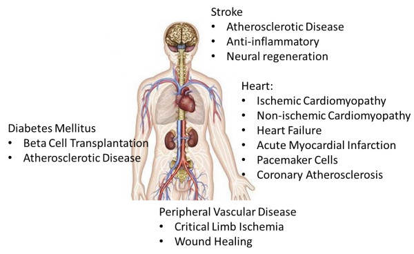
Stem cell therapies can be envisioned to treat a wide variety of cardiovascular diseases ranging from preventing adverse remodeling in ischemic and non-ischemic heart disease, the creation of new pacemaker cells, replacement of beta cells in Diabetes Mellitus, and mitigating atherosclerotic disease leading to peripheral vascular disease as well as stroke.
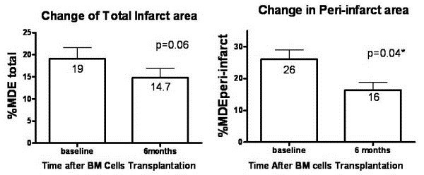
Treatment effect of bone marrow cells (BM) implantation on percentage of total infarct area and peri-infarct area in the BM group as determined by CMR. Data presented as mean ± SD (error bar). Reprinted with permission from Chan et al.[11].
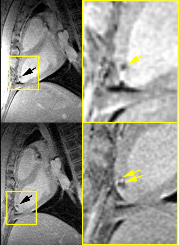
Long-axis CMR showing hypointense lesions (arrows) caused by superparamagnetic iron oxide-labeled mesenchymal stem cells acquired within (top left) 24 h and (bottom left) 1 week of injection. Insets demonstrate expansion of lesion over 1 week. Reprinted with permission from Kraitchman et al.[115].
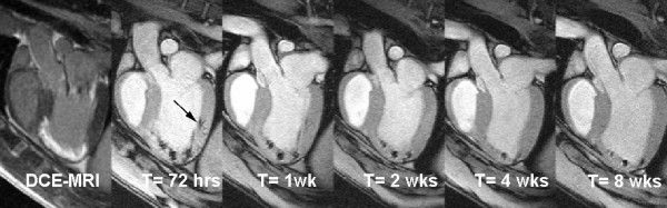
Delayed contrast-enhanced (DCE) long-axis image (left) demonstrating infarcted myocardium (MI). MR-labeled-MSC injections appear as hypointense areas on fast gradient echo images. Serial imaging at 72 hours, 1 week, 2 weeks, 4 weeks, and 8 weeks demonstrates the persistent of the MR-MSC injections. The volume of injections decreases over time. In addition, an injection placed in the normal myocardium (arrow) can no longer be detected at 4 weeks post-injection. Reprinted with permission from Soto et al.[120].
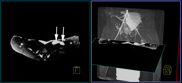
Left: An axial positive contrast image using Inversion-Recovery with On-resonance water suppression (IRON) of SPIO-labeled stem cells injected in a rabbit thigh demonstrates two injection sites (arrows) as bright hyperintensities. Right: A maximum intensity projection of a 3D T2-prepared MR angiogram shows the region of superficial femoral artery occlusion at 24 hours post-occlusion in a rabbit model of peripheral arterial disease can be registered with the IRON images to determine the location of stem cell injections relative to neovasculature. (Adapted with permission from Kraitchman and Bulte [108]).
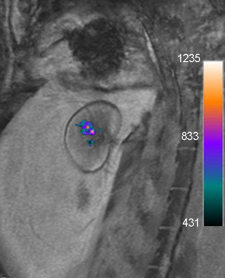
In vivo merged 19 F (color) and proton (grayscale) MRI acquired on a clinical 3T scanner of a rabbit transplanted with 10,000 perfluoropolyether (PFPE)-labeled islets under the kidney capsule demonstrates clear visualization of cell transplants. The signal corresponds to 14,153 μg PFPE. Reprinted with permission from Barnett et al.[153].
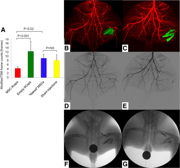
Barium sulfate-labeled microcapsules for X-ray cell tracking (Xcaps) in peripheral arterial disease. (A) A bar graph of the average modified Thrombolysis In Myocardial Infarction (TIMI) frame count, as a measure of collateral vessel development, in the MSC-Xcaps, empty microcapsules, unencapsulated MSCs, and sham injection treated animals demonstrating a significant improvement in distal filling only in the peripheral arterial disease (PAD) rabbits that received microencapsulated cells (*P < 0.001 empty microcapsules vs. MSC-Xcaps; P = NS naked MSCs vs. sham injections). B-G: Representative digital subtraction angiogram (DSA, red) obtained during peak contrast opacification performed at two weeks post injection of encapsulated MSCs-Xcaps (B) and empty microcapsules (C) with an overlay of microcapsules injections (green) obtained from mask image of DSA. The small collateral vessels are somewhat obscured by the Xcap radiopacity. However, the increased collateralization can be appreciated in the MSC-Xcap-treated animal DSA (D) relative to the Xcap-treated animal (E) Native mask digital radiographs demonstrating the location of the MSC-Xcaps (F) and empty Xcaps (G) in the same animals. There was no statistically significant difference in vessel diameter between treatment groups. Reprinted with permission from Kedziorek et al.[171].
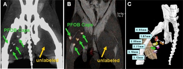
Perfluorocarbon-labeled microcapsules for X-ray visible cell tracking by CT. (A) Cone beam CT acquired on a flat-panel X-ray angiographic system (Axiom Artis, Siemens AG, Forchheim, Germany) demonstrating the detection of four perfluorooctylbromide (PFOB) injection sites in a rabbit medial thigh, while unlabeled capsules in the left thigh are not detectable. (B) 19 F MRI of the same rabbit showing one-to-one correspondence to the injection location on cone beam CT. (C) Co-registering of threshold cone beam CT image of a rabbit with 6 PFOB Caps injection sites (gray) and postmortem 3D rendering volume of each injection sites (color) demonstrating the location of opacities on cone beam CT image is representative of PFOB Caps injections. Registration error for each injection site from a representative rabbit is shown. Reprinted with permission from Fu et al.[172].
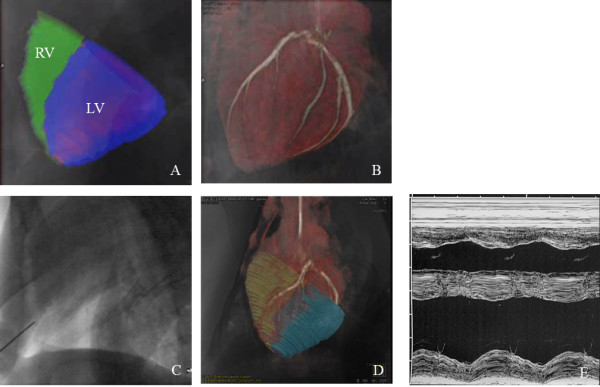
X-ray fused with MRI (XFM) of X-ray-visible microcapsules to the heart. (A) Segmented cine CMR showing epicardial contours (green-RV; blue-LV) overlaid on live X-ray fluoroscopic image. (B) Coronary vasculature from c-arm CT overlaid on live X-ray fluoroscopic image. (C) Live X-ray fluoroscopy demonstrating radiopacity of needle used for pericardial puncture and the lack of ability to visualize the myocardium or coronary vasculature without XFM. (D) Live X-ray fluoroscopy image overlaid on segmented whole heart CMR and c-arm CT volumes showing pericardial puncture. (E) An M-mode echocardiogram at seven days post-injection demonstrating normal cardiac function and no abnormalities to the pericardium. Reprinted with permission from Azene et al.[173].
Similar articles
-
In vivo tracking in cardiac stem cell-based therapy.Prog Cardiovasc Dis. 2007 May-Jun;49(6):414-20. doi: 10.1016/j.pcad.2007.02.005. Prog Cardiovasc Dis. 2007. PMID: 17498521 Review.
-
Stem cell labeling for noninvasive delivery and tracking in cardiovascular regenerative therapy.Expert Rev Cardiovasc Ther. 2010 Aug;8(8):1149-60. doi: 10.1586/erc.10.106. Expert Rev Cardiovasc Ther. 2010. PMID: 20670192 Free PMC article. Review.
-
Stem cell therapy in cardiovascular disorders.Cardiovasc Ther. 2010 Oct;28(5):e101-10. doi: 10.1111/j.1755-5922.2010.00208.x. Cardiovasc Ther. 2010. PMID: 21050418 Review.
-
Magnetic Resonance Imaging of Iron Oxide-Labeled Human Embryonic Stem Cell-Derived Cardiac Progenitors.Stem Cells Transl Med. 2016 Jan;5(1):67-74. doi: 10.5966/sctm.2015-0077. Epub 2015 Nov 18. Stem Cells Transl Med. 2016. PMID: 26582908 Free PMC article.
-
Advances in cardiovascular molecular imaging for tracking stem cell therapy.Thromb Haemost. 2010 Jul;104(1):13-22. doi: 10.1160/TH09-08-0530. Epub 2010 May 10. Thromb Haemost. 2010. PMID: 20458434 Free PMC article. Review.
Cited by
-
Macroscopic fluorescence imaging: a novel technique to monitor retention and distribution of injected microspheres in an experimental model of ischemic heart failure.PLoS One. 2014 Aug 4;9(8):e101775. doi: 10.1371/journal.pone.0101775. eCollection 2014. PLoS One. 2014. PMID: 25089764 Free PMC article.
-
Review of Journal of Cardiovascular Magnetic Resonance 2014.J Cardiovasc Magn Reson. 2015 Nov 20;17:99. doi: 10.1186/s12968-015-0203-4. J Cardiovasc Magn Reson. 2015. PMID: 26589839 Free PMC article. Review.
-
Cardiac shock wave therapy promotes arteriogenesis of coronary micrangium, and ILK is involved in the biomechanical effects by proteomic analysis.Sci Rep. 2018 Jan 29;8(1):1814. doi: 10.1038/s41598-018-19393-z. Sci Rep. 2018. PMID: 29379038 Free PMC article.
-
Magnetically Responsive Bone Marrow Mesenchymal Stem Cell-Derived Smooth Muscle Cells Maintain Their Benefits to Augmenting Elastic Matrix Neoassembly.Tissue Eng Part C Methods. 2016 Apr;22(4):301-11. doi: 10.1089/ten.TEC.2015.0349. Epub 2016 Mar 18. Tissue Eng Part C Methods. 2016. PMID: 26830683 Free PMC article.
-
Epicardium-Derived Cells Formed After Myocardial Injury Display Phagocytic Activity Permitting In Vivo Labeling and Tracking.Stem Cells Transl Med. 2016 May;5(5):639-50. doi: 10.5966/sctm.2015-0159. Epub 2016 Apr 7. Stem Cells Transl Med. 2016. PMID: 27057005 Free PMC article.
References
-
- Schwitter J, Wacker CM, Wilke N, Al-Saadi N, Sauer E, Huettle K, Schonberg SO, Debl K, Strohm O, Ahlstrom H. et al.Superior diagnostic performance of perfusion-cardiovascular magnetic resonance versus SPECT to detect coronary artery disease: The secondary endpoints of the multicenter multivendor MR-IMPACT II (Magnetic Resonance Imaging for Myocardial Perfusion Assessment in Coronary Artery Disease Trial) J Cardiovasc Magn Reson. 2012;14:61. - PMC - PubMed
-
- Schachinger V, Erbs S, Elsasser A, Haberbosch W, Hambrecht R, Holschermann H, Yu J, Corti R, Mathey DG, Hamm CW. et al.Intracoronary bone marrow-derived progenitor cells in acute myocardial infarction. N Engl J Med. 2006;355:1210–1221. - PubMed
-
- Leistner DM, Fischer-Rasokat U, Honold J, Seeger FH, Schachinger V, Lehmann R, Martin H, Burck I, Urbich C, Dimmeler S. et al.Transplantation of progenitor cells and regeneration enhancement in acute myocardial infarction (TOPCARE-AMI): final 5-year results suggest long-term safety and efficacy. Clin Res Cardiol. 2011;100:925–934. - PubMed
Publication types
MeSH terms
LinkOut - more resources
Full Text Sources
Other Literature Sources
Miscellaneous
