. 2014 Jan;12(1):82-9.
doi: 10.1111/jth.12437.

Analysis of inter-subunit contacts reveals the structural malleability of extracellular domains in platelet glycoprotein Ib-IX complex

Affiliations

Analysis of inter-subunit contacts reveals the structural malleability of extracellular domains in platelet glycoprotein Ib-IX complex

L Zhou et al. J Thromb Haemost. 2014 Jan.

Abstract

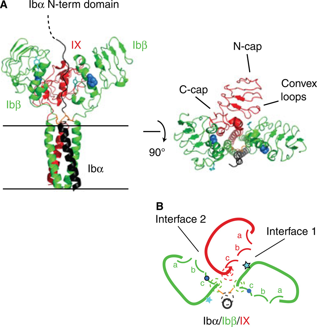
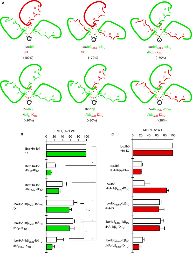
Background: The glycoprotein (GP)Ib-IX complex is critical to hemostasis and thrombosis. Its proper assembly is closely correlated with its surface expression level and requires cooperative interactions among extracellular and transmembrane domains of Ibα, Ibβ and IX subunits. Two interfaces have been previously identified between the extracellular domains of Ibβ and IX.

Objective: To understand how extracellular domains interact in GPIb-IX.

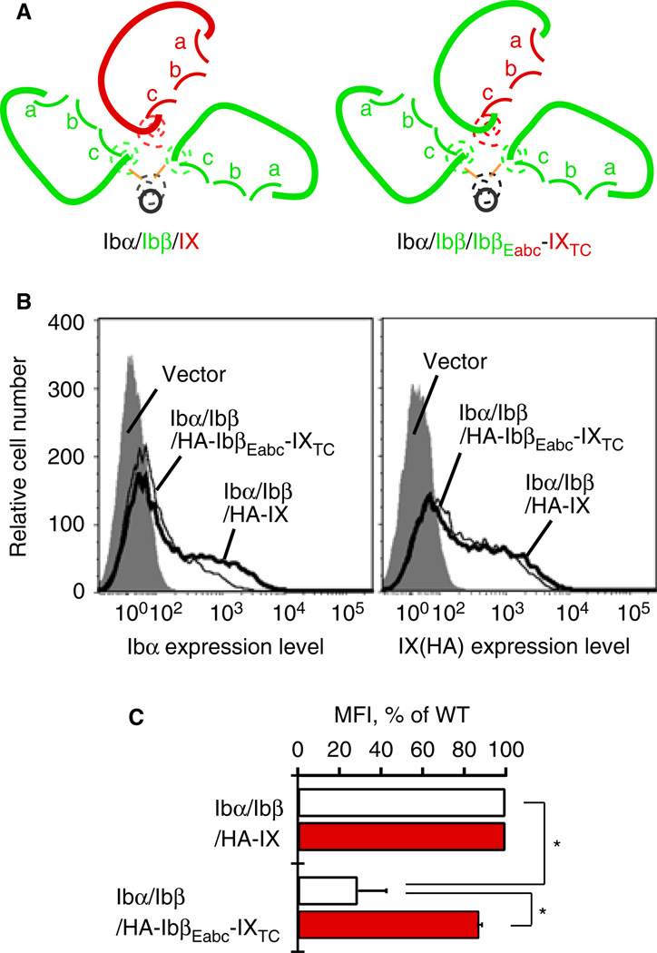
Methods: The Ibβ extracellular domain (IbβE ) or the IX counterpart (IXE ) in GPIb-IX was replaced with a well-folded IbβE /IXE chimera called IbβEabc , and the effect of domain replacement on assembly and expression of the receptor complex in transiently transfected Chinese hamster ovary cells was analyzed.

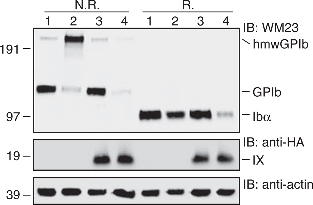
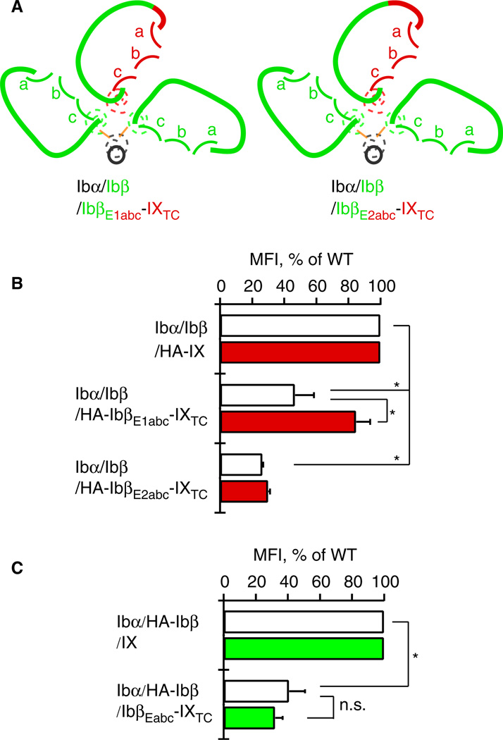
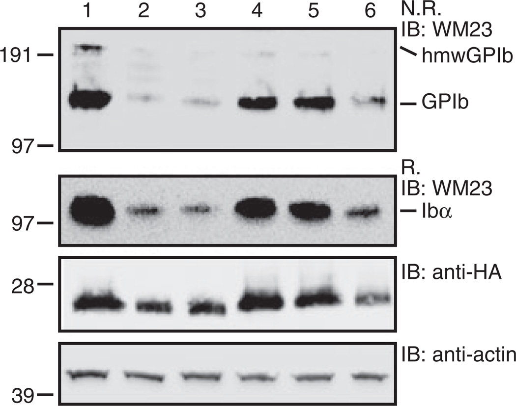
Results: Replacing IXE with IbβEabc in GPIb-IX retained interface 1 but not interface 2 between the extracellular domains. While this domain replacement preserved complex integrity, the expression levels of Ibβ and Ibα were significantly reduced. Additional domain replacement with IbβEabc or IbβE in GPIb-IX produced the complex at disparate expression levels that cannot be simply explained by two separate interfaces. In particular, when IbβE in GPIb-IX was replaced by IbβEabc , Ibα and IX were expressed at approximately 70% of the wild-type level. Their levels were not reduced when IXE was changed further to IbβE .

Conclusions: Our results demonstrate the importance of the association between Ibβ and IX extracellular domains for complex assembly and efficient expression, and provide evidence for the structural malleability of these domains that may accommodate and propagate conformational changes therein.

Keywords: Bernard-Soulier syndrome; leucine-rich repeat proteins; platelet glycoprotein GPIb-IX complex; protein-protein interaction domains; von Willebrand factor receptors.


Conflict of interest statement

Disclosure of Conflict of Interests

The authors state that they have no conflict of interests.

Figures

Fig. 1
Fig. 1
The organization of the GPIb-IX complex, viewed from the extracellular space towards the membrane, is summarized in a sketch for easy visualization and comprehension. (A) The membrane-proximal extracellular and transmembrane domains of GPIb-IX have been modeled in ribbon diagrams, with its side view shown on the left and top view in the middle (adapted from [3]). Ibα, Ibβ and IX subunits are shown in black, green and red color, respectively. The general locations of N- and C-capping regions and convex loops in IXE are marked. The side chains of Tyr106 in Ibβ are shown as ball-and-sticks in teal, and those of Pro74 in Ibβ as spheres in marine. The juxtamembrane Ibα-Ibβ disulfide bonds are shown in orange. These domains are shown in (B) as sketches of the same color. The transmembrane domains are represented by dashed coils. The extracellular domains are drawn in solid curves, with convex loops (a, b, c) marked in each domain. Positions of Tyr106 and Pro74 are marked with a star and dot, respectively.
Fig. 2
Fig. 2
Replacing IXE in GPIb-IX with IbβEabc significantly decreases surface expression of Ibα in transiently transfected CHO cells. (A) Sketches of the mutant GPIb-IX complex containing IbβEabc-IXTC in comparison with the wild type. In GPIbβEabc, three convex loops of IXE are grafted onto the IbβE scaffold [18]. (B) Overlaid histograms showing surface expression of Ibα and IX derivatives in transfected CHO cells measured by flow cytometry using WM23 and anti-HA antibodies, respectively. Each trace is identified by the subunits transfected. Gray peak: cells transfected with empty vector. Thick line: Ibα/Ibβ/HA-IX cells. Thin line: Ibα/Ibβ/HA-IbβEabc-IXTC cells. Each plot is representative of at least four independent experiments. (C) Quantitative representation of relative surface expression levels of Ibα (white bar) and HA-tagged IX derivatives (red) in aforementioned transfected cells. The measured mean fluorescence intensity (MFI), obtained for the entire cell population (10 000 cells per sample), was normalized with the expression level in Ibα/Ibβ/HA-IX (WT) cells being 100% and cells transfected with empty vectors 0% [9,18]. All data are presented as mean ± SD (n = 4 or 6). *P < 0.01.
Fig. 3
Fig. 3
SDS gels showing correct formation of native Ibα-Ibβ disulfide bonds in the Ibα/Ibβ/HA-IbβEabc-IXTC complex. Lysates from various transfected CHO cells were separated in Bis-Tris SDS gels under non-reducing (N.R.) or reducing (R.) conditions, transferred to a PVDF membrane and immunoblotted by noted antibodies. Lane 1, Ibα/Ibβ/IX; 2, Ibα/Ibβ; 3, Ibα/Ibβ/HA-IX; 4, Ibα/Ibβ/HA-IbβEabc-IXTC. This figure is representative of four independent experiments.
Fig. 4
Fig. 4
Reduced surface expression of Ibα is due to the reduced expression of Ibβ in the Ibα/Ibβ/HA-IbβEabc-IXTC complex. (A) Sketches of mutant GPIb-IX complexes used. The style follows that described in Figure 1. (B) Relative surface expression levels of Ibα (white bar) and HA-tagged IX derivatives (red) in transfected CHO cells measured by flow cytometry. Each cell is identified by the subunits transfected. (C) Relative surface expression levels of Ibα (white bar) and HA-tagged Ibβ (green) in transfected CHO cells. The HA tag was appended to Ibβ and removed from IX derivatives to allow detection of Ibβ by flow cytometry. The measurement and quantitation follow the procedure described in Figure 2(C). All data are presented as mean ± SD (n = 3). *P < 0.01.
Fig. 5
Fig. 5
Extracellular domains of Ibβ and IX adopt different conformations to stabilize certain domain-swapped GPIb-IX complexes and enhance their surface expression. (A) Sketches of wild-type GPIb-IX and five chimeric complexes, each of which is identified by the subunits therein in colors as described in Figure 1. The measured mean fluorescence intensity of surface Ibα in transfected CHO cells, expressed as the percentage of that in wild-type cells and indicative of GPIb-IX assembly and stability, is listed in parenthesis for each complex. Note that although the extracellular domains are similarly sketched to denote their high structural homology to one another, they adopt neither the same conformation nor interaction in different complexes. (B) Relative surface expression levels of Ibα (white bar) and HA-tagged Ibβ derivatives (green) in transfected CHO cells. Each CHO cell is identified by the subunits transfected. (C) Relative surface expression levels of Ibα (white bar) and HA-tagged IX derivatives (red) in transfected CHO cells. The measurement and quantitation follow that described in Figure 2(C). All data are presented as mean ± SD (n = 3–5). Only examples of statistical analysis results are shown. *P < 0.01.
Fig. 6
Fig. 6
SDS gels showing the effect of domain replacement on the expression and Ibα-Ibβ disulfide formation in various transfected CHO cells. Lysates from various CHO cells transfected with GPIb-IX subunits were separated in Bis-Tris SDS gels under non-reducing (N.R.) or reducing (R.) conditions, transferred to PVDF membrane and immunoblotted by noted antibodies. Lane 1, Ibα/HA-Ibβ/IX; 2, Ibα/HA-Ibβ/IbβE-IXTC; 3, Ibα/HA-Ibβ/IbβEabc-IXTC; 4, Ibα/HA-IbβEabc-IbβTC/IX; 5, Ibα/HA-IbβEabc-IbβTC/IbβE-IXTC; 6, Ibα/HAIbβEabc-IbβTC/IbβEabc-IXTC. This figure is representative of three independent experiments.

References

    1. Berndt MC, Gregory C, Kabral A, Zola H, Fournier D, Castaldi PA. Purification and preliminary characterization of the glycoprotein Ib complex in the human platelet membrane. Eur J Biochem. 1985;151:637–649. - PubMed
    1. Luo S-Z, Mo X, Afshar-Kharghan V, Srinivasan S, Lopez JA, Li R. Glycoprotein Ibα forms disulfide bonds with 2 glycoprotein Ibβ subunits in the resting platelet. Blood. 2007;109:603–609. - PMC - PubMed
    1. Li R, Emsley J. The organizing principle of the platelet glycoprotein Ib-IX-V complex. J Thromb Haemost. 2013;11:605–614. - PMC - PubMed
    1. Huizinga EG, Tsuji S, Romijn RA, Schiphorst ME, de Groot PG, Sixma JJ, Gros P. Structures of glycoprotein Ibα and its complex with von Willebrand factor A1 domain. Science. 2002;297:1176–1179. - PubMed
    1. Dumas JJ, Kumar R, Seehra J, Somers WS, Mosyak L. Crystal structure of the GpIbα-thrombin complex essential for platelet aggregation. Science. 2003;301:222–226. - PubMed

Publication types

Substances