. 2014 Jan:135:1-14.
doi: 10.1016/j.mad.2013.12.008. Epub 2014 Jan 7.

Cockayne Syndrome group B protein stimulates NEIL2 DNA glycosylase activity

Affiliations

Cockayne Syndrome group B protein stimulates NEIL2 DNA glycosylase activity

Maria D Aamann et al. Mech Ageing Dev. 2014 Jan.

Abstract

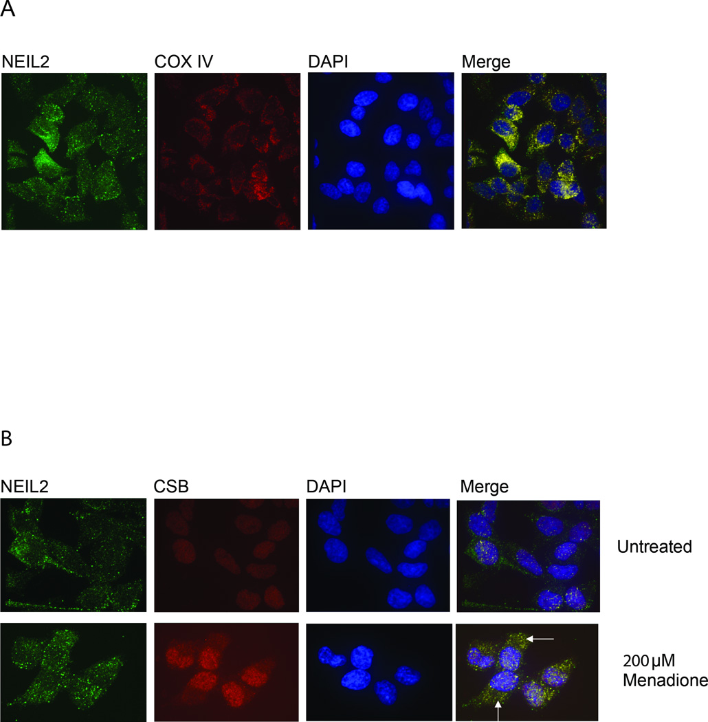
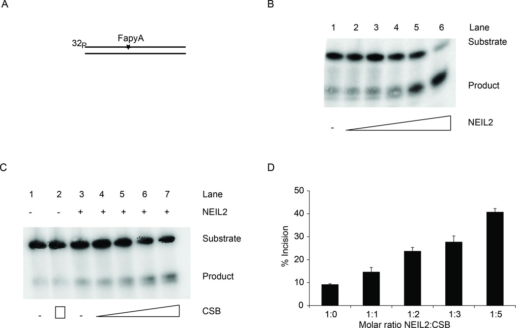
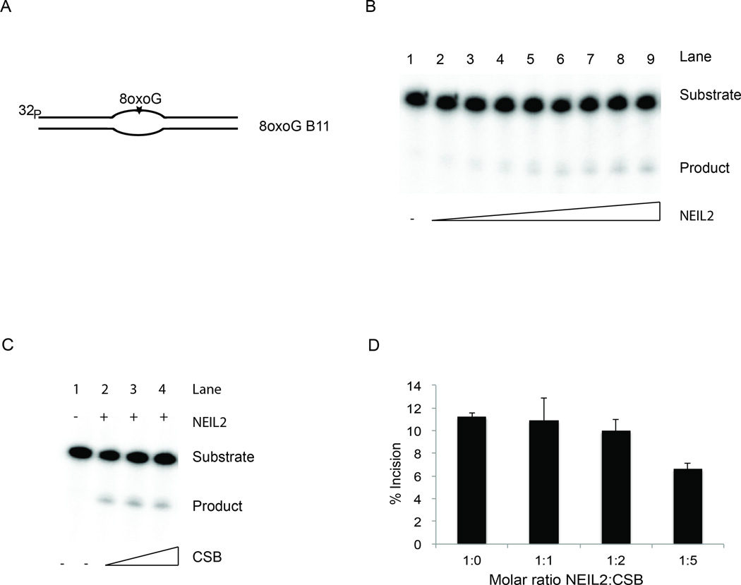
Cockayne Syndrome is a segmental premature aging syndrome, which can be caused by loss of function of the CSB protein. CSB is essential for genome maintenance and has numerous interaction partners with established roles in different DNA repair pathways including transcription coupled nucleotide excision repair and base excision repair. Here, we describe a new interaction partner for CSB, the DNA glycosylase NEIL2. Using both cell extracts and recombinant proteins, CSB and NEIL2 were found to physically interact independently of DNA. We further found that CSB is able to stimulate NEIL2 glycosylase activity on a 5-hydroxyl uracil lesion in a DNA bubble structure substrate in vitro. A novel 4,6-diamino-5-formamidopyrimidine (FapyA) specific incision activity of NEIL2 was also stimulated by CSB. To further elucidate the biological role of the interaction, immunofluorescence studies were performed, showing an increase in cytoplasmic CSB and NEIL2 co-localization after oxidative stress. Additionally, stalling of the progression of the transcription bubble with α-amanitin resulted in increased co-localization of CSB and NEIL2. Finally, CSB knockdown resulted in reduced incision of 8-hydroxyguanine in a DNA bubble structure using whole cell extracts. Taken together, our data supports a biological role for CSB and NEIL2 in transcription associated base excision repair.

Keywords: Base excision repair; CSB; Cockayne Syndrome; NEIL2; Oxidative damage.


Figures

FIGURE 1
FIGURE 1. Physical interaction between NEIL2 and CSB
A, Nuclear extracts from mock or menadione treated (200µM) HeLa cells were used for co-IP with NEIL2, CSB or IgG antibodies. The immunoprecipitated complexes and 5% input were run on SDS-PAGE and blotted against NEIL2, CSB or XPB (p89) protein. B, Dot blot of NEIL2 CSB interaction. Purified recombinant CSB, NEIL2 and control protein, BSA, were blotted on PVDF membranes and incubated with purified NEIL2. The membrane was then incubated with primary mouse anti-NEIL2 antibody followed by secondary anti-mouse IgG peroxidase conjugated antibody. C, Far Western of NEIL2 and CSB interaction. Recombinant NEIL2 and BSA protein separated on a 10% SDS-PAGE gel and transferred to PVDF membrane followed by incubation with purified CSB protein. Bound CSB protein was detected with CSB antibody.
FIGURE 2
FIGURE 2. Co-localization of NEIL2 and CSB
A, Cellular distribution of NEIL2. HeLa cells were fixed and stained for NEIL2 (Mouse monoclonal, Sigma) and COX IV (Goat Polyclonal, Santa Cruz). COX IV was used for mitochondrial staining. B, Co-localization of NEIL2 and CSB. HeLa cells were treated with 200µM menadione, fixed and stained for NEIL2 (Sigma) and CSB (Santa Cruz), respectively.
FIGURE 3
FIGURE 3. Stimulation of NEIL2 incision activity on FapyA substrate with CSB
A, Substrate containing FapyA lesion. B, Activity of NEIL2 on FapyA substrate. 1nM FapyA substrate was incubated with increasing amounts of purified recombinant NEIL2 protein. Lane 1; 1nM FapyA substrate, no NEIL2. Lanes 2–6; 10, 20, 50, 100 and 150nM NEIL2. C, Representative autoradiogram showing stimulation of NEIL2 incision activity on FapyA substrate by CSB. Lane 1; 1nM FapyA substrate alone. Lane 2; 200nM CSB alone. Lane 3: 40nM NEIL2 alone, Lanes 4–7: 40nM NEIL2 with increasing concentrations of CSB; 40, 80, 120 and 200nM respectively. D, Stimulation of NEIL2 (40nM) incision activity on 1nM FapyA substrate with 0, 40, 80, 120 and 200nM CSB. The diagram shows quantification and average of three independent experiments. Error bars represent standard deviations.
FIGURE 4
FIGURE 4. Stimulation of NEIL2 glycosylase activity on 5OHU in a bubble structure by CSB
A, 5-Hydroxyuracil substrate in an 11nt bubble (5OHU B11) used for incision experiments. B, Increasing amounts of NEIL2 protein were incubated with 5OHU B11. Lane 1–9; 0.0, 0.0125, 0.025, 0.05, 0.1, 0.2, 0.5, 1 and 2nM NEIL2 respectively with 0.2nM 5OHU B11. C, Representative autoradiogram showing stimulation of NEIL2 by CSB. Lane 1; substrate alone. Lane 2–7; 0.05nM NEIL2 and 0.0, 0.025, 0.05, 0.1, 0.25 and 0.5nM CSB, respectively. Lane 8; 0.5nM CSB. D, Average of three independent DNA glycosylase experiments with 0.05nM NEIL2 protein, 0.2nM 5OHU B11 substrate and 0.0, 0.025, 0.05, 0.1 and 0.25nM CSB, respectively. E, Graph for time dependent 5OHU B11 incision activity of NEIL2 alone or NEIL2 and CSB in a 1:1 molar ratio at time 0, 1, 2, 5, and 10min using 100nM 5OHU B11 and 10nM NEIL2 with or without 10nM CSB. Representative for two independent experiments. Error bars represent standard deviation.
FIGURE 5
FIGURE 5. NEIL2 glycosylase activity on 5OHU in a DNA duplex in the presence of CSB
A, 5OHU substrate in fully annealed DNA used for incision experiment. B, Representative gel for NEIL2 incision with CSB. Lane 1; Substrate alone. Lane 2–7; 4nM NEIL2 and 0, 2, 3, 4, 8 and 16nM CSB, respectively. C, Average of three independent DNA glycosylase experiments with 4nM NEIL2 protein, 0.2nM 5OHU duplex substrate, and 0, 2, 3, 4, 8 and 16nM CSB, respectively. Error bars represent standard deviations.
FIGURE 6
FIGURE 6. NEIL2 glycosylase activity on 8oxoG in bubble structure in presence of CSB
A, 8oxoG substrate in an 11nt bubble (8oxoG B11) used for incision experiment. B, Increasing amounts of NEIL2 protein were incubated with an 8oxoG B11 substrate. Lane 1–9; 0.0, 0.025, 0.05, 0.1, 0.2, 0.5, 1, 2 and 5nM NEIL2 respectively with 1nM 8oxoG B11. C, Representative gel for NEIL2 incision with CSB. Lane 1; Substrate alone. Lane 2–4; 1nM NEIL2, and 0, 1 and 2nM CSB, respectively. D, Average of three independent DNA glycosylase experiments with 1nM NEIL2 protein, 8oxoG B11 substrate (1nM) and 0, 1, 2, and 5nM CSB, respectively. Error bars represent standard deviations.
FIGURE 7
FIGURE 7. Molecular mechanism for CSB stimulation of NEIL2 on 5OHU B11 substrate by trapping transient Schiff-base intermediate during incision
A, Representative autoradiogram of NEIL2 forming Schiff base with the substrate. Lane 1; no protein. Lane 2; 0.25 nM CSB. Lane 3–7; 0.05nM NEIL2 with increasing amounts of CSB (0.0, 0.025, 0.05, 0.1, 0.25nM,respectively). Lane 8; 0.05nM NEIL2 and 0.25nM CSB, no NaBH4. Lane 9; control B11 substrate with 0.05nM NEIL2. Lane 10; control B11 with 0.05nM NEIL2 and 0.25nM CSB. B, Quantification of three independent experiments with 5OHU B11, 0.05nM NEIL2 and 0.0, 0.025, 0.05, 0.1, 0.25nM CSB, respectively, together with NaBH4. Error bars indicate standard deviations.
FIGURE 8
FIGURE 8. Detection of NEIL2 and CSB by in situ PLA
A, In situ PLA (Duolink) demonstration of the NEIL2 and CSB association in mock (upper row) and α-amanitin (lower row) treated HeLa cells. Left side, PLA specific signals detected by FITC. Right side, merge of FITC, DAPI and DIC signals. B, Merge of FITC, DAPI and DIC from PLA with CSB Ab (rabbit) and NEIL2 ab (mouse) vs. IgG, respectively which served as controls for unspecific PLA signals.
FIGURE 9
FIGURE 9. 8oxoG B11 incision capacity in WCE from mock and H2O2 treated cells
A; 8oxoG substrate in an 11nt bubble (8oxoG B11) and control substrate, respectively, used for incision experiments. B, Representative gel for 8oxoG B11 and control B11 incision reactions with WCE from mock treated NEIL1/CSB KD and NEIL1/control KD cells. Lane 1; single stranded oligonucleotide marker. Lane 2–6; 8oxoG B11 incision with WCE from NEIL1/CSB KD cells. Lane 7–11: 8oxoG B11 incision with WCE from NEIL1/control KD cells. Reactions were stopped after 0, 30, 60, 120 and 180min, respectively. Lane 12–14; control B11 substrate with WCE from NEIL1/CSB KD cells stopped at 180, 120 and 0min, respectively D, Representative gel for 8oxoG B11 incision reaction with WCE from 50µM H2O2 treated NEIL1/CSB KD and NEIL1/control KD cells. Lane 1; single stranded oligonucleotide marker. Lane 2–7; 8oxoG B11 incision with WCE from NEIL1/CSB KD cells. Lane 8–13: 8oxoG B11 incision with WCE from NEIL1/control KD cells. Reactions were stopped at 0, 10, 30, 60, 120 and 180min respectively. C and E: Quantification of three independent experiments as shown in B and D respectively. Error bars indicate standard deviation. * indicates P<0.05 in Student t-test for difference between control siRNA and CSB siRNA KD WCE.

References

    1. Aamann MD, Sorensen MM, Hvitby C, Berquist BR, Muftuoglu M, Tian J, de Souza-Pinto NC, Scheibye-Knudsen M, Wilson DM, 3rd, Stevnsner T, Bohr VA. Cockayne syndrome group B protein promotes mitochondrial DNA stability by supporting the DNA repair association with the mitochondrial membrane. FASEB J. 2010;24:2334–2346. - PMC - PubMed
    1. Aamann MD, Muftuoglu M, Bohr VA, Stevnsner T. Multiple interaction partners for Cockayne syndrome proteins: implications for genome and transcriptome maintenance. Mechanisms of ageing and development. 2013;134:212–224. - PMC - PubMed
    1. Balajee AS, May A, Dianov GL, Friedberg EC, Bohr VA. Reduced RNA polymerase II transcription in intact and permeabilized Cockayne syndrome group B cells. Proceedings of the National Academy of Sciences of the United States of America. 1997;94:4306–4311. - PMC - PubMed
    1. Banerjee D, Mandal SM, Das A, Hegde ML, Das S, Bhakat KK, Boldogh I, Sarkar PS, Mitra S, Hazra TK. Preferential repair of oxidized base damage in the transcribed genes of mammalian cells. The Journal of biological chemistry. 2011;286:6006–6016. - PMC - PubMed
    1. Berquist BR, Wilson DM., 3rd Nucleic acid binding activity of human Cockayne syndrome B protein and identification of Ca(2+) as a novel metal cofactor. J Mol Biol. 2009;391:820–832. - PMC - PubMed

Publication types

MeSH terms

LinkOut - more resources