

B-scan ultrasonography following open globe repair
- PMID: 24406404
- PMCID: PMC3983623
- DOI: 10.1038/eye.2013.289
B-scan ultrasonography following open globe repair
Abstract
Purpose: To examine the accuracy and predictive ability of B-scan ultrasonography in the post-repair assessment of an open globe injury.
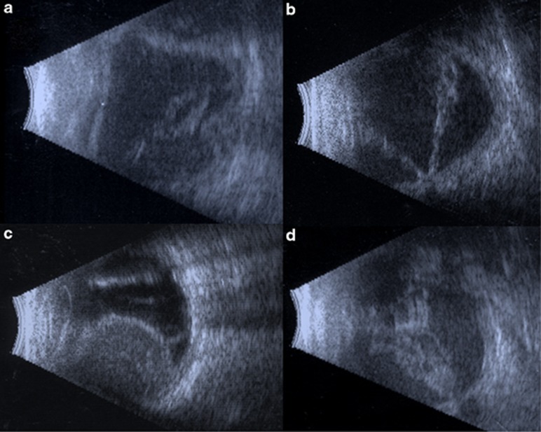
Methods: In all, 965 open globe injuries treated at the Massachusetts Eye and Ear Infirmary between 1 January 2000 and 1 June 2010 were retrospectively reviewed. A total of 427 ultrasound reports on 210 patients were analyzed. Ultrasound reports were examined for the following characteristics: vitreous hemorrhage, vitreous tag, retinal tear, RD (including subcategories total RD, partial RD, closed funnel RD, open funnel RD, and chronic RD), vitreous traction, vitreous debris, serous choroidal detachment, hemorrhagic choroidal detachment, kissing choroidal detachment, dislocated crystalline lens, dislocated intraocular lens (IOL), disrupted crystalline lens, intraocular foreign body (IOFB), intraocular air, irregular posterior globe contour, disorganized posterior intraocular contents, posterior vitreous detachment, choroidal vs retinal detachment, vitreal membranes, and choroidal thickening. The main outcome measure was visual outcome at final follow-up.
Results: Among 427 B-scan reports, there were a total of 57 retinal detachments, 19 retinal tears, 18 vitreous traction, 59 serous choroidal detachments, 47 hemorrhagic choroidal detachments, and 10 kissing choroidal detachments. Of patients with multiple studies, 26% developed retinal detachments or retinal tears on subsequent scans. Ultrasound had 100% positive predictive value for diagnosing retinal detachment and IOFB. The diagnoses of retinal detachment, disorganized posterior contents, hemorrhagic choroidal detachment, kissing choroidal detachment, and irregular posterior contour were associated with worse visual acuity at final follow-up. Disorganized posterior contents correlated with particularly poor outcomes.
Conclusions: B-scan ultrasonography is a proven, cost-effective imaging modality in the management of an open globe injury. This tool can offer both diagnostic and prognostic information, useful for both surgical planning and further medical management.
Figures
References
-
- Sawyer MN. Ultrasound imaging of penetrating ocular trauma. J Emerg Med. 2009;36:181–182. - PubMed
-
- Rubsamen PE, Cousins SW, Winward KE, Byrne SF. Diagnostic ultrasound and pars plana vitrectomy in penetrating ocular trauma. Ophthalmology. 1994;101:809–814. - PubMed
-
- Ritchie JV, Horne ST, Perry J, Gay D. Ultrasound triage of ocular blast injury in the military emergency department. Mil Med. 2012;177:174–178. - PubMed
-
- Gay DA, Ritchie JV, Perry JN, Horne S. Ultrasound of penetrating ocular injury in a combat environment. Clin Radiol. 68:82–84. - PubMed
-
- Joseph DP, Pieramici DJ, Beauchamp NJJr. Computed tomography in the diagnosis and prognosis of open-globe injuries. Ophthalmology. 2000;107:1899–1906. - PubMed
MeSH terms
LinkOut - more resources
Full Text Sources
Other Literature Sources
Medical
Miscellaneous
