. 2014 Mar 28:263:159-80.
doi: 10.1016/j.neuroscience.2013.12.057. Epub 2014 Jan 7.

Connexin36 identified at morphologically mixed chemical/electrical synapses on trigeminal motoneurons and at primary afferent terminals on spinal cord neurons in adult mouse and rat

Affiliations

Connexin36 identified at morphologically mixed chemical/electrical synapses on trigeminal motoneurons and at primary afferent terminals on spinal cord neurons in adult mouse and rat

W Bautista et al. Neuroscience. .

Abstract

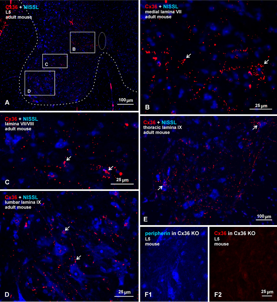
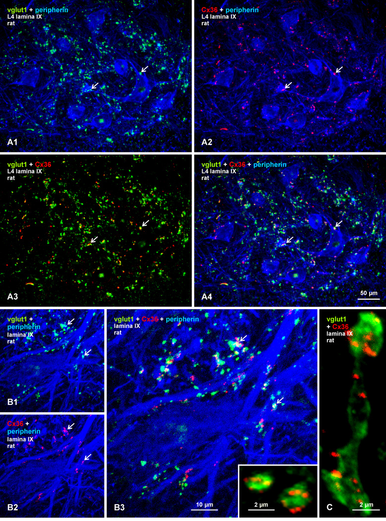
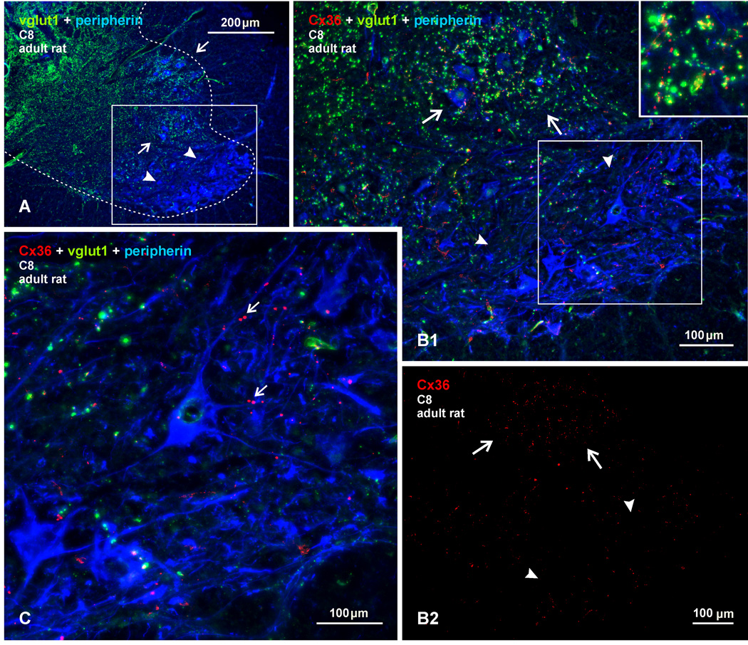
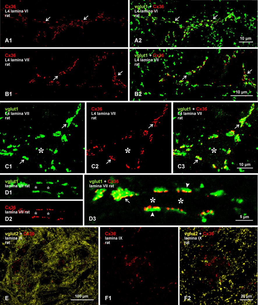
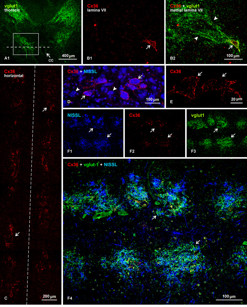
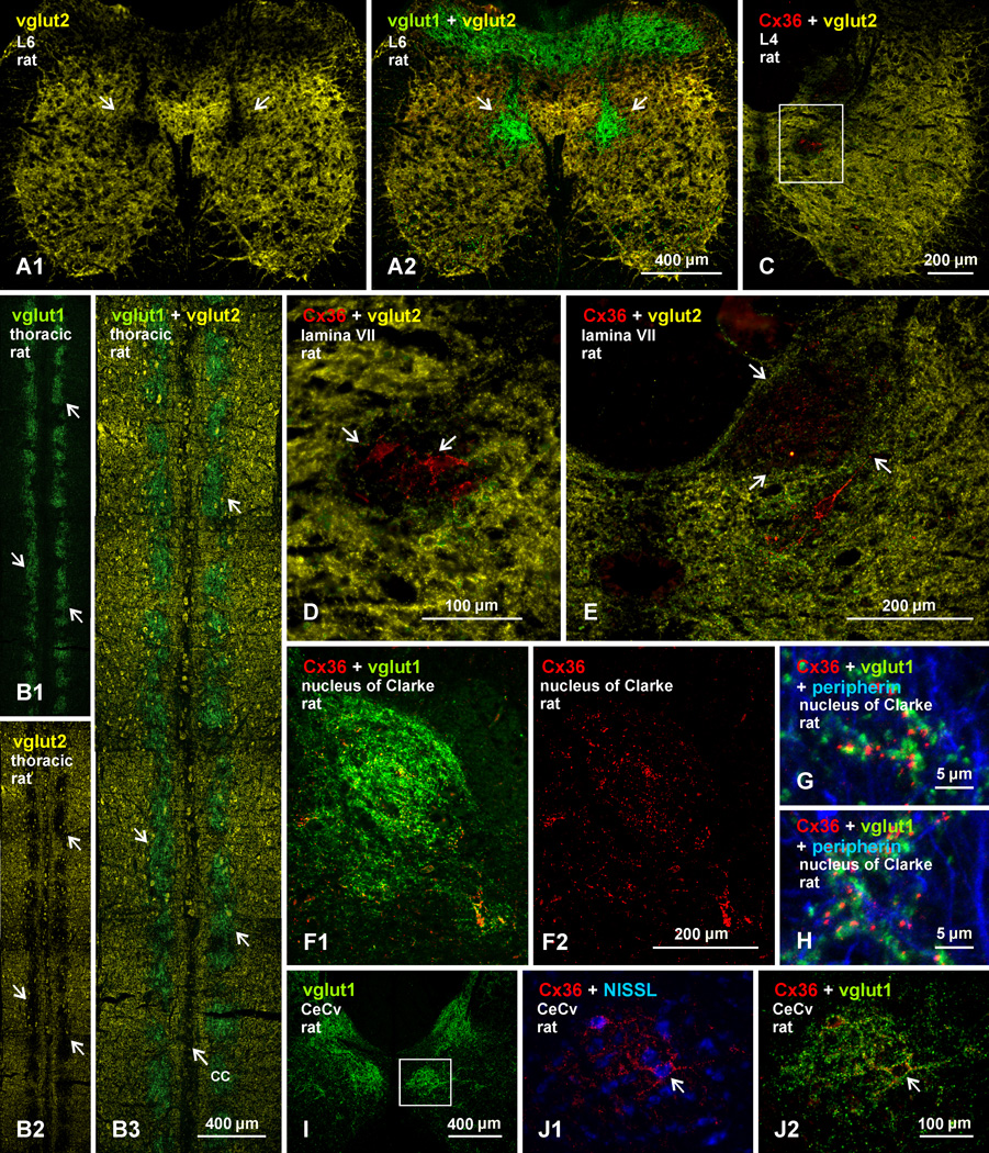
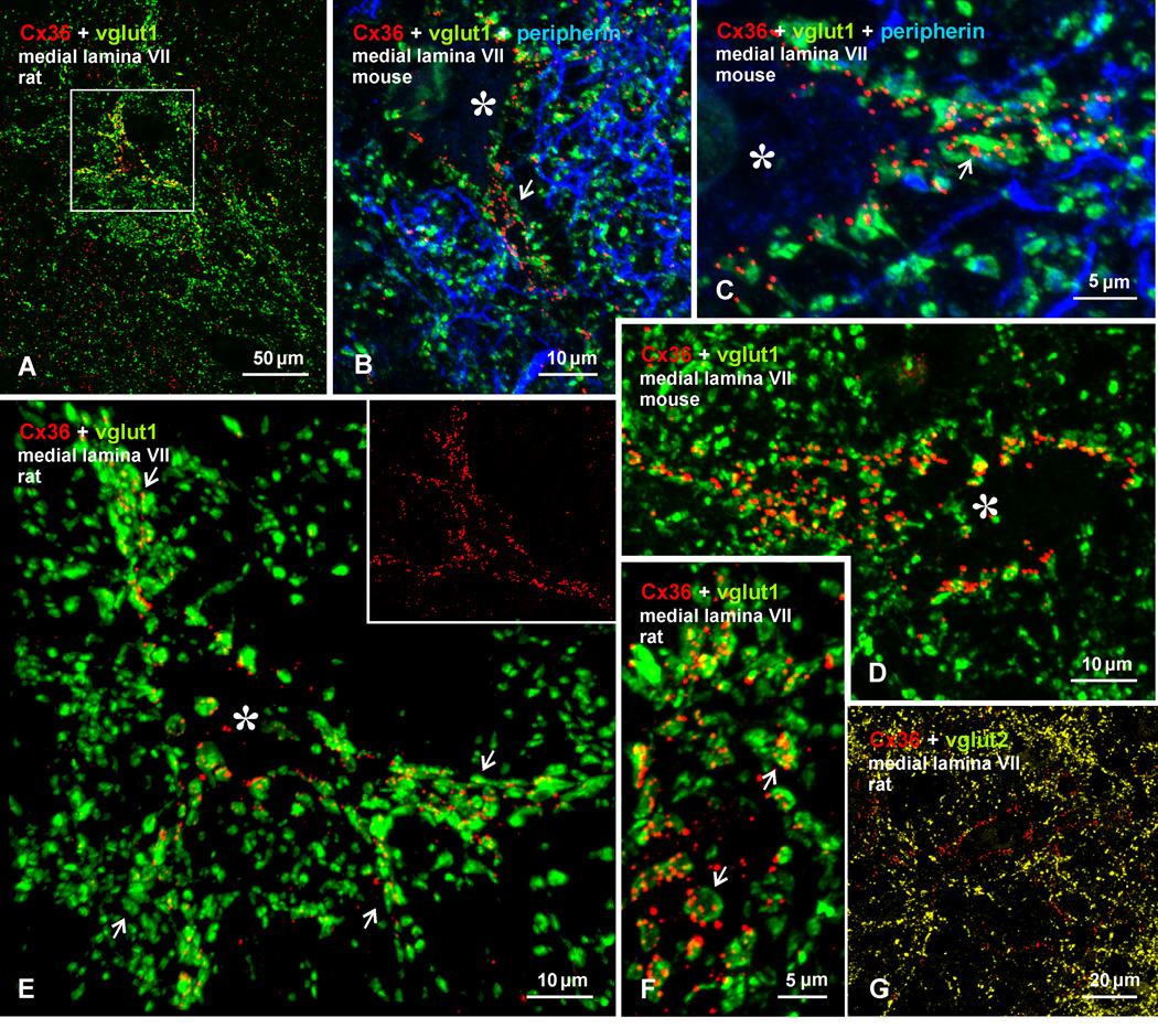
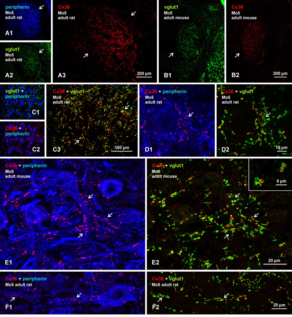
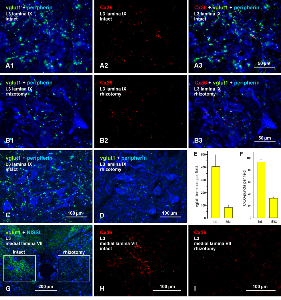
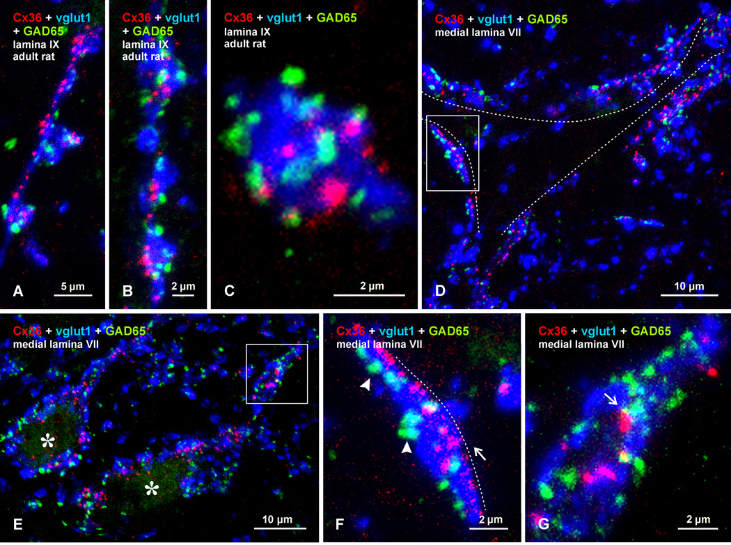
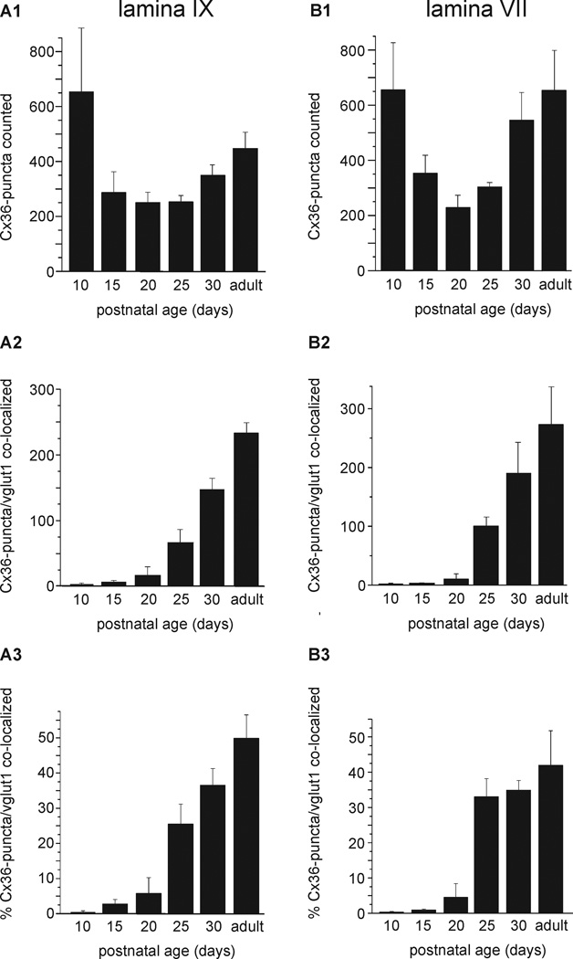
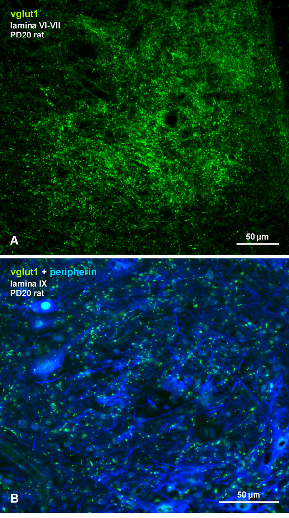
Morphologically mixed chemical/electrical synapses at axon terminals, with the electrical component formed by gap junctions, is common in the CNS of lower vertebrates. In mammalian CNS, evidence for morphologically mixed synapses has been obtained in only a few locations. Here, we used immunofluorescence approaches to examine the localization of the neuronally expressed gap junction forming protein connexin36 (Cx36) in relation to the axon terminal marker vesicular glutamate transporter-1 (vglut1) in the spinal cord and the trigeminal motor nucleus (Mo5) of rat and mouse. In adult rodents, immunolabeling for Cx36 appeared exclusively as Cx36-puncta, and was widely distributed at all rostro-caudal levels in most spinal cord laminae and in the Mo5. A high proportion of Cx36-puncta was co-localized with vglut1, forming morphologically mixed synapses on motoneurons, in intermediate spinal cord lamina, and in regions of medial lamina VII, where vglut1-containing terminals associated with Cx36 converged on neurons adjacent to the central canal. Unilateral transection of lumbar dorsal roots reduced immunolabeling of both vglut1 and Cx36 in intermediate laminae and lamina IX. Further, vglut1-terminals displaying Cx36-puncta were contacted by terminals labeled for glutamic acid decarboxylase65, which is known to be contained in presynaptic terminals on large-diameter primary afferents. Developmentally, mixed synapses begin to emerge in the spinal cord only after the second to third postnatal week and thereafter increase to adult levels. Our findings demonstrate that axon terminals of primary afferent origin form morphologically mixed synapses containing Cx36 in broadly distributed areas of adult rodent spinal cord and Mo5.

Keywords: gap junctions; interneurons; motoneurons; vesicular glutamate transporter-1.


Figures

Fig. 1
Fig. 1
Overview of widely distributed immunofluorescence labelling of Cx36 in transverse sections of adult mouse spinal cord, counterstained by blue fluorescence Nissl or labelled for peripherin. (A) Low magnification at an L5 level, with border between gray and white matter outlined by coarse dotted line, and the central canal outlined by fine dotted line. Boxed areas indicate regions examined in greater detail in subsequent Figures. (B-D) Higher magnifications of the boxed areas in (A), showing dense collections of Cx36-puncta in medial regions of lamina VII near the central canal (B, arrows), a moderate distribution of Cx36-puncta in intermediate lamina VII and VIII (C, arrows), and Cx36-puncta localized to somata and dendrites of motoneurons in lamina IX (D, arrows). (E) Lamina IX at a thoracic level, showing Cx36-puncta (arrows) distributed among motoneurons. (F) Lamina IX at L5 level from spinal cord of a Cx36 knockout mouse, showing peripherin-positive motoneurons (F1) and, in the same field, an absence of labelling for Cx36 (F2).
Fig. 2
Fig. 2
Triple immunofluorescence labelling for Cx36, vglut1 and peripherin in lamina IX at the L4 level in adult rat spinal cord. (A1-A4) Images showing the same field, with overlay of labelling for peripherin and vglut1 (A1), peripherin and Cx36 (A2), Cx36 and vglut1 (A3), and for all three proteins (A4). Red/green overlay appears as yellow, and red/green/blue overlay appears as white. Peripherin-positive motoneurons receiving a moderate innervation by vglut1-containing terminals (A1, arrows) display Cx36-puncta localized on their somata or dendrites (A2, arrows). Most though not all of these Cx36-puncta are co-localized with vglut1-positive terminals (A3 and A4, arrows). (B1-B3) Higher magnification confocal triple immunofluorescence showing immunolabels (arrows) for vglut1 (B1) and Cx36 (B2) associated with initial dendrites of peripherin-positive motoneurons. A proportion of Cx36-puncta are seen co-localized with vglut1-positive terminals, as seen in overlay (B3, arrows), but not all of these terminals harbor Cx36-puncta. (C) Individual vglut1-positive boutons or fibers appearing to form en passant terminal contacts display several Cx36-puncta; also shown in inset in (B3).
Fig. 3
Fig. 3
Triple immunofluorescence labelling of Cx36, vglut1 and peripherin at a lower cervical level (C8) in adult rat spinal cord. (A) Low magnification showing the distribution of vglut1-terminals in spinal cord gray matter (outlined by dotted line), and the heterogeneous density of these terminals among groups of motor nuclei in lamina IX (arrows, high levels; arrowheads, low levels). (B1) Higher magnification of boxed area in (A), showing a moderate density of vglut1-terminals among a dorsally located group of motoneurons (arrows), and few vglut1-terminals in a ventrally located motoneuron group (arrowheads). Cx36-puncta in the upper group of motoneurons show extensive co-localization with vglut1-terminals (shown in inset). (B2) The same field as in (B1), with labelling of Cx36 alone, showing a high density of Cx36-puncta in the upper motoneuronal group (arrows) and sparse Cx36-puncta in the lower group (arrowheads). (C) Magnification of the boxed area in (B2), showing only a few Cx36-puncta (arrows) associated with peripherin-positive motoneurons in the lower group and their lack of co-localization with vglut1.
Fig. 4
Fig. 4
Confocal double immunofluorescence labelling of Cx36 with vglut1 or vglut2 in adult rat lumbar spinal cord. (A,B) Images show the same field in lamina VI (A1,A2) and in lamina VII (B1,B2), with labelling for Cx36 alone (A1,B1) and after overlay with labelling for vglut1 (A2,B2). Arrays of Cx36-puncta are seen streaming across mid regions of these lamina (A1,B1, arrows). Labelling for vglut1 follows similar trajectories as the Cx36-puncta, and nearly all Cx36-puncta are either co-localized with, or lie close to, vglut1-terminals (A2,B2, arrows). (C1-C3) Images show the same field of a single medium sized neuron in lamina VII (not counterstained, but marked with asterisk). The neuron is contacted by vglut1-terminals (C1, arrows) displaying clusters of Cx36-puncta (C2 and C3, arrows). (D1-D3) Images of the same field in lamina VII, showing label for vglut1 (D1) and Cx36 (D2) on a large initial dendrite (not counterstained, but marked with asterisks). Terminals labelled for vglut1 are viewed on edge flattened against the dendrite (D3, arrowheads) or en face (D3, arrow). In edge views, Cx36-puncta are localized to the side of the terminal contacting the dendrite, and straddle sites of terminal/dendrite apposition. (E,F) Double immunofluorescence labelling of Cx36 (red) and vglut2 (yellow) in lamina IX at low (E) and higher confocal magnification (F), showing clusters of Cx36-puncta (F1) that in the same field lack association with vglut2-positive terminals (F2).
Fig. 5
Fig. 5
Immunofluorescence localization of Cx36 in relation to vglut1-terminals in medial lamina VII at a thoracic level in adult rat spinal cord. (A) Low magnification transverse section, showing vglut1-positive fibers and terminals (outlined by boxed area) lateral to the central canal (cc, arrow). (B) Magnification of a similar area as the boxed region in (A), showing a single neuron decorated with Cx36-puncta (B1, arrow) and, in the same field, a confluence of vglut1-positive terminals (arrowheads) terminating on that neuron (B2, arrow). (C) Photomontage of a field 2 mm in length, showing immunolabelling for Cx36 in a horizontal section taken at a dorso-ventral plane indicated by the dotted line in (A), with the central canal lying at midline (dashed line). Cx36-puncta are sufficiently concentrated on neurons to reveal intermittent clusters of Cx36 laden cells (arrows) flanking the central canal. (D,E) Higher magnifications of one of these clusters in a section counterstained with blue fluorescence Nissl, showing Cx36-puncta on some neurons (D, arrows) intermingled with those lacking Cx36-puncta (D, arrowheads). A confocal image shows the punctate appearance of labelling associated with and delineating two neurons within a cluster (E, arrows). (F1-F4) Horizontal section showing the same field double-labelled and blue Nissl counterstained, revealing that clusters of these neurons (F1, arrows), with their associated Cx36-puncta (F2, arrows), are all targeted by vglut1-terminals (F3, arrows), producing patches of labelling for vglut1 that demarcate locations of the clusters (F4, arrows).
Fig. 6
Fig. 6
Comparison of immunofluorescence labelling for vglut1 and vglut2 in relation to Cx36-puncta on neurons in medial lamina VII in adult rat spinal cord. (A1-A2) The same field of a double-labelled transverse section at L6, showing immunofluorescence for vglut2 (yellow) nearly throughout spinal cord gray matter, except in a restricted region of medial lamina VII devoid of vglut2-terminals (A1, arrows), which corresponds to an area rich in vglut1-terminals (A2, arrows). (B1-B3) Horizontal thoracic section at the level of the central canal (cc), showing more broadly the correspondence between patches of dense vglut1-terminals (B1, arrows) and voids in labelling for vglut2 (B, arrows), as seen in overlay (B3, arrows). (C-E) Transverse sections at mid lumbar levels, showing two voids in labelling for vglut2 in medial lamina VII: one adjacent to the central canal (C, boxed area), containing neurons densely decorated with Cx36-puncta (D, arrows; magnification of box in C); and the other corresponding to the nucleus of Clarke (E, arrows). (F-H) Images showing nucleus of Clarke outlined by dense labelling for vglut1 (F1) and containing a moderate distribution of scattered Cx36-puncta (F2) that display co-localization with vglut1, as shown by examples in two regions of Clark’s nucleus (G,H). (I) Image of adult rat spinal cord at C8, showing dense labelling for vglut1 in the central cervical nucleus (CeCv) (boxed area). (J) Higher magnification of CeCv showing neurons decorated with Cx36-puncta (J1, arrow) display co-localization with vglut1-terminals (J2, arrow), similar to those seen in medial lamina VII at other spinal levels.
Fig. 7
Fig. 7
Confocal immunofluorescence of vglut1 and Cx36 in medial lamina VII of adult rat and mouse lumbar spinal cord. (A) Image from rat cord, showing a vglut1-positive patch of terminals outlining a neuron decorated with Cx36-puncta (boxed area). (B-D) Higher magnification immunofluorescence images from medial lamina VII of mouse cord, showing a dense meshwork of peripherin-positive primary afferent fibers (blue) within a vglut1-positive patch, and individual neurons (marked by asterisks) with Cx36-puncta concentrated along their initial dendritic segments (B, arrows), where many are associated with vglut1-terminals (C, arrows), or with Cx36-puncta associated with vglut1-terminals distributed around the entire neuronal somata (D). (E) Higher magnification of the neuron (marked by asterisk) in the boxed area in (A), showing Cx36-puncta co-distributed with vglut1-terminals contacting the neuronal somata and several initial dendrites (arrows). The inset shows the same field as in (E), with labelling for Cx36 alone delineating the entire neuronal somata. (F) Magnification showing multiple Cx36-puncta localized to most vglut1-terminals (arrows). (G) Double immunofluorescence of Cx36 and vglut2 in a field similar to that in (A), showing lack of Cx36-puncta co-localization with vglut2-positive terminals.
Fig. 8
Fig. 8
Triple immunofluorescence labelling of Cx36, vglut1 and peripherin in the trigeminal motor nucleus (Mo5) of adult rat and mouse brainstem. (A,B) Low magnification images of the same field of the Mo5 in rat (A1-A3) and in mouse (B1,B2), showing peripherin-positive motoneurons (A1, arrow), dense labelling for vglut1 (A2,B1, arrows) and Cx36 (A3,B2, arrows) throughout the nucleus, and sparse labelling for these proteins outside the nucleus. (C) Higher magnification showing the distribution of labelling for Cx36 (C1) and vglut1 (C2) among peripherin-positive motoneurons and, in the same field, co-localization of labelling for Cx36 with vglut1-terminals (C3, arrows). (D,E) Higher magnification confocal images, where the fields in D1 and D2 are the same and those in E1 and E2 are the same. Images show punctate labelling of Cx36, distribution of Cx36-puncta around a motoneuronal soma (D1, arrow), association of Cx36-puncta with initial dendrite segments (E1, arrows) as well as more distally along dendrites (F1, arrow), and overlap of the vast majority of Cx36-puncta with vglut1-terminals (D2,E2,F2, arrows). Inset in E2 shows multiple Cx36-puncta localized to individual vglut1-terminals.
Fig. 9
Fig. 9
Association of Cx36 with vglut1-containing axon terminals of primary afferent origin in lamina IX and VII of adult rat. (A,B) Triple immunofluorescence labelling for vglut1, peripherin and Cx36 in lamina IX after unilateral dorsal rhizotomy at the L1-L4 levels. Images at L3 on the intact side (A1-A3, same field) and rhizotomy side (B1-B3, same field) show vglut1/peripherin overlay (A1,B1), Cx36 alone (A2,B2) and overlay of labelling for all three proteins (A3,B3). The intact side shows normal labelling of vglut1 (A1) and Cx36 (A2). The rhizotomy side shows reduced labelling of vglut1 (B1) and Cx36 (B2). Overlay shows vglut1/Cx36 co-localization at some of the remaining vglut1-terminals on the rhizotomy side (B3). (C,D) Labelling for vglut1 and peripherin under optimum fixation conditions for vglut1 (4% formaldehyde), showing vglut1 on the intact side (C) and extensive loss of vglut1-terminals on the rhizotomy side (D). (E,F) Quantitation of labelling in fields of lamina IX on the intact and rhizotomy side, showing an 80% reduction of vglut1-terminals and a 65% reduction of Cx36-puncta on the rhizotomy side. (G) Low magnification blue fluorescence Nissl counterstained transverse section showing areas adjacent to the central canal after unilateral dorsal rhizotomy. Dense labelling for vglut1 is seen on the intact side (boxed region at left), and depletion of labelling is seen on the rhizotomy side (boxed region at right). (H,I) Immunolabelling for Cx36 in the boxed areas shown in (G), with the left box magnified in (H) showing clusters of Cx36-puncta on the intact side, and the right box magnified in (I) showing only a few puncta remaining on the rhizotomy side.
Fig. 10
Fig. 10
Confocal triple immunofluorescence labelling for Cx36 (red), vglut1 (blue) and GAD65 (green), showing localization of GAD65-positive P boutons on vglut1-terminals displaying Cx36-puncta in lamina IX and VII of adult rat. Terminals labelled for vglut1 are pseudo colored blue to avoid obscuring GAD65 P boutons and Cx36-puncta. (A-C) Images from lamina IX, showing GAD65-positive P boutons associated with what appear to be en passant type vglut1-terminals (A) or clusters of these terminals (B), with often multiple P boutons and Cx36-puncta associated with individual vglut1-terminals (C). (D-G) Images from lamina VII showing a single neuron (D, outlined by dotted line), and two tangentially cut dendrites (E, asterisks). In each image, Cx36-puncta are seen co-localized with large vglut1-terminals that contact neuronal somata and dendrites, and that are themselves contacted by GAD65-positive P boutons. (F,G) Boxed areas in (D) and (E) (magnified in F and G, respectively), showing Cx36-puncta (F, arrow) localized along portions of vglut1-terminals nearest the neuronal soma (F, dotted line), and GAD65-positive P boutons (F, arrowheads) at more distal portions of the vglut1-terminals. Cx36-puncta are occasionally in close proximity to GAD65-positive P boutons (G, arrow).
Fig. 11
Fig. 11
Developmental profile of Cx36 association with vglut1-terminals in lamina IX and VII in rat lumbar spinal cord. (A,B) Histograms show the average of total Cx36-puncta counted in fields of lamina IX and VII from four animals are each age indicated (A1,B1), the total Cx36-puncta colocalized with vglut1-terminals averaged from the four animals (A2,B2), and the average percentage of Cx36/vglut1 co-localization (A3,B3). The values at different ages in (A1,B1) reflect differences in the number of fields/per animal taken for counts and/or heterogeneities in numbers of Cx36-puncta in fields chosen for counts, rather than absolute differences in density of Cx36-puncta at the ages examined. In lamina XI, the average percentage of Cx36-puncta/vglut1 co-localization was 0.4% at PD10, 2.7% at PD15, 5.7% at PD20, 25% at PD25, 36% at PD30 and 49% in adult. In lamina VII, the values were 0.3% at PD10, 0.9% at PD15, 4% at PD20, 33% at PD25, 34% at PD30 and 41% in adult. Values are means ± s.e.m. (n = 4).
Fig. 12
Fig. 12
(A,B) Immunofluorescence labelling of vglut1 (green) in laminae VI - VII (A) and in lamina IX (B) at the L4 level of rat spinal cord at PD20. Motoneurons in (B) are immunolabelled for peripherin (blue). A high density of axon terminals are labelled for vglut1 in lamina VI and VII, and abundant vlgut1-positive terminals are present among motoneurons in lamina IX at this postnatal age when Cx36-puncta/vglut1 co-localization in these lamina was a small fraction of that seen in these lamina of adult spinal cord.

References

    1. Alvarez FJ, Villalba RM, Zerda R, Schneider SP. Vesicular glutamate transporters in the spinal cord, with special reference to sensory primary afferent synapses. J Comp Neurol. 2004;472:257–280. - PubMed
    1. Arasaki K, Kudo N, Nakanishi T. Firing of spinal motoneurons due to electrical interactions in the rat: an in vitro study. Exp Brain Res. 1984;54:437–445. - PubMed
    1. Bautista W, Nagy JI, Dai Y, McCrea DA. Requirement of neuronal connexin36 in pathways mediating presynaptic inhibition of primary afferents in functionally mature mouse spinal cord. J Physiol. 2012;590:3821–3839. - PMC - PubMed
    1. Bautista W, Rash JE, Vanderpool KG, Yasumura T, Nagy JI. Re-evaluation of connexin (Cx26, Cx32, Cx36, Cx37, Cx40, Cx43, Cx45) association with motoneurons in rodent spinal cord, sexually dimorphic motor nuclei and trigeminal motor nucleus. 2013a Submitted. - PMC - PubMed
    1. Bautista W, Nagy JI. Connexin36 forms purely electrical synapses in sexually dimorphic lumbosacral motor nuclei in spinal cord of developing and adult rat and mouse. 2013b Submitted. - PMC - PubMed

Publication types

LinkOut - more resources