Review
. 2014 May;146(6):1534-1546.e3.
doi: 10.1053/j.gastro.2014.01.001. Epub 2014 Jan 7.

Gastrointestinal malignancy and the microbiome

Affiliations
Review

Gastrointestinal malignancy and the microbiome

Maria T Abreu et al. Gastroenterology. 2014 May.

Abstract

Microbial species participate in the genesis of a substantial number of malignancies-in conservative estimates, at least 15% of all cancer cases are attributable to infectious agents. Little is known about the contribution of the gastrointestinal microbiome to the development of malignancies. Resident microbes can promote carcinogenesis by inducing inflammation, increasing cell proliferation, altering stem cell dynamics, and producing metabolites such as butyrate, which affect DNA integrity and immune regulation. Studies in human beings and rodent models of cancer have identified effector species and relationships among members of the microbial community in the stomach and colon that increase the risk for malignancy. Strategies to manipulate the microbiome, or the immune response to such bacteria, could be developed to prevent or treat certain gastrointestinal cancers.

Keywords: Bacteria; Cancer; Inflammation.


Figures

Figure 1
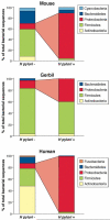
Figure 1. Differences in the composition of human and rodent gastric microbiome, based on H pylori status
There are substantial variations in the gastric microbiota of humans, mice, and gerbils. Nonetheless, the presence of H pylori alters the constituents of these ecosystems. This diagram shows the relative abundance of phyla, determined by high-throughput sequencing, in the stomach of humans, gerbils, and mice,,,. The risk of cancer increases with the presence of H pylori.
Figure 2
Figure 2. Potential effects of the gastroesophageal microbiome on complications of gastroesophageal reflux disease
The healthy esophagus has a type I microbiome, dominated by Gram-positive bacteria. In contrast, patients with GERD or Barrett's esophagus have a different population of esophageal microbes (a type II microbiome), comprised primarily of Gram-negative bacteria. There are several mechanisms through which these different microbial populations might affect gastroesophageal reflux. Gram-negative organisms produce immune-activating molecules such as LPS, which induces innate immune responses that lead to disease. LPS binds to and activates TLR4, leading to activation of NFκB; levels of activated NFκB are increased in esophageal samples from patients with reflux esophagitis, Barrett's esophagus, and esophageal adenocarcinoma. Increased activation of NFκB leads to increased production of inflammatory cytokines such as IL1β, IL6, IL8, and TNFα. In this manner, the type II esophageal microbiome contributes to esophagitis. LPS signaling also increases levels of iNOS, which reduces the basal tone of the lower esophageal sphincter. Over prolonged periods, this increases risk for reflux and its sequelae. LPS has also been shown to delay gastric emptying in rodents, which increases the amount of gastric contents refluxed into the esophagus. Production of iNOS can therefore lower the threshold for reflux of gastric contents into the esophagus.
Figure 3
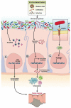
Figure 3. Bacteria associated colon cancer
In a homeostatic colonic environment, bacteria are separated from the colonic epithelial layer by mucus, anti-microbial peptides, and secreted immunoglobulin A (IgA). Certain environmental factors induce microbial dysbiosis, leading to interactions between microbes and intestinal epithelial cells. Dysbiotic microbiota induces colon carcinogenesis by (A) releasing reactive oxygen species or reactive nitrogen species (ROS/RNS) and causing DNA damage, , , (B) releasing toxins that directly damage DNA or induce cell proliferation genes (C) generating MAMPs, which activate the innate immune response, recruit immune cells, and induce inflammation.
Figure 4
Figure 4. Bacteria activate TLR signaling to promote colon carcinogenesis
Microbial dysbiosis or mutations can activate TLR signaling, leading to phosphorylation of β–catenin, cell proliferation, and tumor progression. It is not clear whether other TLRs promote colon carcinogenesis via activation of β-catenin.

References

    1. Cho I, Blaser MJ. The human microbiome: at the interface of health and disease. Nature Rev Genetics. 2012;13:260–70. - PMC - PubMed
    1. Plottel CS, Blaser MJ. Microbiome and malignancy. Cell Host & Microbe. 2011;10:324–35. - PMC - PubMed
    1. de Martel C, Ferlay J, Franceschi S, Vignat J, Bray F, Forman D, Plummer M. Global burden of cancers attributable to infections in 2008: a review and synthetic analysis. Lancet Oncology. 2012;13:607–15. - PubMed
    1. Polk DB, Peek RM., Jr. Helicobacter pylori: gastric cancer and beyond. Nat Rev Cancer. 2010;10:403–14. - PMC - PubMed
    1. Wroblewski LE, Peek RM. H. pylori in gastric carcinogenesis: mechanisms. Gastroenterology Clinics of North America. 2013;42:285–298. - PMC - PubMed

Publication types

MeSH terms