

Morphometric analysis of small arteries in the human retina using adaptive optics imaging: relationship with blood pressure and focal vascular changes
- PMID: 24406779
- PMCID: PMC3966915
- DOI: 10.1097/HJH.0000000000000095
Morphometric analysis of small arteries in the human retina using adaptive optics imaging: relationship with blood pressure and focal vascular changes
Abstract
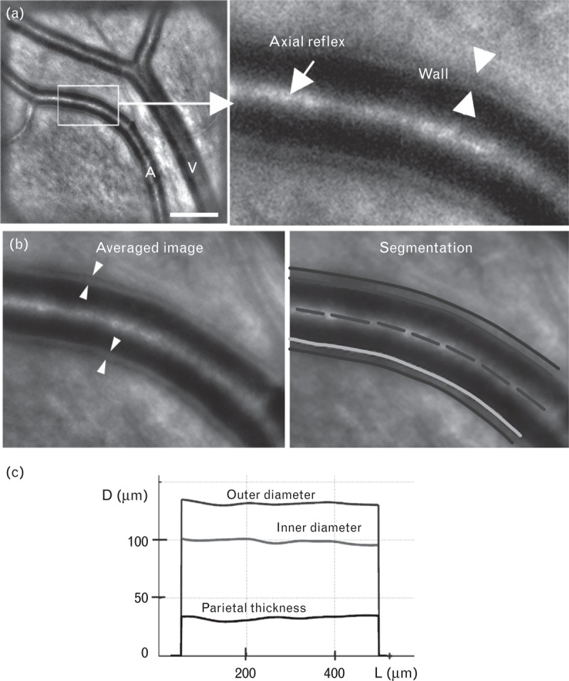
Objectives: The wall-to-lumen ratio (WLR) of retinal arteries is a recognized surrogate of end-organ damage due to aging and/or arterial hypertension. However, parietal morphometry remains difficult to assess in vivo. Recently, it was shown that adaptive optics retinal imaging can resolve parietal structures of retinal arterioles in humans in vivo. Here, using adaptive optics retinal imaging, we investigated the variations of parietal thickness of small retinal arteries with blood pressure and focal vascular damage.
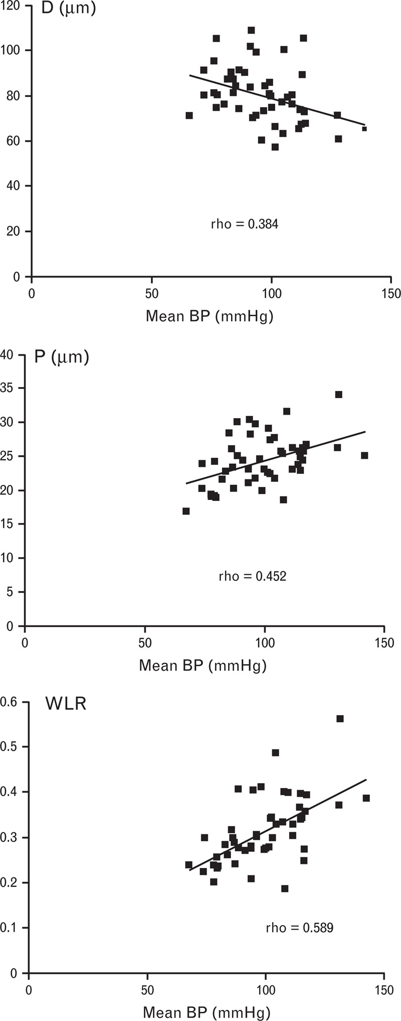
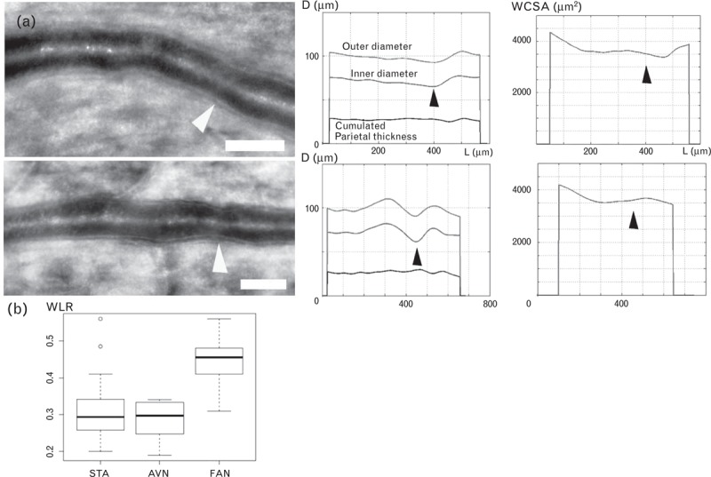
Methods: Adaptive optics imaging of the superotemporal retinal artery was done in 49 treatment-naive individuals [mean age (±SD) 44.9 years (±14); mean systolic pressure 132 mmHg (±22)]. Semi-automated segmentation allowed extracting parietal thickness and lumen diameter. In a distinct cohort, adaptive optics images of arteriovenous nicking (AVN; n = 12) and focal arteriolar narrowing (FAN; n = 10) were also analyzed qualitatively and quantitatively.
Results: In the cohort of treatment-naive individuals, by multiple regression taking into account age, body mass index, mean, systolic, diastolic and pulse blood pressure, the WLR was found positively correlated to mean blood pressure and age which in combination accounted for 43% of the variability of WLR. In the cohort of patients with focal vascular damage, neither FANs or AVNs showed evidence of parietal growth; instead, at sites of FANs, decreased outer diameter suggestive of vasoconstriction was consistently found, while at sites of AVNs venous narrowing could be seen in the absence of arteriovenous contact.
Conclusion: High resolution imaging of retinal vessels by adaptive optics allows quantitative microvascular phenotyping, which may contribute to a better understanding and management of hypertensive retinopathy.
Trial registration: ClinicalTrials.gov NCT01546181.
Figures

Similar articles
-
Measurement of retinal wall-to-lumen ratio by adaptive optics retinal camera: a clinical research.Graefes Arch Clin Exp Ophthalmol. 2015 Nov;253(11):1985-95. doi: 10.1007/s00417-015-3115-y. Epub 2015 Aug 13. Graefes Arch Clin Exp Ophthalmol. 2015. PMID: 26267750
-
Morphometric analysis of retinal arterioles in control and hypertensive population using adaptive optics imaging.Indian J Ophthalmol. 2019 Oct;67(10):1673-1677. doi: 10.4103/ijo.IJO_253_19. Indian J Ophthalmol. 2019. PMID: 31546506 Free PMC article.
-
Effects of age, blood pressure and antihypertensive treatments on retinal arterioles remodeling assessed by adaptive optics.J Hypertens. 2016 Jun;34(6):1115-22. doi: 10.1097/HJH.0000000000000894. J Hypertens. 2016. PMID: 27065002
-
Remodeling of retinal small arteries in hypertension.Am J Hypertens. 2011 Dec;24(12):1267-73. doi: 10.1038/ajh.2011.166. Epub 2011 Sep 29. Am J Hypertens. 2011. PMID: 21956527 Review.
-
How does hypertension affect your eyes?J Hum Hypertens. 2012 Feb;26(2):71-83. doi: 10.1038/jhh.2011.37. Epub 2011 Apr 21. J Hum Hypertens. 2012. PMID: 21509040 Review.
Cited by
-
Retinal Vascular Geometry in Hypertension: cSLO-Based Method.Ophthalmol Ther. 2023 Apr;12(2):939-952. doi: 10.1007/s40123-022-00642-4. Epub 2022 Dec 30. Ophthalmol Ther. 2023. PMID: 36583807 Free PMC article.
-
Pearls and Pitfalls of Adaptive Optics Ophthalmoscopy in Inherited Retinal Diseases.Diagnostics (Basel). 2023 Jul 19;13(14):2413. doi: 10.3390/diagnostics13142413. Diagnostics (Basel). 2023. PMID: 37510157 Free PMC article. Review.
-
Identification of Subclinical Microvascular Biomarkers in Coronary Heart Disease in Retinal Imaging.Transl Vis Sci Technol. 2021 Nov 1;10(13):24. doi: 10.1167/tvst.10.13.24. Transl Vis Sci Technol. 2021. PMID: 34787666 Free PMC article.
-
Retinal Arteriolar Morphometry Based on Full Width at Half Maximum Analysis of Spectral-Domain Optical Coherence Tomography Images.PLoS One. 2015 Dec 9;10(12):e0144437. doi: 10.1371/journal.pone.0144437. eCollection 2015. PLoS One. 2015. PMID: 26650940 Free PMC article. Clinical Trial.
-
Adaptive optics imaging of the human retina.Prog Retin Eye Res. 2019 Jan;68:1-30. doi: 10.1016/j.preteyeres.2018.08.002. Epub 2018 Aug 27. Prog Retin Eye Res. 2019. PMID: 30165239 Free PMC article. Review.
References
-
- Heagerty AM, Aalkjaer C, Bund SJ, Korsgaard N, Mulvany MJ. Small artery structure in hypertension: dual processes of remodeling and growth. Hypertension 1993; 21:391–397 - PubMed
-
- Heagerty AM. Predicting hypertension complications from small artery structure. J Hypertens 2007; 25:939–940 - PubMed
-
- Buus NH, Mathiassen ON, Fenger-Grøn M, Præstholm MN, Sihm I, Thybo NK, et al. Small artery structure during antihypertensive therapy is an independent predictor of cardiovascular events in essential hypertension. J Hypertens 2013; 31:791–797 - PubMed
-
- Mulvany MJ. Small artery remodeling in hypertension. Curr Hypertens Rep 2002; 4:49–55 - PubMed
-
- Feihl F, Liaudet L, Levy BI, Waeber B. Hypertension and microvascular remodelling. Cardiovasc Res 2008; 78:274–278 - PubMed
Publication types
MeSH terms
Associated data
LinkOut - more resources
Full Text Sources
Other Literature Sources
Medical
