. 2014 Apr 4;1841(4):630-44.
doi: 10.1016/j.bbalip.2013.12.017. Epub 2014 Jan 7.

Coenzyme Q supplementation or over-expression of the yeast Coq8 putative kinase stabilizes multi-subunit Coq polypeptide complexes in yeast coq null mutants

Affiliations

Coenzyme Q supplementation or over-expression of the yeast Coq8 putative kinase stabilizes multi-subunit Coq polypeptide complexes in yeast coq null mutants

Cuiwen H He et al. Biochim Biophys Acta. .

Abstract

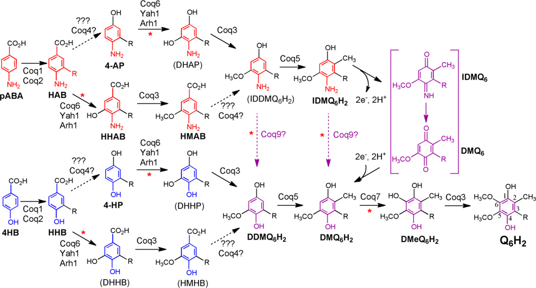
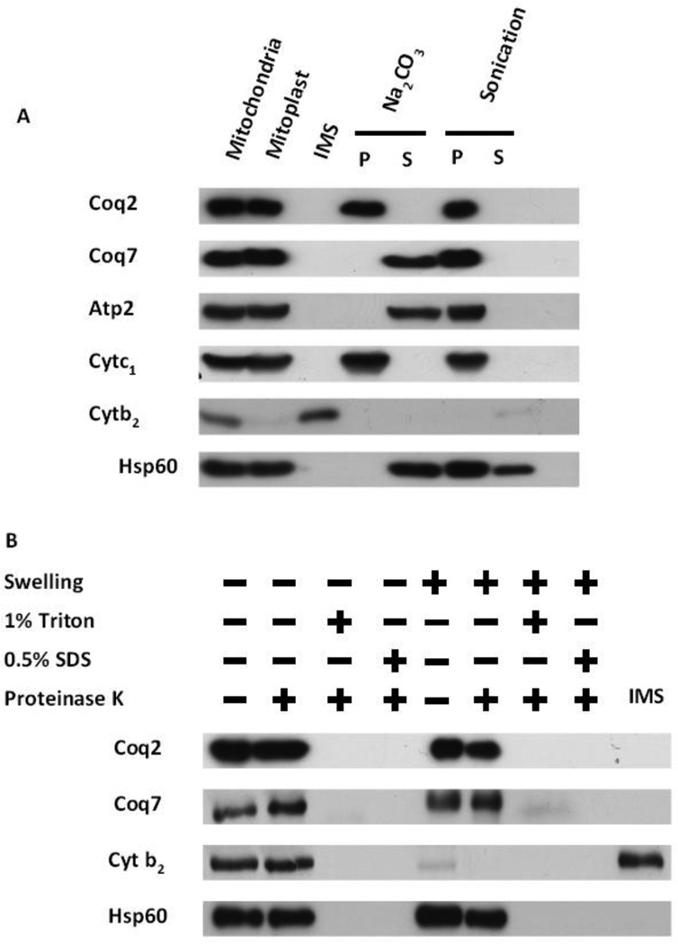
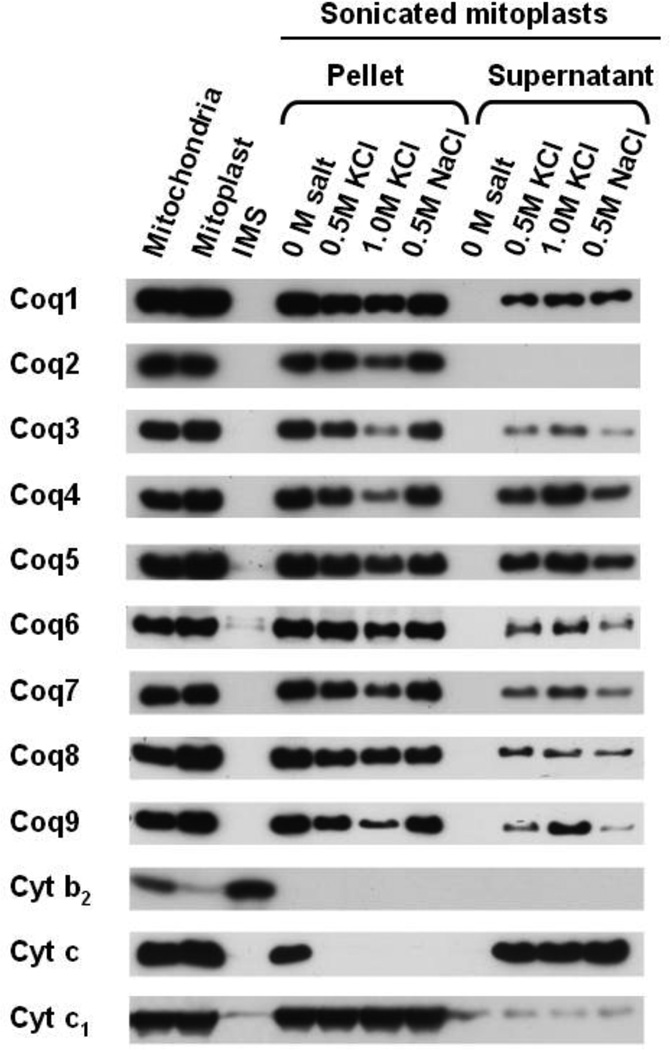
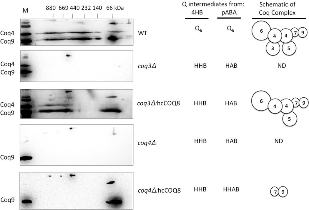
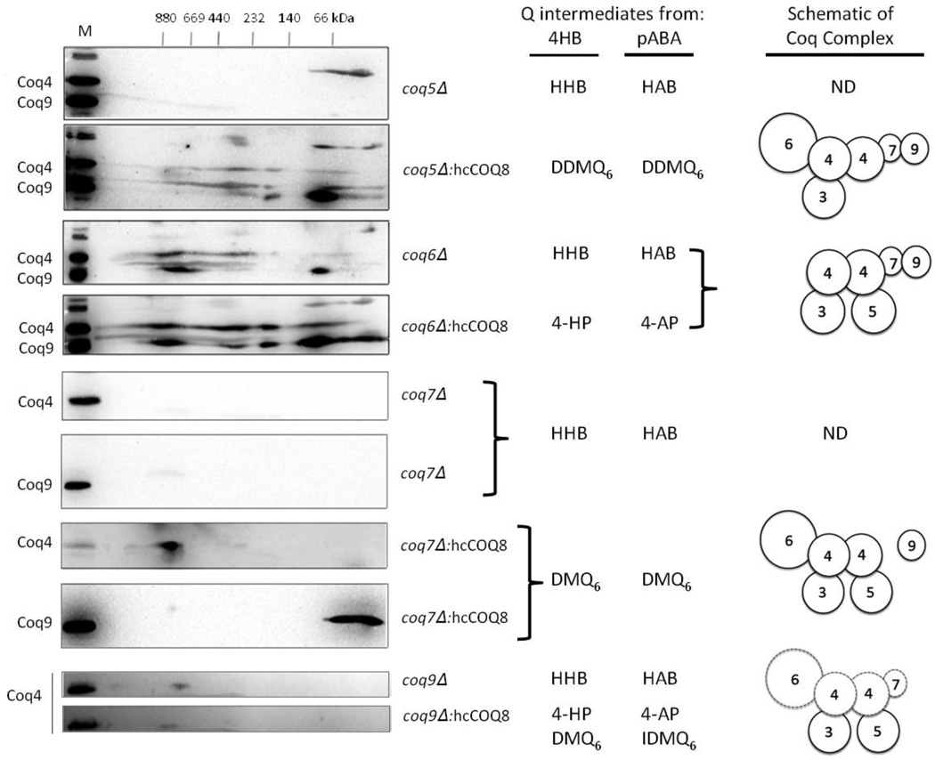
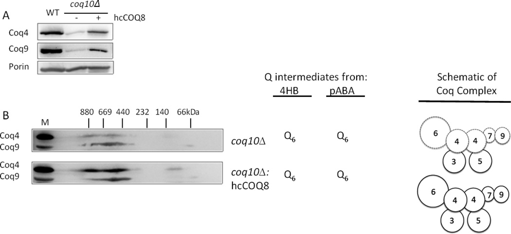
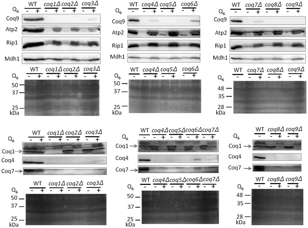
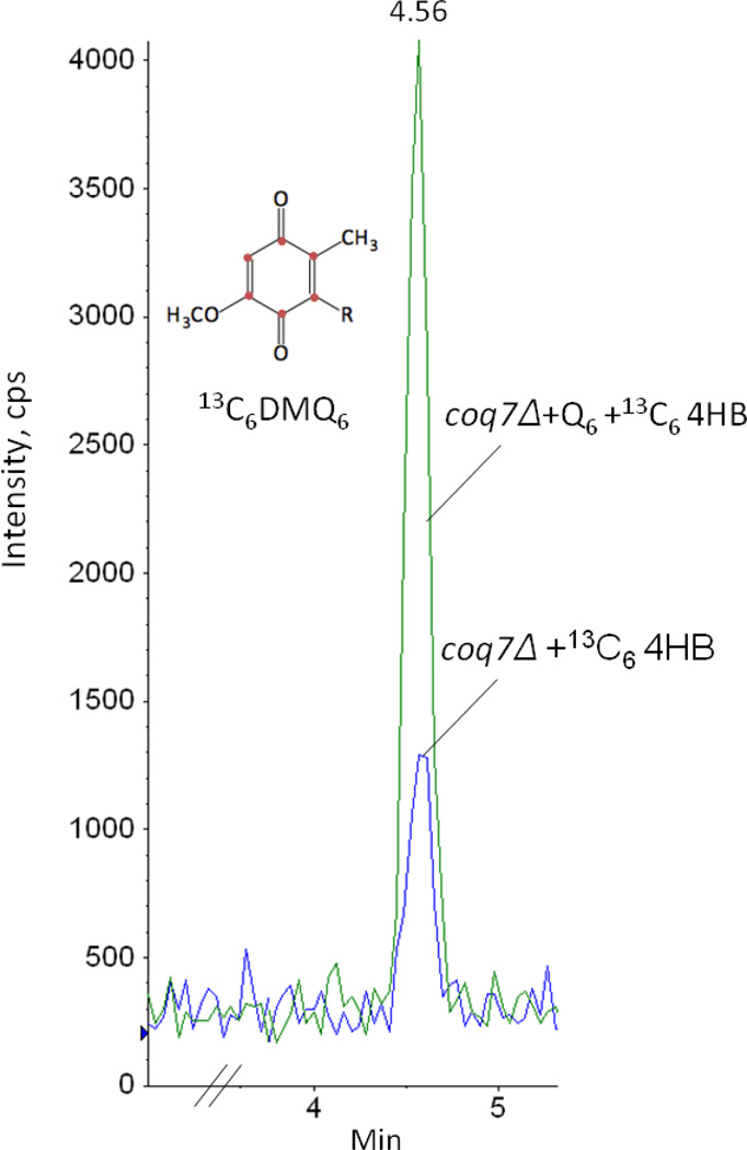
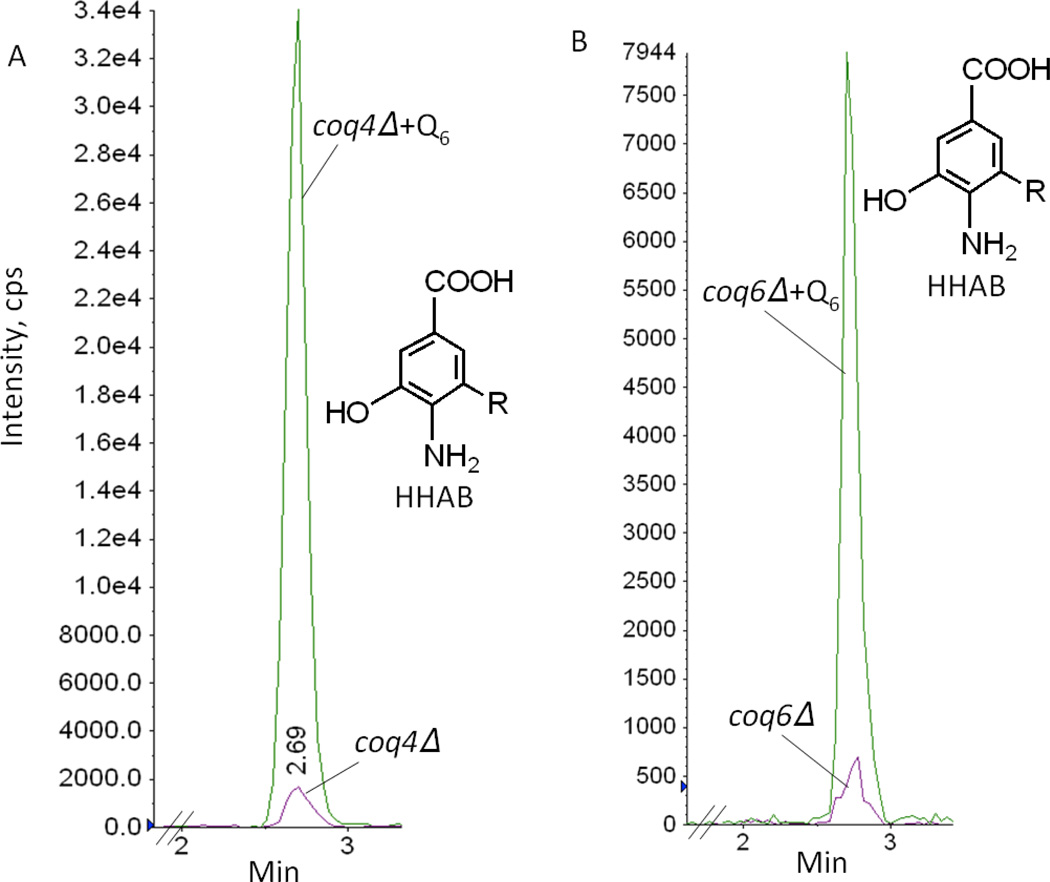
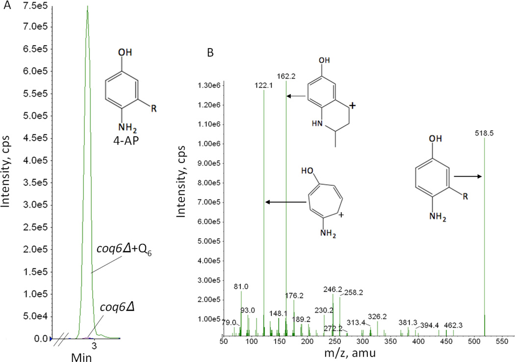
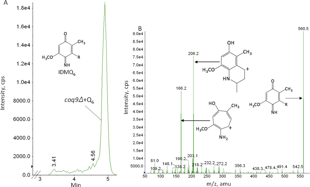
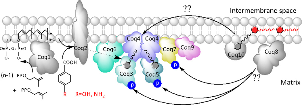
Coenzyme Q biosynthesis in yeast requires a multi-subunit Coq polypeptide complex. Deletion of any one of the COQ genes leads to respiratory deficiency and decreased levels of the Coq4, Coq6, Coq7, and Coq9 polypeptides, suggesting that their association in a high molecular mass complex is required for stability. Over-expression of the putative Coq8 kinase in certain coq null mutants restores steady-state levels of the sensitive Coq polypeptides and promotes the synthesis of late-stage Q-intermediates. Here we show that over-expression of Coq8 in yeast coq null mutants profoundly affects the association of several of the Coq polypeptides in high molecular mass complexes, as assayed by separation of digitonin extracts of mitochondria by two-dimensional blue-native/SDS PAGE. The Coq4 polypeptide persists at high molecular mass with over-expression of Coq8 in coq3, coq5, coq6, coq7, coq9, and coq10 mutants, indicating that Coq4 is a central organizer of the Coq complex. Supplementation with exogenous Q6 increased the steady-state levels of Coq4, Coq7, and Coq9, and several other mitochondrial polypeptides in select coq null mutants, and also promoted the formation of late-stage Q-intermediates. Q supplementation may stabilize this complex by interacting with one or more of the Coq polypeptides. The stabilizing effects of exogenously added Q6 or over-expression of Coq8 depend on Coq1 and Coq2 production of a polyisoprenyl intermediate. Based on the observed interdependence of the Coq polypeptides, the effect of exogenous Q6, and the requirement for an endogenously produced polyisoprenyl intermediate, we propose a new model for the Q-biosynthetic complex, termed the CoQ-synthome.

Keywords: Coenzyme Q supplementation; Mitochondrial metabolism; Protein complex; Q-biosynthetic intermediate; Saccharomyces cerevisiae; Ubiquinone.


Figures

Figure 1
Figure 1
Q6 biosynthesis in S. cerevisiae proceeds from either 4HB or pABA. The classic Q biosynthetic pathway is shown in blue emanating from 4HB (4-hydroxybenzoic acid). R represents the hexaprenyl tail present in Q6 and all intermediates. The numbering of the aromatic carbon atoms used throughout this study is shown on the reduced form of Q6, Q6H2. Coq1p synthesizes the hexaprenyldiphosphate tail, which is transferred by Coq2p to 4HB to form HHB (3-hexaprenyl-4-hydroxybenzoic acid). Alternatively, the red pathway indicates that pABA (para-aminobenzoic acid) is prenylated by Coq2p to form HAB (3-hexaprenyl-4-aminobenzoic acid). Both HHB and HAB are early Q6-intermediates, readily detected in each of the coq null strains (Δcoq3-Δcoq9). Subsequent ring modification steps are thought to occur in the sequences shown, including hydroxylation by Coq6 in concert with ferredoxin (Yah1) and ferredoxin reductase (Arh1). Coq3 performs the two O-methylation steps, Coq5 the C-methylation step, and Coq7 performs the penultimate hydroxylation step. The functional roles of the Coq4, Coq8, and Coq9 polypeptides are elaborated in this study. Coq8p over-expression (hcCOQ8) in certain Δcoq strains leads to the accumulation of novel intermediates [17], suggesting these branched pathways. For example, in the presence of hcCOQ8, coq6 or coq9 mutants accumulate 4-AP (derived from pABA), and 4-HP (derived from 4HB) [17], indicating that in some cases decarboxylation and hydroxylation at position 1 of the ring may precede the Coq6 hydroxylation step. Purple dotted arrows designate the replacement of the C4-amine with a C4-hydroxyl and correspond to a proposed C4-deamination/deimination reaction, resulting in a convergence of the 4HB and pABA pathways. A putative mechanism to replace the C4-imino group with the C4-hydroxy group is shown in purple brackets for IDMQ6 but could also occur on IDDMQ6 (not shown). Several steps defective in the Δcoq9 strain are designated with red asterisks. Intermediates previously detected are shown in bold: 4-AP (3-hexaprenyl-4-aminophenol); DDMQ6H2, the reduced form of demethyl-demethoxy-Q6; DMQ6, demethoxy-Q6; DMQ6H2, demethoxy-Q6H2 (the reduced form of DMQ6); HHAB, 3-hexaprenyl-5-hydroxy-4-aminobenzoic acid; HMAB, 3-hexaprenyl-5-methoxy-4-aminobenzoic acid; 4-HP (3-hexaprenyl-4-hydroxyphenol); IDMQ6, (4-imino-demethoxy-Q6); IDMQ6H2, 4-amino-demethoxy-Q6H2 (the reduced form of IDMQ6). Intermediates shown in parentheses indicate that they have not yet been detected: DHAP, 5-hydroxy-4-aminohexaprenylphenol; DHHB, 3-hexaprenyl-4,5-dihydroxybenzoic acid; DHHP, 4,5-dihydroxy-hexaprenylphenol; HMHB, 3-hexaprenyl-4-hydroxy-5-methoxybenzoic acid; IDDMQ6H2, 4-amino-demethyl-demethoxyQ6H2.
Figure 2
Figure 2
Coq2 is an integral membrane protein while Coq7 is peripherally associated to the inner mitochondrial membrane, facing toward the matrix side. (A), Mitochondria were subjected to a hypotonic swelling and centrifugation to separate intermembrane space protein (IMS) and mitoplasts. The mitoplasts were treated with 0.1 M Na2CO3, pH 11.5, or sonicated, then separated by centrifugation (100,000× g for 1 h) into supernatant (S) or pellet (P) fractions. (B), Intact mitochondria or mitoplasts were treated with 100 µg/ml Proteinase K for 30 min on ice, with or without detergent. Equal aliquots of pellet and TCA-precipitated soluble fractions were analyzed. Mitochondrial control markers are: Atp2, peripheral inner membrane protein; Cytb2, inter-membrane space protein; Cytc1, integral inner membrane protein; and Hsp60, soluble matrix protein.
Figure 3
Figure 3
Coq2 is resistant to salt extraction of sonicated mitoplasts while other Coq proteins are partially disassociated from the mitochondrial inner membrane. Purified mitochondria were subjected to hypotonic swelling to generate mitoplasts. Equal volumes of sonication buffer with or without salt were added to sonicated mitoplasts. Samples were incubated for 15 min on ice then separated by centrifugation (100,000 × g for 1 h) into supernatant or pellet fractions. Equal aliquots of pellet and TCA-precipitated supernatant fractions were analyzed.
Figure 4
Figure 4
Over-expression of Coq8 in the coq3 null mutant, but not in the coq4 null mutant, stabilizes the multi-subunit Coq polypeptide complex. Mitochondria were isolated from WT (W303-1A), coq3 null or coq4 null with and without the over-expression of Coq8 (hcCOQ8). Purified mitochondria (200 µg protein), were separated by two-dimensional blue native/SDS PAGE, and the immunoblots were probed with antibodies against Coq4 and Coq9. A sample of wild-type mitochondria separated just in the SDS-second dimension served as a positive control and is designated by M. Q or Q-intermediates derived from either 4HB or pABA that accumulated in the yeast strains were determined in the study by Xie et al. [17]. The coq mutants over-expressing Coq8 continue to produce HHB and HAB, but in addition the coq4 mutant also accumulates HHAB. Schematics show interactions of the Coq polypeptides and illustrate interactions potentially favored by over-expression of Coq8; ND, Coq polypeptides not detected.
Figure 5
Figure 5
Over-expression of Coq8 in coq5, coq6, coq7 or coq9 null mutant strains stabilizes the multi-subunit Coq polypeptide complex. Mitochondria were isolated from yeast strains harboring a deletion in one of the coq5, coq6, coq7, or coq9 genes with and without the over-expression of Coq8 (hcCOQ8). Purified mitochondria (200 µg protein) were separated by two-dimensional blue native/SDS PAGE, and the immunoblots were probed with antibodies against Coq4 and Coq9. A sample of wild-type mitochondria separated only in the SDS-second dimension served as a positive control and is designated by M. Q-intermediates derived from either 4HB or pABA that accumulated in the yeast mutants were determined in the study by Xie et al. [17]. The coq mutants over-expressing Coq8 continue to produce HHB and HAB, but in addition the designated late-stage Q-intermediates are also observed. Schematics show interactions of the Coq polypeptides and illustrate interactions potentially favored by over-expression of Coq8; dotted lines indicate that steady state-Coq polypeptides are present but are decreased relative to wild type; ND, Coq polypeptides not detected.
Figure 6
Figure 6
Over-expression of Coq8 in coq10 null mutant strain stabilizes the Coq4 and Coq9 polypeptide levels. Mitochondria were purified from coq10 null mutant yeast strain with and without the over-expression of Coq8 (hcCOQ8). (A), Purified mitochondria (20 µg protein) were subject to SDS-PAGE and Western blot probing with antibodies against Coq4, Coq9, and Porin. (B), Purified mitochondria (200 µg protein), were subjected to two-dimensional Blue Native/SDS PAGE, and immunoblots were probed with antibodies against Coq4 and Coq9. A sample of wild-type mitochondria separated only in the SDS-second dimension served as a positive control and is designated by M. The coq10 mutant produces Q6 from 4HB and pABA and retains high molecular mass complexes of the Coq polypeptides as indicated by the schematic of the Coq complex; dotted lines indicate that steady state-Coq polypeptides are present but are decreased relative to wild type.
Figure 7
Figure 7
Inclusion of exogenous Q6 during culture of coq1-coq9 null yeast mutants stabilizes certain Coq and mitochondrial polypeptides. Wild type or coq1-coq9 null mutant yeast were grown in 18 ml of YPD with either 1.5 µl ethanol/ml medium (no Q6 addition) or the same volume of Q6 dissolved in ethanol giving a final concentration of 10 µM Q6 (+Q6) for 42 hours. Yeast cells (10 A600nm) were collected as pellets. Protein extracts were prepared from the pellets and analyzed by SDS-PAGE and immunoblot. Immunoblots were performed with antibodies against Coq1, Coq4, Coq7, Coq9, Atp2, malate dehydrogenase (Mdh1), or Rieske iron-sulfur protein (Rip1). Ponceau staining was used to detect the total proteins transferred to the membrane and served as the loading control.
Figure 8
Figure 8
Exogenous Q6 increases synthesis of demethoxy-Q6 (DMQ6) in coq7 null mutant. Yeast coq7 null mutant was cultured in YPD with either 10 µg/ml 13C6 -4HB and 1.5 µl ethanol/ml medium (no Q6 addition) or 10 µg/ml 13C6 -4HB and the same volume of Q6 dissolved in ethanol giving a final concentration of 10 µM Q6 (+Q6) for 42 hours. Yeast cells (30 A600nm) were collected as pellets and washed twice with distilled water. Q4 (145.4 pmol) was added as internal standard. Lipid extracts were prepared from the pellets and analyzed by RP-HPLC-MS/MS. Multiple reaction monitoring (MRM) detected precursor-to-product ion transitions 567.0/173.0 (13C6 -DMQ6). The green trace designates the 13C6-DMQ6 signal in the +Q6 condition, and the blue trace indicates the 13C6-DMQ6 signal in the absence of added Q6. The peak areas of 13C6-DMQ6 normalized by peak areas of Q4 are 0.0665 in coq7Δ +13C6-4HB and 0.215 in coq7Δ+ Q6 +13C6-4HB.
Figure 9
Figure 9
Exogenous Q6 increases the accumulation of 3-hexaprenyl-4-amino-5-hydroxybenzoic acid (HHAB) in coq4 and coq6 null mutants. Yeast coq4 and coq6 null mutants were cultured in YPD with either 1.5 µl ethanol/ml medium (no Q6 addition) or the same volume of Q6 dissolved in ethanol giving a final concentration of 10 µM Q6 (+Q6) for 42 hours. Yeast cells (30 A600nm) were collected as pellets and washed twice with distilled water. Q4 (145.4 pmol) was added as internal standard. Lipid extracts were prepared from the pellets and analyzed by RP-HPLC-MS/MS. Multiple reaction monitoring (MRM) detected precursor-to-product ion transitions 562.0/166.0 (HHAB) and 455.4/197.0 (Q4). The arbitrary units (cps) and the scale is the same for all the traces within the same panel. In panels A and B, green traces designate the HHAB signal in the +Q6 condition, and purple traces the HHAB signal in the absence of added Q6. The peak areas of HHAB normalized by peak areas of Q4 are 0.01 in coq4Δ (A), 0.14 in coq4Δ+ Q6 (A), 0.003 in coq6Δ (B), and 0.03 in coq6Δ+Q6 (B). The retention times of HHAB are 2.69 min in coq4Δ+Q6 (A), and 2.71 min in coq6Δ+Q6 (B).
Figure 10
Figure 10
Exogenous Q6 increases the accumulation of 3-hexaprenyl-4-aminophenol (4-AP) in the coq6 null mutant. Lipid extracts were prepared from the cell pellets of coq6 null mutant yeast following growth in YPD with either the presence (+Q6) or absence of Q6 and analyzed by RP-HPLC-MS/MS as described in Fig.9. MRM detected precursor-to-product ion transitions 518.4/122.0 (4-AP) and 455.4/197.0 (Q4). In panel A, the green trace designates the 4-AP signal in the +Q6 condition, and the purple trace designates the 4-AP signal in the absence of added Q6 (coq6Δ). The peak areas of 4AP normalized by peak areas of Q4 are 0.008 in coq6Δ and 2.68 in coq6Δ+Q6. Panel B shows the fragmentation spectrum for the 4-AP [M+H]+ precursor ion (C36H56NO+; monoisotopic mass 518.4), the 4-AP tropylium ion [M]+ (C7H8NO+; 122.06), and the 4-AP chromenylium ion [M]+ (C10H12NO+; 162.1).
Figure 11
Figure 11
Exogenous Q6 leads to the accumulation of imino-demethoxy-Q6 (IDMQ6) in the coq9 null mutant. Lipid extracts were prepared from cell pellets of the coq9 null mutant yeast following growth in YPD with either the presence (+Q6) or absence of Q6 and analyzed by RP-HPLC-MS/MS as described in Fig. 8. Panel A shows the MRM detected precursor-to-product ion transition 560.5/166.2 (IDMQ6). Panel B, shows the fragmentation spectrum for the IDMQ6 [M+H]+ precursor ion (C38H58NO+; monoisotopic mass 560.4), the IDMQ6 tropylium ion [M]+ (C9H12NO2 +; 166.1), and the IDMQ6 chromenylium ion [M]+ (C12H16NO2 +; 206.1).
Figure 12
Figure 12
Proposed model for the yeast CoQ-synthome. This model is consistent with co-precipitation studies in S. cerevisiae with tagged Coq polypeptides, and the association of several of the Coq polypeptides in high molecular mass complexes, as assayed in digitonin extracts of mitochondria separated by two-dimensional blue native/SDS PAGE. The over-expression of Coq8, a putative kinase, is required to observe phosphorylated forms of Coq3, Coq5, and Coq7 [31]. Coq10, a START domain polypeptide, binds to Q and is postulated to act as a Q chaperone that delivers Q to the CoQ-synthome and/or the bc1 complex [38]. Coq4 is denoted as a scaffolding protein, with binding sites for Q or polyisoprenyl-intermediates and serves to organize the high molecular mass Q biosynthetic complexes. See text for additional explanation.

Similar articles

Cited by

  • Cerebellar Ataxia and Coenzyme Q Deficiency through Loss of Unorthodox Kinase Activity.
    Stefely JA, Licitra F, Laredj L, Reidenbach AG, Kemmerer ZA, Grangeray A, Jaeg-Ehret T, Minogue CE, Ulbrich A, Hutchins PD, Wilkerson EM, Ruan Z, Aydin D, Hebert AS, Guo X, Freiberger EC, Reutenauer L, Jochem A, Chergova M, Johnson IE, Lohman DC, Rush MJP, Kwiecien NW, Singh PK, Schlagowski AI, Floyd BJ, Forsman U, Sindelar PJ, Westphall MS, Pierrel F, Zoll J, Dal Peraro M, Kannan N, Bingman CA, Coon JJ, Isope P, Puccio H, Pagliarini DJ. Stefely JA, et al. Mol Cell. 2016 Aug 18;63(4):608-620. doi: 10.1016/j.molcel.2016.06.030. Epub 2016 Aug 4. Mol Cell. 2016. PMID: 27499294 Free PMC article.
  • Structure and functionality of a multimeric human COQ7:COQ9 complex.
    Manicki M, Aydin H, Abriata LA, Overmyer KA, Guerra RM, Coon JJ, Dal Peraro M, Frost A, Pagliarini DJ. Manicki M, et al. Mol Cell. 2022 Nov 17;82(22):4307-4323.e10. doi: 10.1016/j.molcel.2022.10.003. Epub 2022 Oct 27. Mol Cell. 2022. PMID: 36306796 Free PMC article.
  • Coenzyme Q biochemistry and biosynthesis.
    Guerra RM, Pagliarini DJ. Guerra RM, et al. Trends Biochem Sci. 2023 May;48(5):463-476. doi: 10.1016/j.tibs.2022.12.006. Epub 2023 Jan 24. Trends Biochem Sci. 2023. PMID: 36702698 Free PMC article. Review.
  • Coenzyme Q10 Supplementation in Aging and Disease.
    Hernández-Camacho JD, Bernier M, López-Lluch G, Navas P. Hernández-Camacho JD, et al. Front Physiol. 2018 Feb 5;9:44. doi: 10.3389/fphys.2018.00044. eCollection 2018. Front Physiol. 2018. PMID: 29459830 Free PMC article. Review.
  • Coenzyme Q Biosynthesis: An Update on the Origins of the Benzenoid Ring and Discovery of New Ring Precursors.
    Fernández-Del-Río L, Clarke CF. Fernández-Del-Río L, et al. Metabolites. 2021 Jun 14;11(6):385. doi: 10.3390/metabo11060385. Metabolites. 2021. PMID: 34198496 Free PMC article. Review.

References

    1. Crane FL, Barr R. Chemical structure and properties of coenzyme Q and related compounds. In: Lenaz G, editor. Coenzyme Q: Biochemistry, bioenergetics and clinical applications of ubiquinone. New York, NY: John Wiley & Sons; 1985. pp. 1–37.
    1. Turunen M, Olsson J, Dallner G. Metabolism and function of coenzyme Q. Biochim. Biophys. Acta. 2004;1660:171–199. - PubMed
    1. Overkamp KM, Bakker BM, Kotter P, van Tuijl A, de Vries S, van Dijken JP, Pronk JT. In vivo analysis of the mechanisms for oxidation of cytosolic NADH by Saccharomyces cerevisiae mitochondria. J. Bacteriol. 2000;182:2823–2830. - PMC - PubMed
    1. Lenaz G, De Santis A. A survey of the function and specificity of ubiquinone in the mitochondrial respiratory chain. In: Lenaz G, editor. Coenzyme Q Biochemistry, Bioenergetics and Clinical Applications of Ubiquinone. New York: John Wiley & Sons; 1985. pp. 165–199.
    1. Hildebrandt TM, Grieshaber MK. Three enzymatic activities catalyze the oxidation of sulfide to thiosulfate in mammalian and invertebrate mitochondria. The FEBS journal. 2008;275:3352–3361. - PubMed

Publication types

MeSH terms