Clinical Trial
. 2014 Apr;42(4):282-293.e4.
doi: 10.1016/j.exphem.2013.12.007. Epub 2014 Jan 6.

Identification of Ponatinib as a potent inhibitor of growth, migration, and activation of neoplastic eosinophils carrying FIP1L1-PDGFRA

Affiliations
Clinical Trial

Identification of Ponatinib as a potent inhibitor of growth, migration, and activation of neoplastic eosinophils carrying FIP1L1-PDGFRA

Irina Sadovnik et al. Exp Hematol. 2014 Apr.

Abstract

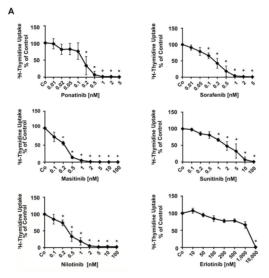
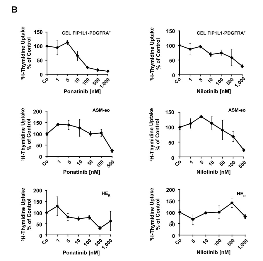
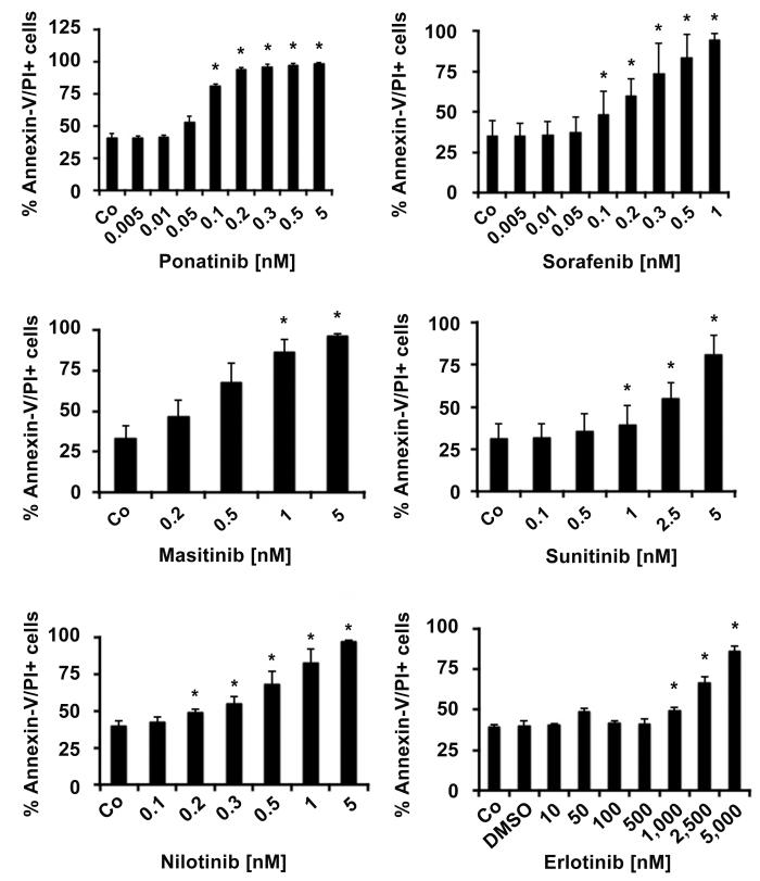
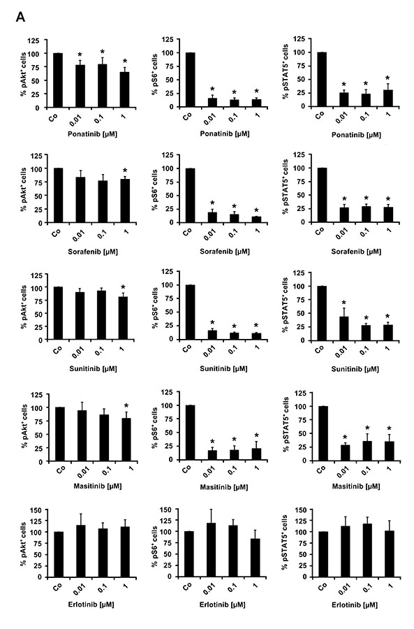
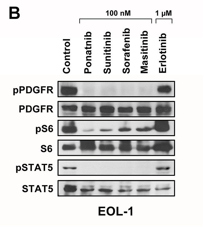
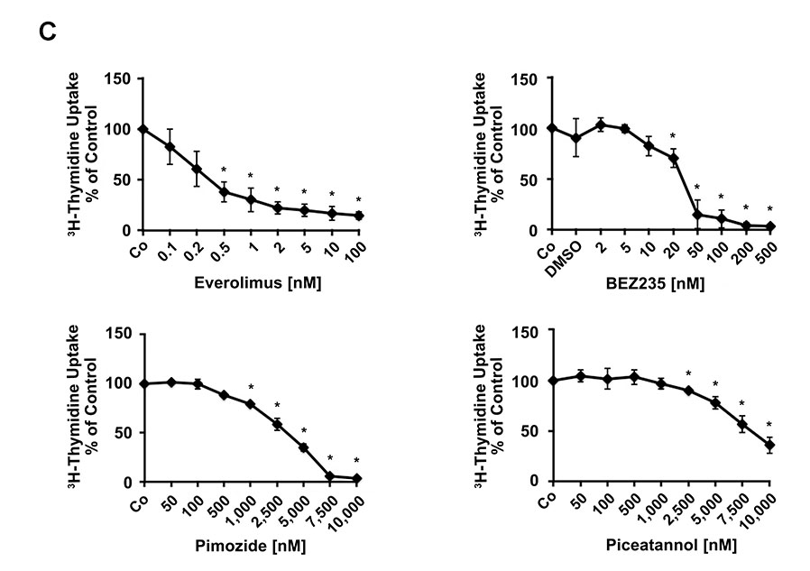
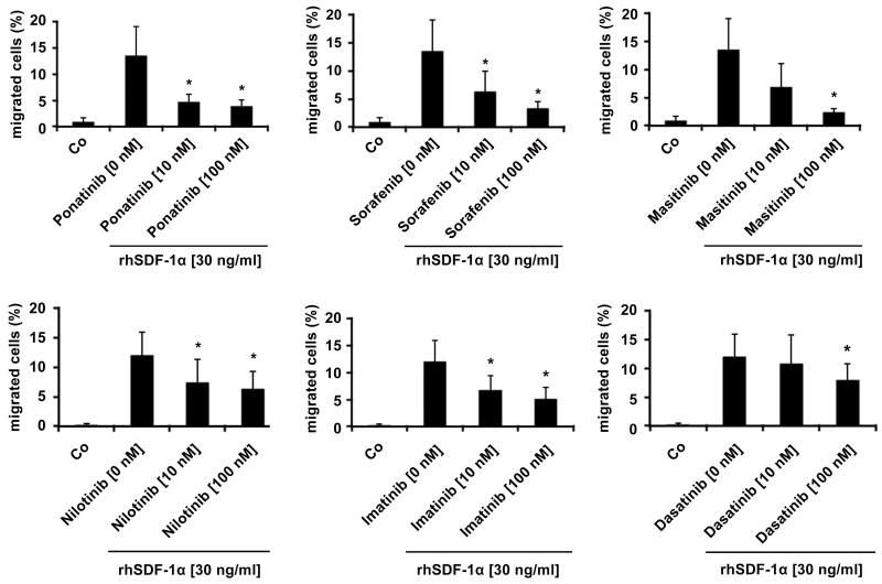
In chronic eosinophilic leukemia, the transforming oncoprotein FIP1L1-PDGFRA is a major target of therapy. In most patients, the tyrosine kinase inhibitor (TKI) imatinib induces complete remission. For patients who are intolerant or resistant, novel TKIs have been proposed. We examined the in vitro effects of 14 kinase blockers on growth and function of EOL-1 cells, a FIP1L1-PDGFRA(+) eosinophil cell line. Major growth-inhibitory effects were seen with all PDGFR-blocking agents, with IC50 values in the low nanomolar range: ponatinib, 0.1-0.2 nmol/L; sorafenib, 0.1-0.2 nmol/L; masitinib, 0.2-0.5 nmol/L; nilotinib, 0.2-1.0 nmol/L; dasatinib, 0.5-2.0 nmol/L; sunitinib, 1-2 nmol/L; midostaurin, 5-10 nmol/L. These drugs were also found to block activation of PDGFR-downstream signaling molecules, including Akt, S6, and STAT5 in EOL-1 cells. All effective TKIs produced apoptosis in EOL-1 cells as determined by microscopy, Annexin-V/PI, and caspase-3 staining. In addition, PDGFR-targeting TKIs were found to inhibit cytokine-induced migration of EOL-1 cells. In all bioassays used, ponatinib was found to be the most potent compound in EOL-1 cells. In addition, ponatinib was found to downregulate expression of the activation-linked surface antigen CD63 on EOL-1 cells and to suppress the growth of primary neoplastic eosinophils. We also examined drug effects on Ba/F3 cells expressing two clinically relevant, imatinib-resistant, mutant forms of FIP1L1-PDGFRA, namely T674I and D842V. Strong inhibitory effects on both mutants were seen only with ponatinib. In summary, novel PDGFR-targeting TKIs may be alternative agents for the treatment of patients with imatinib-resistant chronic eosinophilic leukemia. Although several different PDGFR-targeting agents are effective, the most potent drug appears to be ponatinib.


Figures

Figure 1
Figure 1. Effects of various kinase blockers on proliferation of neoplastic eosinophils
(A) EOL-1 cells were incubated in control medium (Co) or in various concentrations of targeted drugs as indicated at 37°C for 48 hours. After incubation, 3H-thymidine uptake was measured. Results are expressed as percent of control and represent the mean±SD from 3 independent experiments. Asterisk: p<0.05. (B) Primary eosinophils obtained from a patient with FIP1L1-PDGFRA+ CEL (upper panels), one with aggressive systemic mastocytosis with eosinophilia (ASM-eo; middle panels) and one with reactive hypereosinophilia (HER, lower panels), were incubated in control medium (Co) or in various concentrations of targeted drugs as indicated at 37°C for 48 hours. After incubation, 3H-thymidine uptake was measured. Results are expressed as percent of control and represent the mean±SD of triplicates.
Figure 2
Figure 2. PDGFR-targeting drugs induce apoptosis in EOL-1 cells
EOL-1 cells were cultured in the absence (Co) or presence of various concentrations of targeted drugs as indicated at 37°C for 48 hours. Apoptosis was determined by Annexin-V/PI-staining by flow cytometry. Results are expressed as percent of control and represent the mean±S.D. of 3 or 4 independent experiments. Asterisk: p<0.05. In the lower right panel, the DMSO control is also shown.
Figure 3
Figure 3. Effects of various targeted drugs on PDGFR-downstream signaling molecules in EOL-1 cells
(A) EOL-1 cells were incubated in control medium in the absence (Co) or presence of ponatinib, sorafenib, masitinib, sunitinib or erlotinib (each 0.01, 0.1 or 1 μM) at 37°C for 4 hours. After incubation, cells were analyzed for expression of pAKT, pS6, and pSTAT5, by flow cytometry as described in the text. Results are expressed as percent of control and represent the mean±SD from at least 3 independent experiments. DMSO (solvent) per se did not show any effect on phosphorylation of tested molecules (not shown). Asterisk, p<0.05. (B) EOL-1 cells were incubated in control medium in the absence (Control) or presence of ponatinib, sunitinib, sorafenib, masitinib or erlotinib (each 0.1 or 1 μM) at 37°C for 4 hours. After incubation, cells were analyzed for expression of pPDGFR, pS6 and pSTAT5 by western blotting as described in the text. (C) EOL-1 cells were incubated in control medium (Co) or in various concentrations of everolimus, BEZ235, pimozide, or piceatannol at 37°C for 48 hours. After incubation, 3H-thymidine uptake was measured. Results are expressed as percent of control and represent the mean±SD from 3 independent experiments. Asterisk: p<0.05.
Figure 4
Figure 4. Effects of targeted drugs on SDF-1α-induced migration of EOL-1 cells
EOL-1 cells were pre-incubated in control medium (Co) or in medium containing ponatinib, sorafenib, masitinib, nilotinib, imatinib, or dasatinib (each 10 or 100 nM) at 37°C for 1 hour. Then, migration of cells against SDF-1α (30 ng/ml) was determined in a double-chamber chemotaxis assay (4 hours) as described in the text. Numbers of viable migrated cells are expressed as percent of total (100% input) cells. Results represent the mean±S.D. of 4 independent experiments. Asterisk: p<0.05.

References

    1. Gotlib J. Molecular classification and pathogenesis of eosinophilic disorders: 2005 update. Acta Haematol. 2005;114:7–25. - PubMed
    1. Tefferi A, Patnaik MM, Pardanani A. Eosinophilia: secondary, clonal and idiopathic. Br J Haematol. 2006 Jun;133:468–492. - PubMed
    1. Bain BJ, Fletcher SH. Chronic eosinophilic leukemias and the myeloproliferative variant of the hypereosinophilic syndrome. Immunol Allergy Clin North Am. 2007;27:377–388. - PubMed
    1. Gotlib J. Chronic eosinophilic leukemia/hypereosinophilic syndrome. Cancer Treat Res. 2008;142:69–106. - PubMed
    1. Valent P. Pathogenesis, classification, and therapy of eosinophilia and eosinophil disorders. Blood Rev. 2009;23:157–165. - PubMed

Publication types

MeSH terms