. 2014 Jan 9;5(1):e984.
doi: 10.1038/cddis.2013.513.

Mitochondrial dysfunction, oxidative stress, and neurodegeneration elicited by a bacterial metabolite in a C. elegans Parkinson's model

Affiliations

Mitochondrial dysfunction, oxidative stress, and neurodegeneration elicited by a bacterial metabolite in a C. elegans Parkinson's model

A Ray et al. Cell Death Dis. .

Abstract

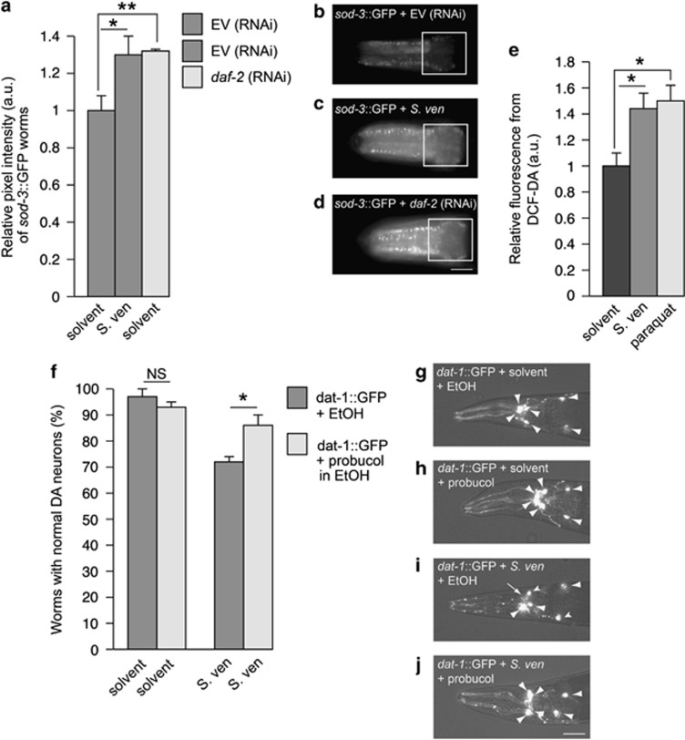
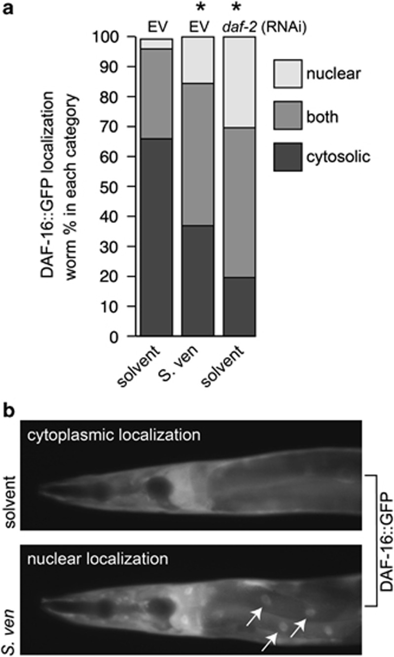
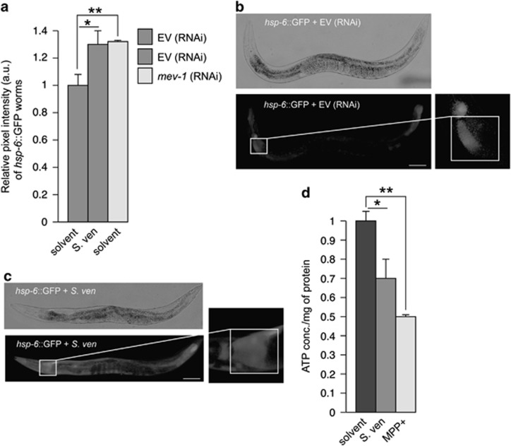
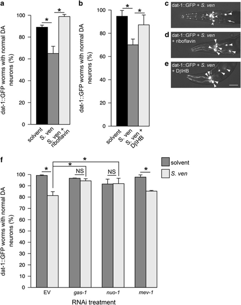
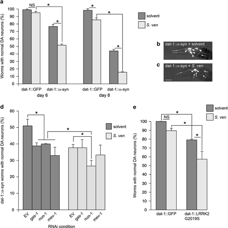
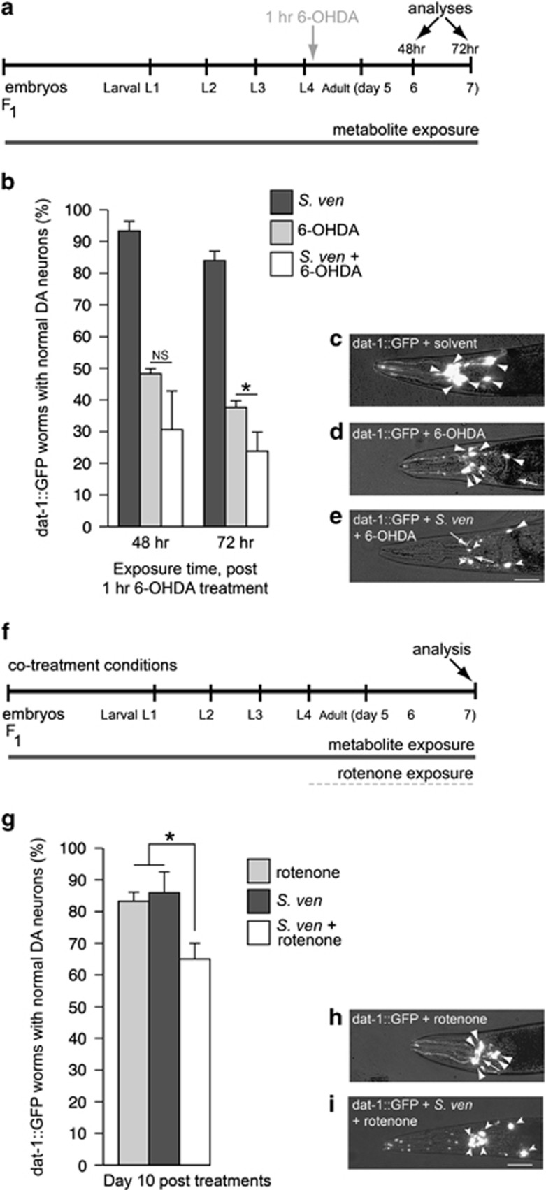
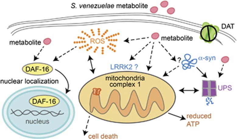
Genetic and idiopathic forms of Parkinson's disease (PD) are characterized by loss of dopamine (DA) neurons and typically the formation of protein inclusions containing the alpha-synuclein (α-syn) protein. Environmental contributors to PD remain largely unresolved but toxins, such as paraquat or rotenone, represent well-studied enhancers of susceptibility. Previously, we reported that a bacterial metabolite produced by Streptomyces venezuelae caused age- and dose-dependent DA neurodegeneration in Caenorhabditis elegans and human SH-SY5Y neurons. We hypothesized that this metabolite from a common soil bacterium could enhance neurodegeneration in combination with PD susceptibility gene mutations or toxicants. Here, we report that exposure to the metabolite in C. elegans DA neurons expressing human α-syn or LRRK2 G2019S exacerbates neurodegeneration. Using the PD toxin models 6-hydroxydopamine and rotenone, we demonstrate that exposure to more than one environmental risk factor has an additive effect in eliciting DA neurodegeneration. Evidence suggests that PD-related toxicants cause mitochondrial dysfunction, thus we examined the impact of the metabolite on mitochondrial activity and oxidative stress. An ex vivo assay of C. elegans extracts revealed that this metabolite causes excessive production of reactive oxygen species. Likewise, enhanced expression of a superoxide dismutase reporter was observed in vivo. The anti-oxidant probucol fully rescued metabolite-induced DA neurodegeneration, as well. Interestingly, the stress-responsive FOXO transcription factor DAF-16 was activated following exposure to the metabolite. Through further mechanistic analysis, we discerned the mitochondrial defects associated with metabolite exposure included adenosine triphosphate impairment and upregulation of the mitochondrial unfolded protein response. Metabolite-induced toxicity in DA neurons was rescued by complex I activators. RNA interference (RNAi) knockdown of mitochondrial complex I subunits resulted in rescue of metabolite-induced toxicity in DA neurons. Taken together, our characterization of cellular responses to the S. venezuelae metabolite indicates that this putative environmental trigger of neurotoxicity may cause cell death, in part, through mitochondrial dysfunction and oxidative stress.


Figures

Figure 1
Figure 1
S. venezuelae metabolite causes oxidative stress in C. elegans. (a) S. venezuelae (S. ven) metabolite caused an upregulation of sod-3::GFP expression, an indicator of oxidative stress, in empty vector (EV) RNAi-treated worms when compared with worms exposed to solvent only, as quantitated using pixel intensities as described in (bd). When compared with metabolite exposure, RNAi knockdown of daf-2, used as a positive control, expressed similar levels of sod-3::GFP. Values are the mean±S.D. of 3 experiments where 30 animals were analyzed per replicate (*P<0.05; **P<0.01; one-way ANOVA). The values were normalized to the untreated solvent control. (bd) Representative worm images for each of the treatments described in (a) where pixel intensities were measured in a 100 × 100 μm region at the anterior bulb of the pharynx. The white box shows the region of GFP measured in all animals. (e) S. venezuelae metabolite and 100 μM paraquat (positive control) significantly increased the amount of intracellular ROS compared with solvent control. Worms were evaluated using a DCF-DA assay by examining extracts (*P<0.01; one-way ANOVA; n=3 independent experiments). The values were normalized to the untreated solvent control. (f) Treatment with 1 mM probucol (dissolved in ethanol), an anti-oxidant, significantly rescued the S. venezuelae-induced neurotoxicity in DA neurons compared with metabolite treatment alone (*P<0.01; Student's t-test; n=3 independent experiments). (gj) Representative images of the probucol experiment described in (g). All C. elegans (strain BY200) express GFP specifically in the six anterior DA neurons. In all images, large arrowheads show intact dopaminergic neuron cell bodies. Arrows indicate areas where dopaminergic neurons have degenerated. Small arrowheads indicate cell body degeneration. (g) Exposure to EtAc and ethanol (solvent for probucol) did not result in DA neuron loss. (h) The addition of probucol did not cause neurotoxicity, as evidenced by intact DA neurons. (i) S. venezuelae metabolite exposure caused substantial degeneration of cell processes, as displayed throughout the processes. Further, two of the cell bodies are degenerating and one is missing in this representative worm. (j) Probucol rescues S. venezuelae-induced DA neuronal toxicity, as shown in this C. elegans example. Magnification bars=50 μm
Figure 2
Figure 2
Effect of S. venezuelae metabolite on DAF-16 localization. (a) Stacked graph representing the percentage of C. elegans with DAF-16::GFP localization in the nucleus, cytoplasm, or both. S. venezuelae exposure promotes nuclear translocation of DAF-16::GFP in a manner similar to daf-2 (RNAi), where both treatments were significantly different from EV solvent control (*P<0.05; one-way ANOVA; n=3 independent experiments with 30 animals/experiment). (b) Representative images of DAF-16::GFP localization in the cytoplasm and nucleus (arrows) following treatment with EtAc solvent and metabolite, respectively
Figure 3
Figure 3
S. venezuelae metabolite causes mitochondrial dysfunction. (a) S. venezuelae metabolite caused an upregulation of hsp-6::GFP, an indicator of UPRmt stress response. In this experiment, expression of hsp-6::GFP in EV RNAi-treated worms exposed to metabolite was significantly increased compared with worms exposed to solvent only, as quantitated using pixel intensities as visualized in (b and c). When compared with metabolite exposure, RNAi knockdown of mev-1, used as a positive control, expressed similar levels of hsp-6::GFP. Values are the mean±S.D. of 3 experiments where 30 animals were analyzed per replicate (*P<0.05; **P<0.01; one-way ANOVA). The values were normalized to the untreated solvent control. (b and c) Representative worm images (brightfield (top) and fluorescence (bottom)) for EtAc and metabolite treatments described in (a). Pixel intensities were measured in a 100 × 100 μm region in the intestinal lumen immediately posterior to the grinder of the pharynx. The region highlighted in the white box shows GFP expression driven by the hsp-6 gene promoter that was measured in all animals and is magnified to the right. (d) C. elegans exposed to the S. venezuelae metabolite showed reduced ATP production compared with the solvent control; worms treated with 1 mM MPP+, which is known to reduce ATP levels, also demonstrated a similar effect (*P<0.05; **P<0.01; one-way ANOVA). Values are the mean±S.D. of three independent experiments and were normalized to the untreated control. Magnification bar=100 μm
Figure 4
Figure 4
The S. venezuelae metabolite impacts mitochondrial complex I. (ae) Metabolite-induced DA neurotoxicity was rescued by riboflavin and DβHB, drugs that rescue mitochondria complex I deficiency. (a) S. venezuelae and 1 μg/ml riboflavin significantly rescued DA neurons compared with the metabolite alone. (b) When 50 mM DβHB is co-administered with the metabolite, C. elegans DA neurons are rescued from neurotoxicity. (ce) Representative images of riboflavin and DβHB rescuing DA neurotoxicity induced by the metabolite. All C. elegans (strain BY200) express GFP specifically in the six anterior DA neurons. In the images, large arrowheads show intact dopaminergic neuron cell bodies. Arrows indicate areas where dopaminergic neurons have degenerated. Small arrowheads indicate cell body degeneration. (c) Exposure to the S. venezuelae metabolite caused neuronal loss in this worm where five of the six DA neurons are degenerating. (d) The addition of riboflavin completely rescued DA neurons in an animal exposed to the metabolite. (e) DβHB also rescued S. venezuelae-induced DA neuronal toxicity, as shown in this C. elegans example. Magnification bar=50 μm. (f) RNAi knockdown of complex I components gas-1 and nuo-1 resulted in rescue of DA neurodegeneration when treated with metabolite while complex II component mev-1 and EV RNAi showed enhanced DA neurodegeneration with metabolite exposure (*P<0.05; one-way ANOVA; n=90 worms). C. elegans (strain UA202) were analyzed at day 12 where data were analyzed as the mean±S.D.
Figure 5
Figure 5
Gene and environment interaction enhances DA neurodegeneration. A combination of exposure to S. venezuelae and overexpression of known Parkinson's gene products, α-syn (ad) or LRRK2 G2019S (e) enhances DA neurodegeneration. (a) C. elegans expressing GFP alone exhibit metabolite-induced age-dependent DA neurodegeneration that is evident when examining day 6 versus day 8 animals. Worms overexpressing α-syn display age-dependent DA neurodegeneration and are also more susceptible to metabolite-induced DA neurotoxicity when compared with populations of α-syn-expressing worms treated with solvent only. These data are represented as the mean±S.D. n=90 per data point (*P<0.05 by one-way ANOVA). (b and c) Representative images of C. elegans (strain UA44) expressing α-syn and GFP specifically in the six anterior DA neurons in solvent (b) or in combination with metabolite exposure (c). In the images, large arrowheads show intact dopaminergic neuron cell bodies. Arrows indicate areas where dopaminergic neurons have degenerated. (d) RNAi knockdown of gas-1, nuo-1, and mev-1 showed enhanced α-syn-induced DA neurodegeneration compared with α-syn alone without metabolite exposure. C. elegans (strain UA196) were analyzed at day 6 where data were analyzed as the mean±S.D. After metabolite exposure, nuo-1 RNAi-treated worms showed a significant sensitivity in worms expressing α-syn compared with EV (RNAi) metabolite-treated worms and nuo-1 (RNAi) solvent control worms, whereas RNAi of gas-1 and mev-1 failed to cause a significant degeneration with metabolite treatment when compared with untreated controls (*P<0.05; one-way ANOVA). (e) Metabolite-treated worms expressing GFP alone do not display a significant DA neurodegeneration when compared with solvent control at 7 days of exposure. Susceptibility to metabolite-induced DA neurotoxicity is enhanced when C. elegans overexpress human LRRK2 G2019S when compared with populations of worms treated with solvent only. Data are represented as the mean±S.D., n=90 per independent transgenic line; where three separate transgenic lines were analyzed (*P<0.05 by one-way ANOVA). Magnification bar=50 μm
Figure 6
Figure 6
Hypersensitivity to S. venezuelae DA neurotoxicity when worms are treated with rotenone or 6-OHDA. (a) A timeline representing an experimental paradigm depicting the length of S. venezuelae metabolite exposure and 6-OHDA treatment. The abbreviations L1–L4 are the larval stages of C. elegans, while the ‘adult' designations represent days post hatching. The 48 and 72 h represents times, post 1 h 6-OHDA (30 mM) treatment when DA neurons were analyzed. (b) At 48 h after 6-OHDA treatment, co-treatment with metabolite was not significantly different from individual treatments alone. Whereas, after 72 h, C. elegans co-exposed with the metabolite and 6-OHDA displayed significantly more susceptibility to DA neurodegeneration than either treatment alone (*P<0.05; one-way ANOVA; n=90 per treatment). (ce) Representative images of C. elegans (strain BY200) expressing GFP specifically in the six anterior DA neurons. In the images, large arrowheads show intact dopaminergic neuron cell bodies. Arrows indicate areas where dopaminergic neurons have degenerated. Small arrowheads indicate cell body degeneration. (c) A control worm exposed to EtAc solvent only (no 6-OHDA or metabolite) has six normal DA neurons in the anterior region. (d) This representative worm exposed to 6-OHDA is missing one neuron. (e) In this example, a worm exposed to both 6-OHDA and the metabolite is missing two neurons while another three neurons display cell body rounding, indicative of degeneration. Magnification bar=50 μm. (f) A timeline representing the experimental paradigm for a combination of S. venezuelae metabolite and rotenone exposure scored for DA neurodegeneration. The abbreviations are described in (a). (g) C. elegans co-exposed with the metabolite and 5 μM rotenone (in 0.05% DMSO) display significantly more susceptibility to DA neurodegeneration than either treatment alone (*P<0.01; Student's t-test; n=90 per treatment). (h and i) Representative images of C. elegans (strain BY200) expressing GFP specifically in the six anterior DA neurons. In the images, large arrowheads show intact dopaminergic neuron cell bodies. Arrows indicate areas where dopaminergic neurons have degenerated. Small arrowheads indicate cell body degeneration. (h) This representative worm exposed to rotenone was missing one neuron. (i) In this representative worm exposed to both rotenone and the metabolite, all neurons display cell body rounding, indicative of degeneration. Magnification bar=50 μm
Figure 7
Figure 7
Experimental model for S. venezuelae metabolite-induced toxicity. This tentative model depicts our current understanding of the cellular mechanisms impacted. We previously determined that this metabolite does not enter cell membranes through the DA transporter, DAT. It is possible that the metabolite diffuses through the cellular membrane because the initial biochemical structural characterization and purification efforts have shown that the molecule is small and highly lipophilic. However, an unknown receptor could also be transducing a signal cascade. Regardless, we observe enhanced oxidative stress in response to metabolite exposure. The excessive production of ROS by the metabolite could be a direct result of redox cycling in vivo or it could be the result of mitochondrial dysfunction. There could also be a reciprocal relationship between ROS and mitochondrial dysfunction following metabolite exposure. Alternatively, the metabolite could directly target mitochondrial complex I, leading to ATP production impairment, upregulation of the UPRmt. These cellular responses could trigger cell death. Additionally, the metabolite could activate the FOXO transcription factor DAF-16 directly or as a secondary cellular response to ROS production. Furthermore, based on our DA neurodegeneration studies, there might be a direct or indirect association between the S. venezuelae metabolite and α-synuclein or LRRK2 in targeting mitochondria. We have previously reported that the metabolite inhibits the UPS. Considering that proteasome function intersects with the various mechanisms described in this model, future studies will explore this functional association.

Similar articles

Cited by

References

    1. Spillantini MG, Schmidt ML, Lee VM, Trojanowski JQ, Jakes R, Goedert M. Alpha-synuclein in Lewy bodies. Nature. 1997;388:839–840. - PubMed
    1. Tanner CM. Is the cause of Parkinson's disease environmental or hereditary? Evidence from twin studies. Adv Neurol. 2003;91:133–142. - PubMed
    1. Priyadarshi A, Khuder SA, Schaub EA, Priyadarshi SS. Environmental risk factors and Parkinson's disease: a meta-analysis. Environ Res. 2001;86:122–127. - PubMed
    1. Costello S, Cockburn M, Bronstein J, Zhang X, Ritz B. Parkinson's disease and residential exposure to maneb and paraquat from applications in the central valley of California. Am J Epidemiol. 2009;169:919–926. - PMC - PubMed
    1. Liou HH, Tsai MC, Chen CJ. Environmental risk factors and Parkinson's disease: a case-control study in Taiwan. Neurology. 1997;48:1583–1588. - PubMed

Publication types

MeSH terms

Substances