. 2014 Feb 28;289(9):6199-211.
doi: 10.1074/jbc.M113.537480. Epub 2014 Jan 9.

A novel unsaturated β-glucuronyl hydrolase involved in ulvan degradation unveils the versatility of stereochemistry requirements in family GH105

Affiliations

A novel unsaturated β-glucuronyl hydrolase involved in ulvan degradation unveils the versatility of stereochemistry requirements in family GH105

Pi Nyvall Collén et al. J Biol Chem. .

Abstract

Ulvans are cell wall matrix polysaccharides in green algae belonging to the genus Ulva. Enzymatic degradation of the polysaccharide by ulvan lyases leads to the production of oligosaccharides with an unsaturated β-glucuronyl residue located at the non-reducing end. Exploration of the genomic environment around the Nonlabens ulvanivorans (previously Percicivirga ulvanivorans) ulvan lyase revealed a gene highly similar to known unsaturated uronyl hydrolases classified in the CAZy glycoside hydrolase family 105. The gene was cloned, the protein was overexpressed in Escherichia coli, and enzymology experiments demonstrated its unsaturated β-glucuronyl activity. Kinetic analysis of purified oligo-ulvans incubated with the new enzyme showed that the full substrate specificity is attained by three subsites that preferentially bind anionic residues (sulfated rhamnose, glucuronic/iduronic acid). The three-dimensional crystal structure of the native enzyme reveals that a trimeric organization is required for substrate binding and recognition at the +2 binding subsite. This novel unsaturated β-glucuronyl hydrolase is part of a previously uncharacterized subgroup of GH105 members and exhibits only a very limited sequence similarity to known unsaturated β-glucuronyl sequences previously found only in family GH88. Clan-O formed by families GH88 and GH105 was singular in the fact that it covered families acting on both axial and equatorial glycosidic linkages, respectively. The overall comparison of active site structures between enzymes from these two families highlights how that within family GH105, and unlike for classical glycoside hydrolysis, the hydrolysis of vinyl ether groups from unsaturated saccharides occurs independently of the α or β configuration of the cleaved linkage.

Keywords: Biodegradation; Crystal Structure; Enzyme Catalysis; GH105; Glycoside Hydrolases; Polysaccharide; Ulvan.


Figures
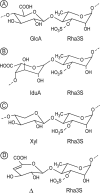
FIGURE 1.
FIGURE 1.
Chemical structure of the three main disaccharide repetition moieties encountered in ulvans and the unsaturated oligosaccharide produced by lyase degradation. A and B, ulvanobiouronic acid A and B, respectively. C, ulvanobiose. D, 4-deoxy-l-threo-hex-4-enopyranosiduronic acid (▵) linked to the sulfated rhamnose occurring at the non-reducing end after cleavage of the glycosidic linkage by ulvan lyase.
FIGURE 2.
FIGURE 2.
Amino acid sequence alignment of the N. ulvanivorans unsaturated β-glucuronyl hydrolase sequence (Nu_GH105, Nu_AFQ98272–4CE7) with the two characterized proteins belonging to the GH105 family (YesR, Bs_CAB12519-YesR; YteR, Bs_CAB14990-1NC5 from B. subtilis) as well as the three other members of GH105 with known three-dimensional structures, Kp_ABR77815-3PMM, Se_AAV76933-3QWT, and Bt_AAO79281-3K11. Conserved amino acids involved in the recognition of the unsaturated uronyl sugar surrounding the −1 binding site are marked by a gray oval; the catalytic acid/base is marked by a star, and two important residues in Nu_GH105 not conserved in other GH105 are marked by a gray open square. The secondary structure elements of Nu_GH105 are symbolized above the sequences, and the numbering corresponds to the mature Nu_GH105 enzyme. The figure was prepared using Espript (40).
FIGURE 3.
FIGURE 3.
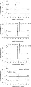
High performance anion-exchange chromatography of purified oligosaccharides incubated with unsaturated β-glucuronyl hydrolase. A, Δ-Rha3S was partially degraded by the enzyme. B and C, the tetrasaccharides Δ-Rha3S-Glc-Rha3S and Δ-Rha3S-Idu-Rha3S were completely degraded. D, partial degradation of the tetrasaccharide Δ-Rha3S-Xyl-Rha3S. *, uronic tetrasaccharides eluting like in B and C. −enz, without unsaturated β-glucuronyl hydrolase. +enz, with unsaturated β-glucuronyl hydrolase.
FIGURE 4.
FIGURE 4.
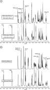
1H NMR of the purified end-products of the unsaturated β-glucuronyl hydrolase. A, spectrum of a mixture of 50:50 Δ-Rha3S-Glc-Rha3S:Δ-Rha3S-Idu-Rha3S (bottom) and trisaccharides obtained after incubation with unsaturated β-glucuronyl hydrolase (top). B, same experiment as in A but with the Δ-R3S-Xyl-R3Sα/β tetrasaccharide as the starting substrate.
FIGURE 5.
FIGURE 5.
Kinetics of degradation of purified oligosaccharides. Structure of the unsaturated oligo-ulvans are indicated with the normalized degradation rates (s−1) in parentheses. The activities were determined as the absorbance decrease at 235 nm determined in a quartz cuvette with a 1-cm light path at 30 °C starting with 100 μm oligosaccharide and 4.4 nm enzyme in a final volume of 500 μl.
FIGURE 6.
FIGURE 6.
The trimeric organization of Nu_GH105 provides the residues responsible for the observed substrate specificity in the +2 sub-binding site. A, the crystallographic trimer organization of Nu_GH105. The schematic representations of the three independent molecules, arranged around the non-crystallographic 3-fold axis, are colored in purple, blue, and gray, respectively. The loops from residues 104–111 of each monomer are colored in green, and the arginine residue Arg-106 that could play a role in binding a iduronic or glucuronic acid at +2 of the Nu_GH105 substrate is colored in orange. B, the superimposition of Nu_GH105 with YteR and Ugl in complex with substrate molecules allowed the modeling of a putative substrate molecule that binds to subsites −1 to +2, highlighting the possibility that Arg-106 from a neighboring molecule could bind to a carboxylic acid function of the sixth position of a sugar unit bound to +2.
FIGURE 7.
FIGURE 7.
Close-up view of the superimposed active sites of Nu_GH105 with the substrate complexes of YteR and Ugl. The figure highlights that both α- and β-configured substrates could be accommodated in the active site pockets. A, the superimposition of Nu_GH105 and YteR in complex with an unsaturated α-rhamnogalacturonyl substrate (2GH4) allows identification of conserved residues and variations due to the different substrate specificities. The residues in the active site pocket of Nu_GH105 are colored with yellow carbon atoms, and the residue labels are black. The residues in contact with the substrate molecule in YteR are colored and labeled in cyan. The largest variations were observed for Ile-331 and Phe-346 (Nu_GH105), that are replaced by Gly-332 and His-352 in YteR, in contact with the +1 sub-binding site. Another difference was observed for Trp-146. In YteR the corresponding residue Trp-141 is in a perpendicular conformation, and the residues His-132 and Tyr-136 of YteR have no equivalents in Nu_GH105. B, the superimposition of Nu_GH105 and Ugl in complex with an unsaturated chondroitin disaccharide (ΔGlcA-GalNac) substrate molecule (2AHG) highlights the more divergent active site pockets, although both enzymes are active on β-configured substrate molecules. The residues in the active site pocket of Nu_GH105 are colored with yellow carbon atoms, and the residue labels are black. The residues in contact with the substrate molecule in Ugl are colored and labeled in light gray. The largest differences were again observed around the +1 binding site; Ile-331 and Phe-346 of Nu_GH105 were replaced by Tyr-338 and Trp-352 in Ugl, and the loop containing the residues Trp-146, His-87, and Arg-84 (Nu_GH105) was arranged differently in Ugl, repositioning divergent residues Ile-147, Trp-134, and His-87 on this side of the substrate binding site.
FIGURE 8.
FIGURE 8.
Ribbon representation of the structural superimposition of all members of family GH105 for which crystal structures have been reported. The backbone of the three-dimensional structure of Nu_GH105 is colored in light yellow, and the divergent loops (numbered from 1 to 3) are highlighted in orange; Bs_1NC5 is colored in light gray, and the loops 1–3 are in magenta; Kp_3PMM is in gray, and loops 1–3 are in bright blue; Se_3QWT is in medium gray, and loops 1–3 are in dark blue; the backbone of Bt_3K11 is colored in dark gray, and loops 1–3 are in purple-blue.
FIGURE 9.
FIGURE 9.
Phylogram representation of the hierarchical clustering of representative GH105 members. Those that have been biochemically or structurally characterized (already found in Fig. 2) are labeled with their known reference GenBankTM and PDB accessions and eventually by their gene name. Three clear distinct subgroups or subfamilies can be identified with branches shown in red, blue, and green, respectively.

References

    1. Gao S., Chen X., Yi Q., Wang G., Pan G., Lin A., Peng G. (2010) A strategy for the proliferation of Ulva prolifera, main causative species of green tides, with formation of sporangia by fragmentation. Plos ONE 5, 1–7 - PMC - PubMed
    1. Lahaye M., Robic A. (2007) Structure and functional properties of ulvan, a polysaccharide from green seaweeds. Biomacromolecules 8, 1765–1774 - PubMed
    1. Percival E., McDowell R. H. (1967) Chemistry and enzymology of marine algal polysaccharides, Academic Press Inc., London
    1. Quemener B., Lahaye M., Bobin-Dubigeon C. (1997) Sugar determination in ulvans by a chemical-enzymatic method coupled to high performance anion exchange chromatography. J. Appl. Phycol. 9, 179–188
    1. Ray B., Lahaye M. (1995) Cell wall polysaccharides from the marine green alga Ulva “rigida” (Ulvales, Chlorophyta). Extraction and chemical composition. Carbohydr. Res. 274, 313–318 - PubMed

Publication types

MeSH terms

Associated data

LinkOut - more resources