. 2014 Feb 28;289(9):5914-24.
doi: 10.1074/jbc.M113.531384. Epub 2014 Jan 8.

Kruppel-like factor 15 is a critical regulator of cardiac lipid metabolism

Affiliations

Kruppel-like factor 15 is a critical regulator of cardiac lipid metabolism

Domenick A Prosdocimo et al. J Biol Chem. .

Abstract

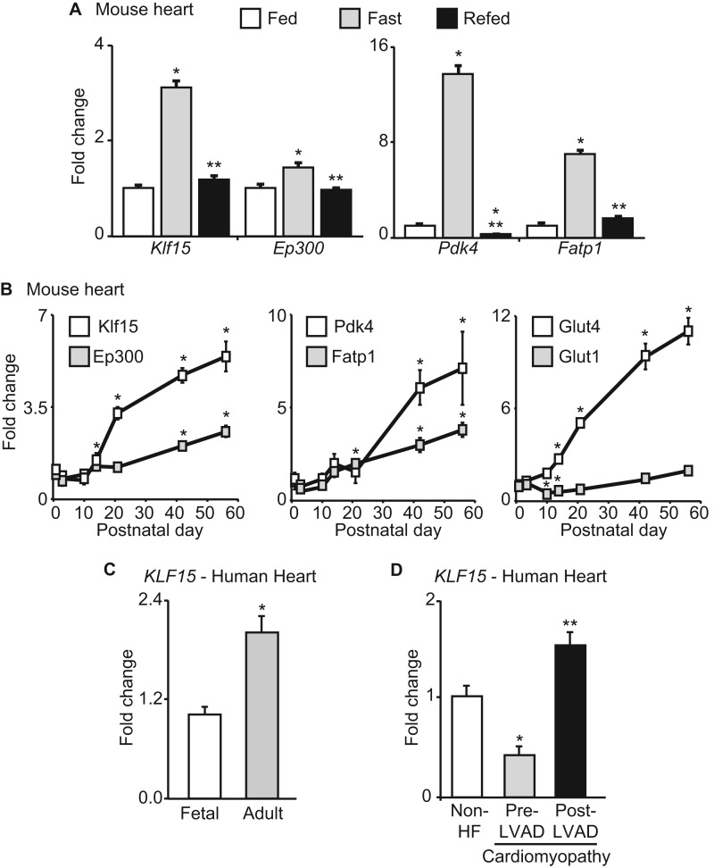
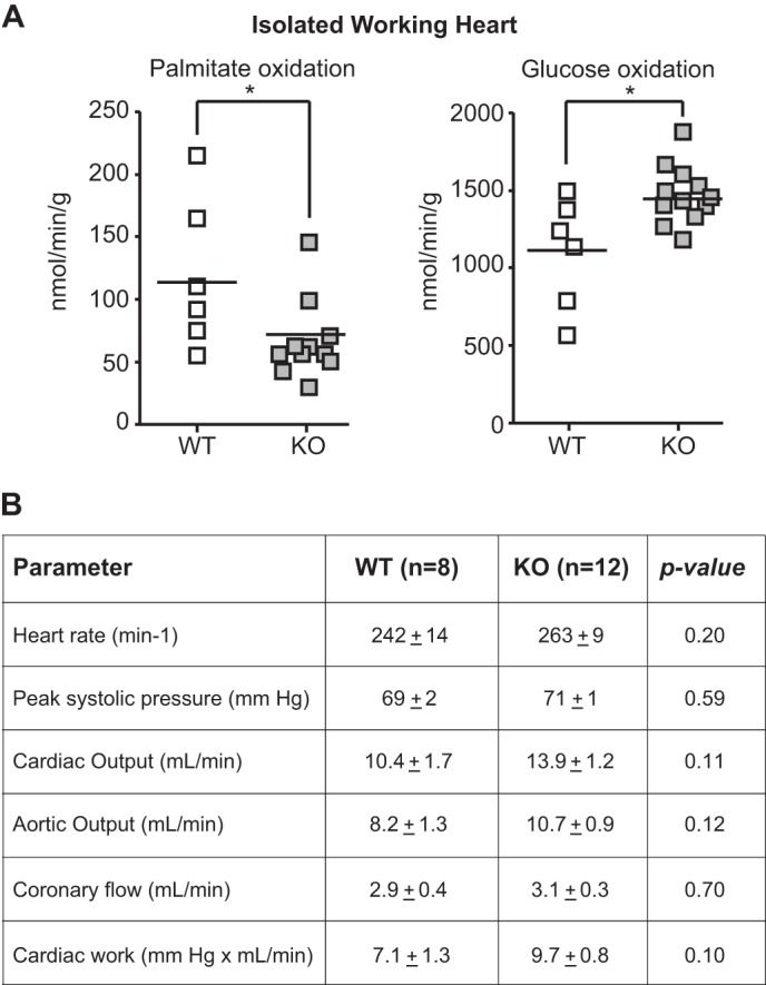
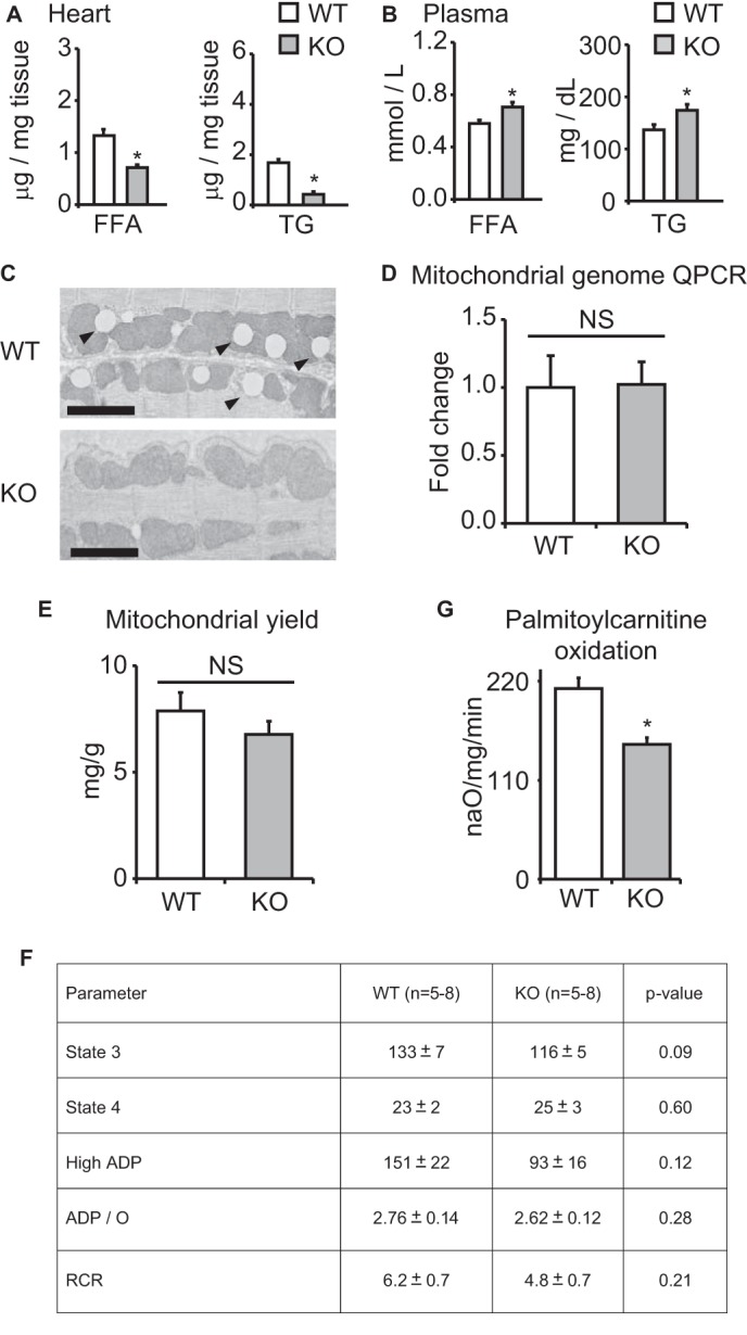
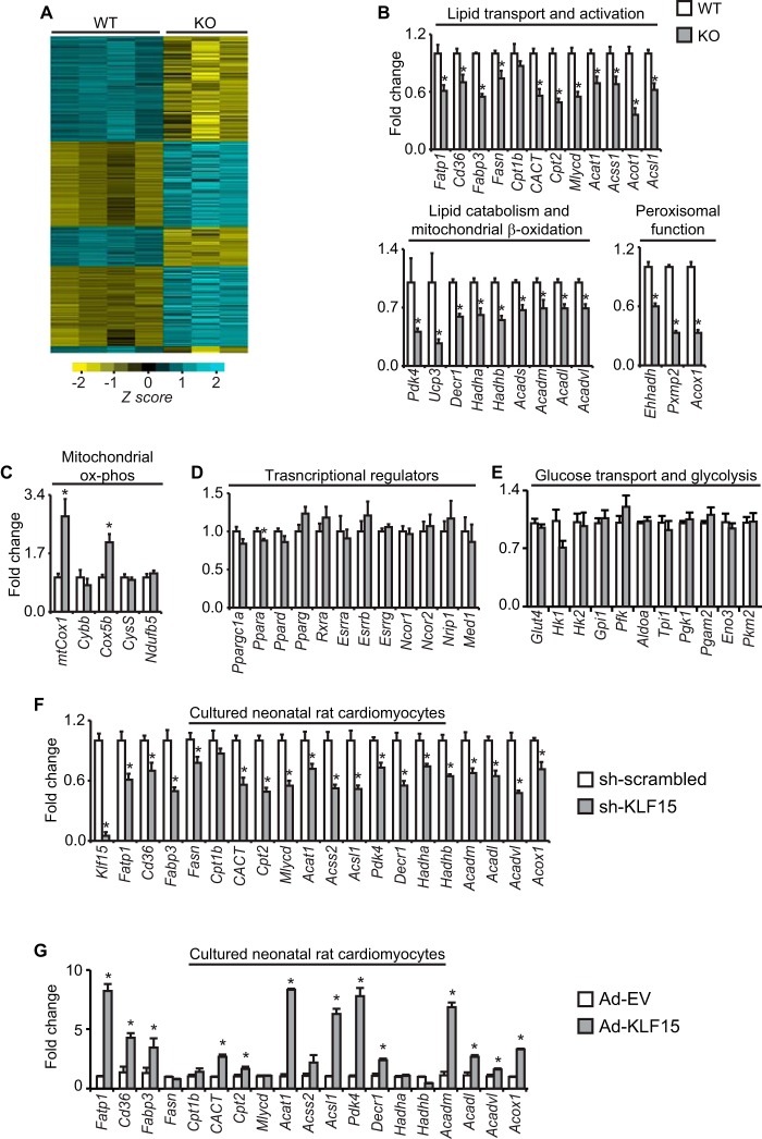
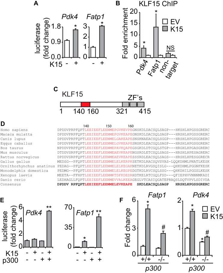
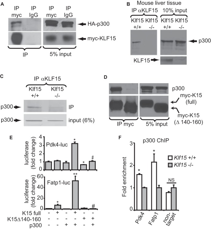
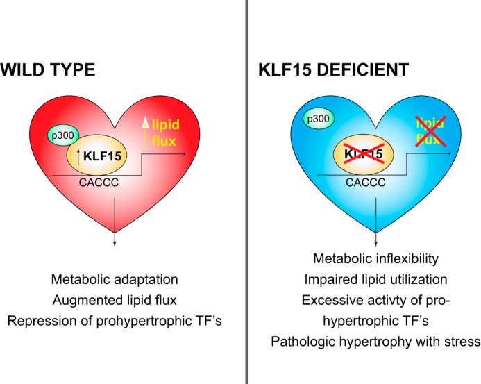
The mammalian heart, the body's largest energy consumer, has evolved robust mechanisms to tightly couple fuel supply with energy demand across a wide range of physiologic and pathophysiologic states, yet, when compared with other organs, relatively little is known about the molecular machinery that directly governs metabolic plasticity in the heart. Although previous studies have defined Kruppel-like factor 15 (KLF15) as a transcriptional repressor of pathologic cardiac hypertrophy, a direct role for the KLF family in cardiac metabolism has not been previously established. We show in human heart samples that KLF15 is induced after birth and reduced in heart failure, a myocardial expression pattern that parallels reliance on lipid oxidation. Isolated working heart studies and unbiased transcriptomic profiling in Klf15-deficient hearts demonstrate that KLF15 is an essential regulator of lipid flux and metabolic homeostasis in the adult myocardium. An important mechanism by which KLF15 regulates its direct transcriptional targets is via interaction with p300 and recruitment of this critical co-activator to promoters. This study establishes KLF15 as a key regulator of myocardial lipid utilization and is the first to implicate the KLF transcription factor family in cardiac metabolism.

Keywords: Cardiac Metabolism; Cardiovascular; Kruppel-like Factor (KLF); Lipid Metabolism; Transcription Factors.


Figures

FIGURE 1.
FIGURE 1.
KLF15 is regulated in physiological and pathological conditions. A, Klf15, Ep300, Pdk4, and Fatp1 expression in mouse heart during fed, fasted (24 h), and refed (24 h of fasting followed by 24 h of refeeding). n = 5. *, p < 0.05 versus Fed. **, p < 0.05 versus fasted. B, Klf15, Ep300, Pdk4, Fatp1, Glut4, and Glut1 expression in mouse heart during postnatal maturation (n = 4). *, p < 0.05 versus day 1. C, KLF15 expression in fetal versus adult human heart tissue (n = 3–4). p < 0.05. D, KLF15 expression in human hearts that are non-failing, with advanced heart failure (prior to LVAD placement), and post-LVAD placement (n = 3–4). *, p < 0.05 versus non-HF. **, p < 0.05 versus Pre-LVAD. Rodent values are normalized to cyclophilin-B (Ppib) and human values to 18 S rRNA. Error bars, S.E.
FIGURE 2.
FIGURE 2.
KLF15 is essential for myocardial lipid utilization. A, fatty acid (palmitate) and glucose oxidation rates in WT versus KO hearts in isolated working heart preparation (n = 8–12). *, p < 0.05 versus WT. B, hemodynamic parameters in isolated working heart studies. WT and KO hearts (n = 8–12) were perfused in isolated working mode, and hemodynamic parameters were measured as listed. There was no statistically significant difference in hemodynamic performance between genotypes.
FIGURE 3.
FIGURE 3.
Mitochondrial parameters in KLF15 KO. Shown are FA and TG content in WT versus KO heart tissue (A) and plasma (B) (n = 5). *, p < 0.05 versus WT. C, representative transmission electron micrographs of WT versus KO heart tissue demonstrating marked paucity of intramyocellular lipid droplets (arrowheads) in KO tissue. Bar, 2 μm. D, qPCR for relative mitochondrial genome content in WT versus KO heart tissue (n = 4). Values are normalized to Rplp0 (36B4) genomic DNA. E, mitochondrial yield from whole heart tissue of WT versus KO mice, expressed as mg of mitochondrial protein/g of heart tissue (n = 4). F, rates of glutamate oxidation in mitochondria freshly isolated from WT versus KO hearts (n = 5–8). G, palmitoylcarnitine oxidation rates in freshly isolated mitochondria from WT versus KO hearts (n = 4). *, p < 0.05. Error bars, S.E.
FIGURE 4.
FIGURE 4.
KLF15 regulates the myocardial lipid flux gene program. A, microarray heat map of transcript expression profiles from WT/KO hearts (n = 3–4). B, expression (qPCR) in WT versus KO hearts of critical transcripts involved in myocardial lipid flux; C, mitochondrial oxidative phosphorylation; D, transcriptional regulation; E, glycolysis (n = 4–5). *, p < 0.05. Expression (qPCR) of a parallel panel of lipid flux transcripts in cultured neonatal rat ventricular cardiomyocytes after shRNA-mediated Klf15 silencing (F) and adenovirus-mediated Klf15 overexpression (G) (n = 5–6). *, p < 0.05. Values normalized to Ppib. Error bars, S.E.
FIGURE 5.
FIGURE 5.
KLF15 directly regulates metabolic targets. A, induction of Pdk4(−1.6kb)-luc and Fatp1(−1.2kb)-luc in transfected NRVM (n = 6). *, p < 0.05. B, ChIP against exogenously expressed KLF15 in NRVM on the proximal promoter regions of the endogenous rat Pdk4 and Fatp1 loci in the vicinity of highly conserved KLF consensus sites (n = 3). *, p < 0.05. C, domain map of KLF15 protein depicting putative p300-interacting transactivation domain (residues 140–160). ZF, zinc finger. D, alignment of KLF15 protein sequence across species in the vicinity of highly conserved p300-interacting transactivation domain (residues 140–160). E, cooperative promoter induction between KLF15 and p300 on Pdk4(−1.6kb)-luc and Fatp1(−1.2kb)-luc in C2C12 cells (n = 5). *, p < 0.05 versus mock transfection. **, p < 0.05 versus K15 full-length. F, expression of the indicated transcripts in p300+/+ versus p300−/− MEFs after KLF15 overexpression (n = 5). Values normalized to Ppib. *, p < 0.05 versus EV p300+/+. #, p < 0.05 versus K15 p300+/+. Error bars, S.E.
FIGURE 6.
FIGURE 6.
KLF15 and p300 interact to regulate metabolic targets. A, co-IP of heterologously expressed, epitope-tagged KLF15 and p300 in 293 cells. B, co-IP of endogenous KLF15 and p300 in mouse liver tissue nuclear extracts. C, co-IP of endogenous KLF15 and p300 in mouse heart tissue nuclear extracts. Klf15−/− tissue was used as a negative control for co-IPs. D, co-IP between overexpressed KLF15 and endogenous p300 in nuclear extracts of 293 cells. E, transfections demonstrating loss of cooperativity with K15Δ140–160 in C2C12 cells. n = 5. *, p < 0.05 versus mock transfection. **, p < 0.05 versus K15 full-length. #, p < 0.05 versus K15 full-length + p300. F, ChIP against p300 in WT versus KO mouse heart tissue in the proximal promoters of Pdk4 and Fatp1 in the vicinity of the KLF15 binding site (n = 3). *, p < 0.05. Error bars, S.E.
FIGURE 7.
FIGURE 7.
Schematic representing KLF15 as a regulator of cardiac lipid flux.

References

    1. Lopaschuk G. D., Ussher J. R., Folmes C. D., Jaswal J. S., Stanley W. C. (2010) Myocardial fatty acid metabolism in health and disease. Physiol. Rev. 90, 207–258 - PubMed
    1. Goldberg I. J., Trent C. M., Schulze P. C. (2012) Lipid metabolism and toxicity in the heart. Cell Metab. 15, 805–812 - PMC - PubMed
    1. Bertrand L., Horman S., Beauloye C., Vanoverschelde J. L. (2008) Insulin signalling in the heart. Cardiovasc. Res. 79, 238–248 - PubMed
    1. Kolwicz S. C., Jr., Tian R. (2009) Metabolic therapy at the crossroad. How to optimize myocardial substrate utilization? Trends Cardiovasc. Med. 19, 201–207 - PMC - PubMed
    1. Neubauer S. (2007) The failing heart. An engine out of fuel. N. Engl. J. Med. 356, 1140–1151 - PubMed

Publication types

MeSH terms