. 2014 Mar;127(3):423-439.
doi: 10.1007/s00401-013-1238-y. Epub 2014 Jan 10.

Sequential distribution of pTDP-43 pathology in behavioral variant frontotemporal dementia (bvFTD)

Affiliations

Sequential distribution of pTDP-43 pathology in behavioral variant frontotemporal dementia (bvFTD)

Johannes Brettschneider et al. Acta Neuropathol. 2014 Mar.

Erratum in

Abstract

We examined regional distribution patterns of phosphorylated 43-kDa TAR DNA-binding protein (pTDP-43) intraneuronal inclusions in frontotemporal lobar degeneration (FTLD). Immunohistochemistry was performed on 70 μm sections from FTLD-TDP autopsy cases (n = 39) presenting with behavioral variant frontotemporal dementia. Two main types of cortical pTDP-43 pathology emerged, characterized by either predominantly perikaryal pTDP-43 inclusions (cytoplasmic type, cFTLD) or long aggregates in dendrites (neuritic type, nFTLD). Cortical involvement in nFTLD was extensive and frequently reached occipital areas, whereas cases with cFTLD often involved bulbar somatomotor neurons and the spinal cord. We observed four patterns indicative of potentially sequential dissemination of pTDP-43: cases with the lowest burden of pathology (pattern I) were characterized by widespread pTDP-43 lesions in the orbital gyri, gyrus rectus, and amygdala. With increasing burden of pathology (pattern II) pTDP-43 lesions emerged in the middle frontal and anterior cingulate gyrus as well as in anteromedial temporal lobe areas, the superior and medial temporal gyri, striatum, red nucleus, thalamus, and precerebellar nuclei. More advanced cases showed a third pattern (III) with involvement of the motor cortex, bulbar somatomotor neurons, and the spinal cord anterior horn, whereas cases with the highest burden of pathology (pattern IV) were characterized by pTDP-43 lesions in the visual cortex. We interpret the four neuropathological patterns in bvFTD to be consistent with the hypothesis that pTDP-43 pathology can spread sequentially and may propagate along axonal pathways.


Figures
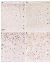
Fig. 1
Fig. 1
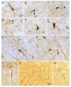
Subtypes of cortical pTDP-43 pathology in FTLD. pTDP-43 IHC shows pTDP-43 pathology in the anterior cingulate gyrus thatp resented either with predominantly perikaryal pTDP-43 inclusions (cytoplasmic type in a, c, e) or with long and extensive aggregates in dendrites (neuritic type in b, d, f). Both the neuritic type and the cytoplasmict ype showed prominent involvement of projection neurons of cortical layers II and III (c, d), although the cytoplasmic type more often showed involvement of Betz pyramidal cells of layer Vb in the primary motor cortex than the neuritic type. Scale in a applies to b; scale bar in c is also valid for df
Fig. 2
Fig. 2
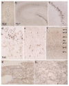
pTDP-43 pathology in dendrites of neuritic type FTLD-TDP. Double-labeling IHC of the middle frontal gyrus using pTDP-43 antibody (dark blue) and an anti-neurofilament marker (SMI-311, ai) or the anti-neurofilament antibody NFH 200 (k, m) (brown). a pTDP-43 aggregates in the neuronal perikaryon and proximal parts of a basal dendrite. b pTDP-43 aggregates at the ramification of basal dendrite showing a focal (arrow) bulb-like dendritic swelling. c Occasionally, neuritic type cases also showed intra-perikaryonal pTDP-43 aggregates. d Elongated pTDP-43 lesion in a basal dendrite that shows curvatures and a focal (arrow) dendritic swelling. e Intraneuronal pTDP-43 aggregate encompassing part of the perikaryon as well as the proximal portions of two dendrites. f, g pTDP-43 aggregates in the perikaryon and proximal portion of an apical dendrite. h Bulbous swelling (arrow) of a basal dendrite caused by pTDP-43 aggregates. im pTDP-43 aggregates in apical dendrites and their ramifications (k, m); framed area in k is shown at higher resolution in m. Scale bar in a also applies to bi and m
Fig. 3
Fig. 3
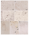
pTDP-43 pathology in the olfactory, mesiotemporal regions, and striatal regions of FTLD-TDP. a the anterior olfactory nucleus contains pTDP-43-immunoreactive inclusions. b pTDP-43 IHC shows extensive pathology in granular cells of the hippocampal fasciad entata and in dendrites of molecular layer originating from granularc ells. c dare higher resolution excerpts from b. e pyramidal neurons of the Ammon's horn (CA1) containing pTDP-43 inclusions. f pTDP-43 pathology in the entorhinal cortex affecting mainly layers pre-β as well as pri-α and pri-β. g, h Striatal pTDP-43 pathology in the accumbens nucleus affecting mainly medium-sized projection neurons; h is a higher resolution excerpt from the framed area in g. Scale bar in a is also valid for c, e, and h; scale bar in b applies to f and g
Fig. 4
Fig. 4
Thalamic, bulbar, spinal cord, and occipital pTDP-43 pathology in FTLD-TDP. a pTDP-43 IHC pTDP-43 pathology in cortically projecting thalamic neurons. b pTDP-43 aggregates in cells of the inferior olive. c Cervical spinal cord anterior horn showing pTDP-43 pathology in α-motoneurons as well as dot-like or circumferential inclusions in oligodendroglia. d pTDP-43 aggregates in hypoglossal nucleus (XII) neurons of the medulla oblongata. e, f Hypoglossal nucleus and intramedullary axons of this nucleus with pTDP-43 aggregates. Framed area in e is shown at higher resolution in f. f Arrows indicate the bundles of intramedullary axons. g Visual cortex pTDP-43 pathology (peristriate region, Brodmann area 17, 18) as observed in pattern IV cases. Scale bar in a is also valid in c, d, f, and g; scale bar in b also applies to e
Fig. 5
Fig. 5
Summary diagram of four regional distribution patterns of pathology in bvFTLD illustrating the hypothesis that pTDP-43 pathology may spread sequentially, possibly propagating (white arrows) along axonal pathways. Preferentially vulnerable cortical and subcortical sites are involved. Pattern I cases with the lowest burden of pTDP pathology display the presence of pTDP-43 immunoreactive inclusions in projection neurons and oligodendrocytes of basal and anterior portions of the prefrontal neocortex (orbital gyri, gyrus rectus, inferior frontal gyrus). Pattern II cases show the same lesion distribution as pattern I, but with pTDP pathology also in prefrontal neocortical areas located further caudal to the frontal pole, including the middle frontal gyrus, insular cortex, and anterior cingulate gyrus, and also in anteromedial areas (transentorhinal region, adjoining allocortical entorhinal region and hippocampal formation), the superior and middle temporal gyri, striatum (accumbens nucleus, putamen, claustrum), parvocellular portion of the red nucleus, medial and lateral portions of the thalamus, and in precerebellar nuclei of the pons and medulla oblongata. Cases with additional pathology beyond that seen in the first two patterns are characterized by the presence of pTDP-43 immunoreactive inclusions in the agranular motor neocortex (Brodmann fields 4 and 6), in parietal neocortical areas (sensory cortex, angular gyrus), in bulbar somatomotor neurons, and in α-motoneruons of the cervical spinal cord anterior horns which is pattern III. Cases with the highest burden of pathology (pattern IV) show the presence of pTDP-43 lesions in the occipital neocortex (visual cortex, Brodmann 17, 18) in addition to involvement of all of the areas involved in patterns I–III. a pc precerebellar nuclei; c XII hypoglossal nucleus, io inferior olive; e ca caudate nucleus, pu putamen, pi internal pallidum, pe external pallidum, fo fornix, cl claustrum, mc mediocentral subnuclei of the bl basolateral subnuclei of amygdala, en entorhinal cortex (en); f pu putamen, md mediodorsal thalamus, rn red nucleus, sn substantia nigra (sn), ca1 Ammon's horn region 1, en entorhinal cortex

Similar articles

Cited by

References

    1. Armstrong MJ, Litvan I, Lang AE, et al. Criteria for the diagnosis of corticobasal degeneration. Neurology. 2013;80:496–503. - PMC - PubMed
    1. Armstrong RA, Carter D, Cairns NJ. A quantitative study of the neuropathology of 32 sporadic and familial cases of frontotemporal lobar degeneration with TDP-43 proteinopathy (FTLD-TDP) Neuropathol Appl Neurobiol. 2012;38:25–38. - PMC - PubMed
    1. Barbas H. Specialized elements of orbitofrontal cortex in primates. Ann N Y Acad Sci. 2007;1121:10–32. - PubMed
    1. Barbas H, De Olmos J. Projections from the amygdala to basoventral and mediodorsal prefrontal regions in the rhesus monkey. J Comp Neurol. 1990;300:549–571. - PubMed
    1. Barbas H, Henion TH, Dermon CR. Diverse thalamic projections to the prefrontal cortex in the rhesus monkey. J Comp Neurol. 1991;313:65–94. - PubMed

Publication types

MeSH terms