. 2014 Feb 14;343(6172):791-795.
doi: 10.1126/science.1247575. Epub 2014 Jan 9.

Somites without a clock

Affiliations

Somites without a clock

Ana S Dias et al. Science. .

Abstract

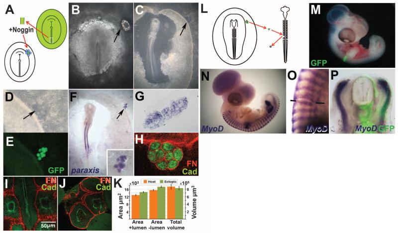
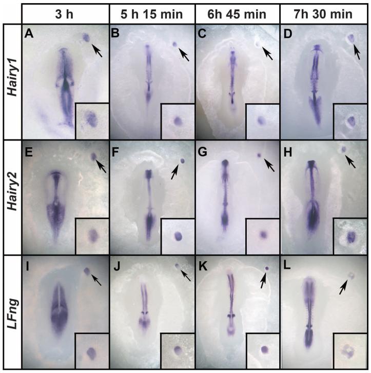
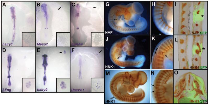
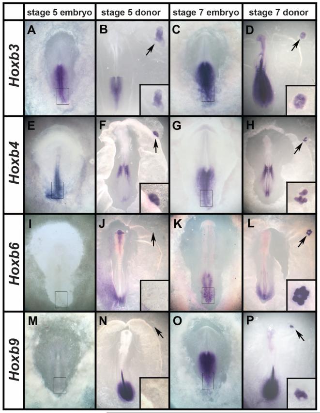
The formation of body segments (somites) in vertebrate embryos is accompanied by molecular oscillations (segmentation clock). Interaction of this oscillator with a wave traveling along the body axis (the clock-and-wavefront model) is generally believed to control somite number, size, and axial identity. Here we show that a clock-and-wavefront mechanism is unnecessary for somite formation. Non-somite mesoderm treated with Noggin generates many somites that form simultaneously, without cyclic expression of Notch-pathway genes, yet have normal size, shape, and fate. These somites have axial identity: The Hox code is fixed independently of somite fate. However, these somites are not subdivided into rostral and caudal halves, which is necessary for neural segmentation. We propose that somites are self-organizing structures whose size and shape is controlled by local cell-cell interactions.


Figures

Fig. 1
Fig. 1. BMP inhibition generates normal somites
(A to E) Experimental design. The PS of a donor quail or GFP-transgenic embryo is excised; exposed to Noggin; and grafted, surrounded by Noggin-beads, to the periphery of a host chick embryo [(A and B), arrows]. After overnight incubation, a group of somite-like structures—arranged as a bunch of grapes—appears [(C and D), arrows]. These structures fluoresce if the donor is a GFP-transgenic embryo (E). (F to P) The ectopic structures are real somites: They express paraxis (F and G) and N-cadherin [green in (H) to (J)] and are surrounded by a Fibronectin matrix [red in (H) to (J)]. Multiphoton confocal sections through normal (I) and ectopic (J) somites were used to estimate somite sizes (K). When an ectopic somite is grafted instead of a somite in an older embryo (L), the graft incorporates well (M). After 2 to 3 days, the grafted somite appropriately expresses MyoD (N to P).
Fig. 2
Fig. 2. Ectopic somites form without cyclic expression of segmentation clock genes
Embryos were fixed at 45-min intervals (examples shown at 3, 5.15, 6.45, and 7.5 hours after grafting to a host embryo) and stained for expression of Hairy1 (A to D), Hairy2 (E to H), and LFng (I to L). The in situ embryos were developed to reveal the segmentation clock in the presomitic cells of the host. Although patterns of expression in the presomitic mesoderm of the host are dynamic, no major differences in expression are seen in the graft (insets). Arrows mark the graft region, which is shown magnified in the insets.
Fig. 3
Fig. 3. Ectopic somites are not subdivided into rostral and caudal halves
(A to F) Ectopic somites were analyzed for expression of caudal (Hairy1, Meso2, LFng, Hairy2, and Uncx4.1) and rostral (EphA4) markers. Hairy1 (A), Meso2 (B), and EphA4 (C) are not expressed; LFng and Hairy2 (D and E) are weak and uniform; and Uncx4.1 is expressed as random patches (F). Insets show a magnified view of the graft. (G to O) As a further test of rostrocaudal patterning, embryos grafted as in Fig. 1L were stained for motor axons [neurofilament-associated protein NAP, (G to I), brown] or neural crest [HNK1, (J to O), brown] and anti-GFP [green in (I), (L), and (O)]. A large gap (G to I), fused roots (J to L), or multiple small ganglia (M to O) form in the ectopic somite (arrows, asterisks). Sections (I) and (L) are coronal, (O) is transverse at the level of the graft.
Fig. 4
Fig. 4. Ectopic somites have trunk identity, fixed according to the Hox genes expressed in the donor PS
(A to P) At stage 5, the posterior PS expresses Hoxb3 (A) and b4 (E), but not b6 (I) or b9 (M). Ectopic somites made from posterior streak explants from these stages show a similar pattern of expression (B, F, J, and N). At stages 7 and 8, the posterior streak expresses all four genes (C, G, K, and O), as do the ectopic somites formed from it (D, H, L, and P). Arrows point to the graft region, shown magnified in the insets.

Comment in

Similar articles

Cited by

References

    1. Muñoz-Sanjuán I, Brivanlou AH. In: Gastrulation: From Cells to Embryo. Stern CD, editor. Cold Spring Harbor Press; New York: 2004. pp. 475–489.
    1. Dubrulle J, McGrew MJ, Pourquié O. Cell. 2001;106:219–232. - PubMed
    1. Dubrulle J, Pourquié O. Nature. 2004;427:419–422. - PubMed
    1. Cooke J, Zeeman EC. J. Theor. Biol. 1976;58:455–476. - PubMed
    1. Harima Y, Takashima Y, Ueda Y, Ohtsuka T, Kageyama R. Cell Rep. 2013;3:1–7. - PubMed

Publication types

MeSH terms