. 2014 Mar 1;306(5):G412-24.
doi: 10.1152/ajpgi.00118.2013. Epub 2014 Jan 9.

Rab1a regulates sorting of early endocytic vesicles

Affiliations

Rab1a regulates sorting of early endocytic vesicles

Aparna Mukhopadhyay et al. Am J Physiol Gastrointest Liver Physiol. .

Abstract

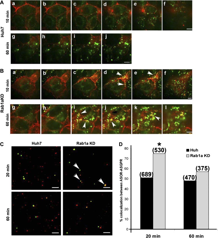
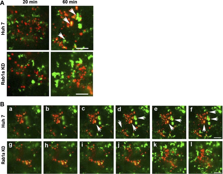
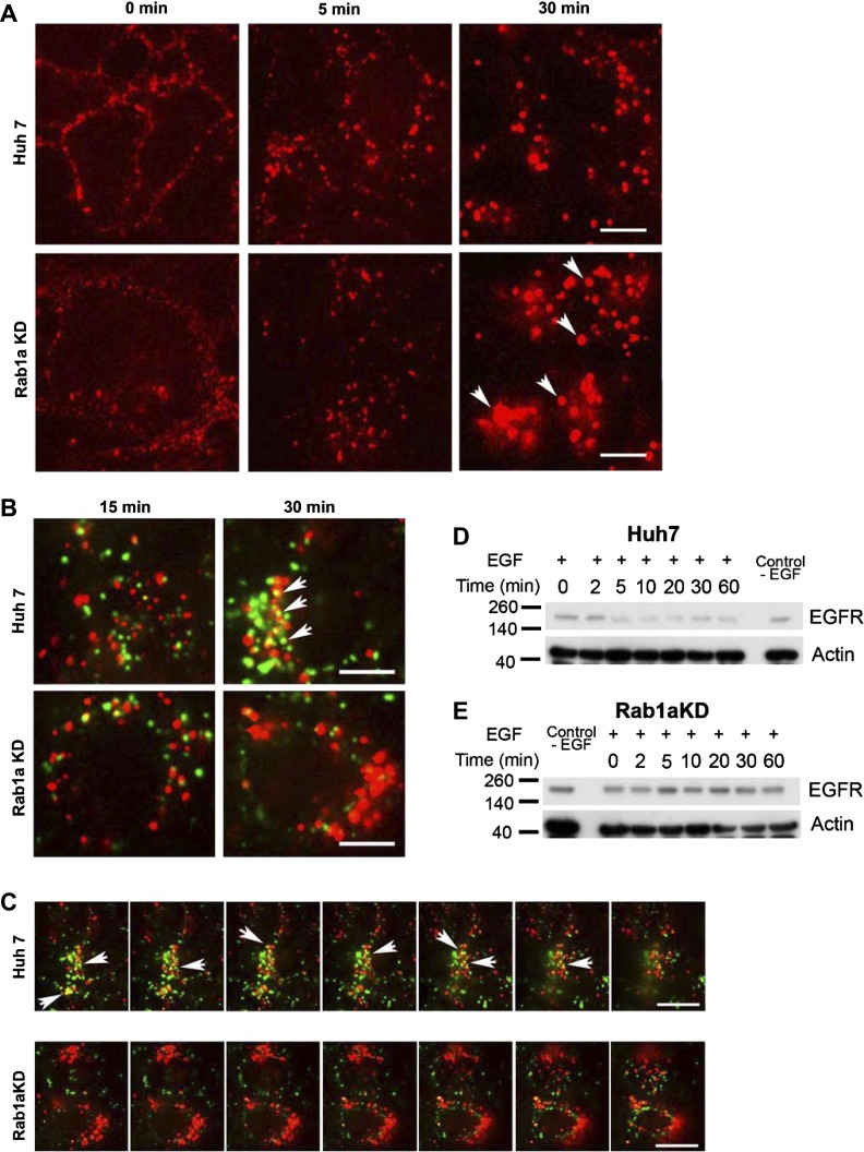
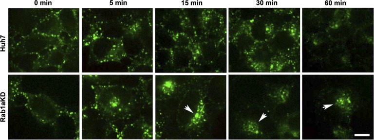
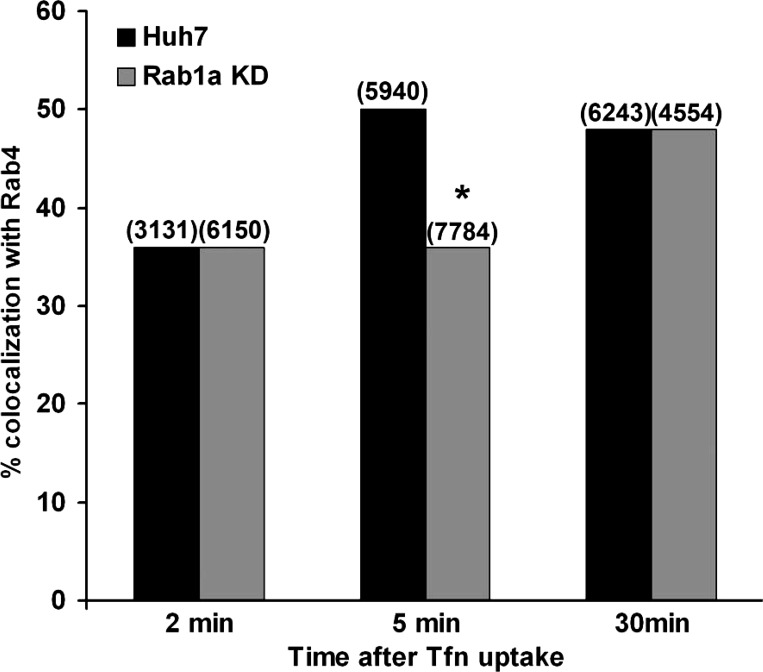
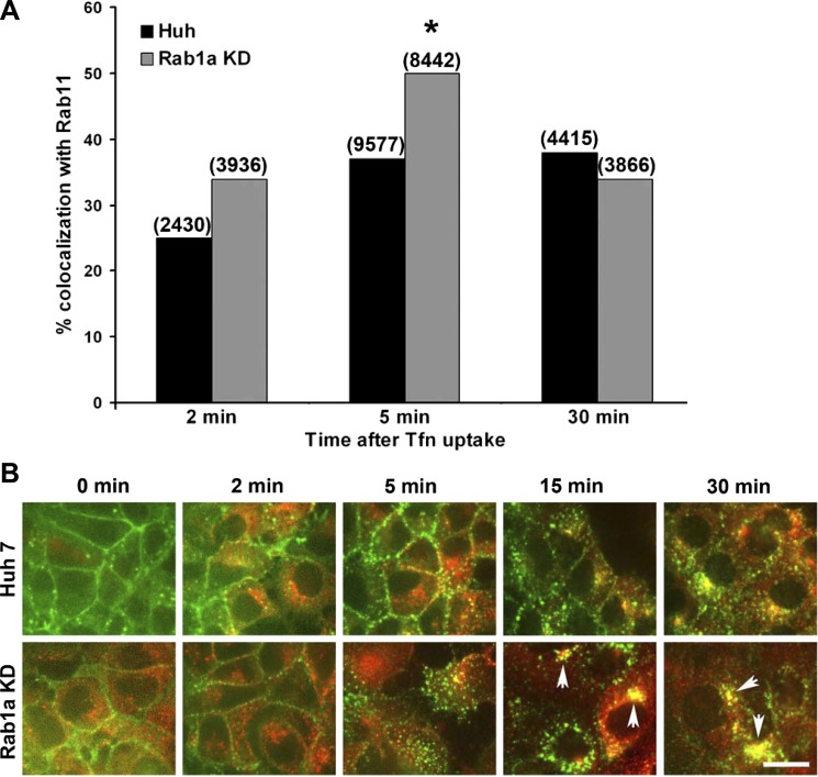
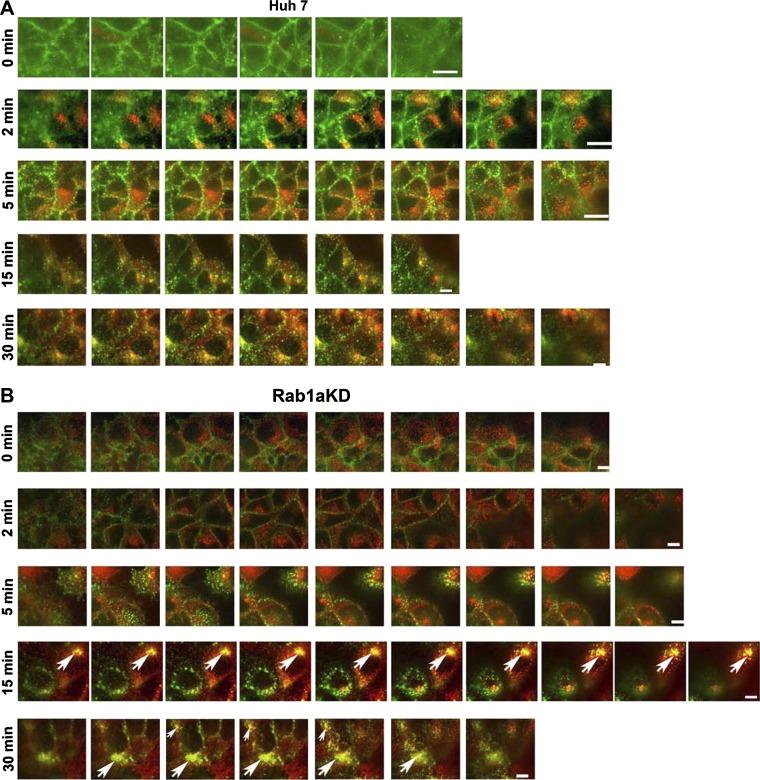
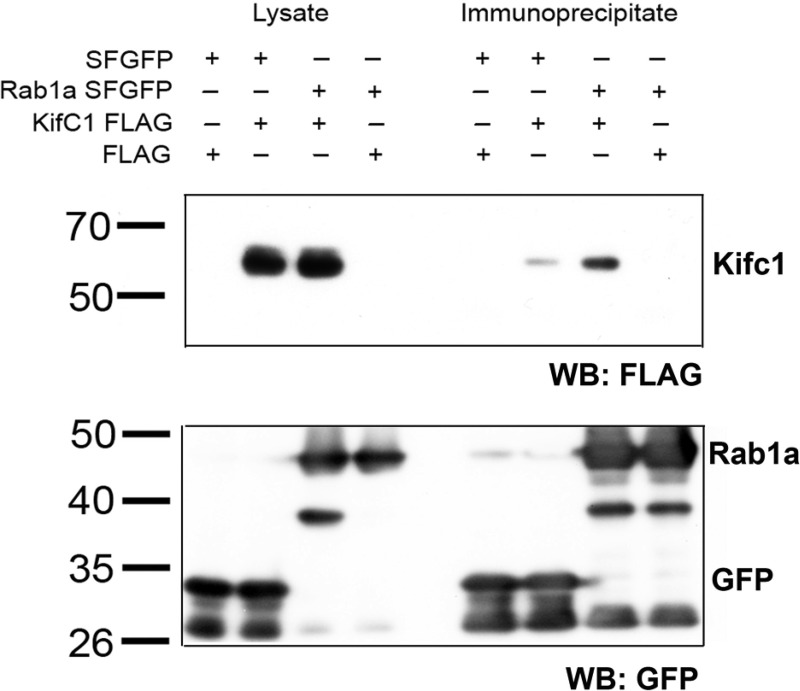
We previously reported that Rab1a is associated with asialoorosomucoid (ASOR)-containing early endocytic vesicles, where it is required for their microtubule-based motility. In Rab1a knockdown (KD) cell lines, ASOR failed to segregate from its receptor and, consequently, did not reach lysosomes for degradation, indicating a defect in early endosome sorting. Although Rab1 is required for Golgi/endoplasmic reticulum trafficking, this process was unaffected, likely due to retained expression of Rab1b in these cells. The present study shows that Rab1a has a more general role in endocytic vesicle processing that extends to EGF and transferrin (Tfn) trafficking. Compared with results in control Huh7 cells, EGF accumulated in aggregates within Rab1a KD cells, failing to reach lysosomal compartments. Tfn, a prototypical example of recycling cargo, accumulated in a Rab11-mediated slow-recycling compartment in Rab1a KD cells, in contrast to control cells, which sort Tfn into a fast-recycling Rab4 compartment. These data indicate that Rab1a is an important regulator of early endosome sorting for multiple cargo species. The effectors and accessory proteins recruited by Rab1a to early endocytic vesicles include the minus-end-directed kinesin motor KifC1, while others remain to be discovered.

Keywords: EGF; Rab1a; endocytosis; sorting; transferrin.


Figures

Fig. 1.
Fig. 1.
Reduced segregation of asialoorosomucoid (ASOR) from its receptor asialoglycoprotein receptor (ASGPR) in Rab1a knockdown (KD) cells. Control Huh7 and Rab1a KD cells were incubated with Alexa 488-ASOR (green) on ice and then transferred to 37°C for 10 or 60 min to internalize Alexa 488-ASOR. A and B: representative z-plane confocal images of fixed Huh7 (control) and Rab1a KD cells immunostained for ASGPR (red). a–l: 1- to 1.5-μm confocal cuts from the top to the bottom of the cells. Arrows indicate areas of colocalization. Scale bars, 10 μm. C: endocytic vesicles purified from cells 20 and 60 min after internalization of Alexa 488-ASOR (green) were stained for ASGPR (red) using specific antibody. Arrows indicate areas of colocalization. Scale bars, 5 μm. D: percent colocalization of ASOR and ASGPR in isolated vesicles in multiple experiments performed as described in C. Numbers in parentheses indicate total number of vesicles counted. *P < 0.001.
Fig. 2.
Fig. 2.
Trafficking of ASOR to lysosomes is delayed in the absence of Rab1a. Control Huh7 and Rab1a KD cells were incubated with Alexa 488-ASOR (green) on ice and then transferred to 37°C for 20 or 60 min to internalize Alexa 488-ASOR. After fixation, cells were immunostained for the lysosomal marker lysosome-associated membrane protein 1 (LAMP-1; red). A: representative single-plane confocal images of ASOR uptake by cells. B: confocal z-plane images of Huh7 and Rab1a KD cells that had taken up ASOR for 60 min. a–l: 1- to 1.5-μm confocal cuts from the top to the bottom of the cells. Arrows indicate areas of colocalization. Scale bars, 10 μm.
Fig. 3.
Fig. 3.
Endocytic processing of EGF is delayed in the absence of Rab1a. A: representative single-plane confocal images of control Huh7 and Rab1a KD cells incubated with Alexa 555-EGF (red) on ice, shifted to 37°C for 0, 5, and 30 min, and fixed. Arrows indicate large aggregates of EGF at 30 min of endocytosis in Rab1a KD cells. B: fixed Huh7 and Rab1a KD cells prepared as described in A were stained with the lysosomal marker LAMP-1 (green). Arrows indicate areas of colocalization with Alexa 555-EGF, primarily in control Huh7 cells. C: confocal z-plane images of Huh7 and Rab1a KD cells that had endocytosed EGF (red) for 60 min before fixation and immunostaining for LAMP-1 (green). From left to right: 1- to 1.5-μm confocal cuts from the top to the bottom of the cells. Arrows indicate areas of colocalization, primarily in control Huh7 cells. Scale bars, 10 μm. D and E: EGF was added to Huh7 and Rab1a KD cells on ice, and cells were incubated at 37°C for 0–60 min to initiate endocytosis. Lysate was prepared and assayed by immunoblotting for EGF receptor (EGFR) and actin (loading control). Control −EGF represents lysates from cells that were not incubated with EGF. Position of molecular weight markers (in kDa) is indicated by lines at left of each immunoblot.
Fig. 4.
Fig. 4.
Rab1a is required for endocytic trafficking of transferrin (Tfn). Control Huh7 and Rab1a KD cells were incubated with Alexa 488-Tfn on ice and then incubated at 37°C for 0, 5, 15, 30, and 60 min to initiate endocytosis. Representative single-plane confocal images are shown. In Huh7 cells, Tfn was progressively taken up into punctate vesicular structures and then gradually disappeared by 60 min, as apotransferrin was recycled back to the cell surface and released. In Rab1a KD cells, Tfn was taken up into vesicular structures and by 15 min appeared in large juxtanuclear aggregates (arrows). Scale bar, 10 μm.
Fig. 5.
Fig. 5.
Tfn is delayed in reaching Rab4-associated compartments in Rab1a KD cells. Huh7 and Rab1a KD cells were incubated with Alexa 488-Tfn on ice and then shifted to 37°C for 2, 5, and 30 min to initiate endocytosis. Endocytic vesicles were purified from these cells and immunostained for Rab4. Colocalization of Rab4 and Tfn in vesicles was quantified as described in materials and methods. By 5 min in control cells, 50% of the Tfn-containing endocytic vesicles were associated with Rab4, and association remained for 30 min. Association with Rab4 was significantly reduced at 5 min in Tfn-containing vesicles prepared from Rab1a KD cells but increased over time, so that, by 30 min after Tfn internalization, percentage of vesicles colocalizing with Rab4 was similar for control and Rab1a KD vesicles. Numbers in parentheses indicate total number of vesicles counted. *P < 0.001.
Fig. 6.
Fig. 6.
Tfn more rapidly enters a Rab11-associated recycling compartment in Rab1a KD cells. Huh7 and Rab1a KD cells were incubated with Alexa 488-Tfn on ice and then shifted to 37°C for 2, 5, and 30 min to initiate endocytosis. A: endocytic vesicles were purified from these cells and immunostained for Rab11. Colocalization of Rab11 and Tfn in vesicles was quantified as described in materials and methods. Numbers in parentheses indicate total number of vesicles counted. *P < 0.001. B: representative single-plane images of cells fixed and immunostained for Rab11 (red). Colocalization of Rab11 (red) with Tfn-containing endocytic vesicles (green) was reduced in control Huh7 cells compared with Rab1a KD cells. As indicated by arrows, juxtanuclear aggregates of Tfn in Rab1a KD cells, which are visible in Fig. 4, are associated with Rab11, likely in a recycling compartment. Scale bar, 20 μm.
Fig. 7.
Fig. 7.
Accumulation of Tfn in a Rab11 compartment in Rab1a KD cells. Huh7 and Rab1a KD cells were incubated with Alexa 488-Tfn (green) on ice and then shifted to 37°C for 0, 2, 5, 15, and 30 min to initiate endocytosis. Cells were fixed and immunostained for Rab11 (red), and confocal z-plane images were obtained. From left to right: 1- to 1.5-μm confocal cuts from the top to the bottom of the cells. Arrows indicate areas of colocalization of Tfn and Rab11, primarily in Rab1a KD cells at 15 and 30 min of endocytosis. Scale bars, 10 μm.
Fig. 8.
Fig. 8.
Interaction of Rab1a with the minus-end-directed kinesin KifC1. 293 cells stably expressing Rab1a-superfolder green fluorescent protein (sfGFP) or sfGFP alone were transiently transfected with KifC1-FLAG or FLAG empty vector. Lysates were prepared, and sfGFP was immunoprecipitated using anti-GFP-agarose beads. Lysate and immunoprecipitates were subjected to SDS-PAGE and immunoblotted for FLAG (top) or GFP (bottom). Migration of molecular weight markers (in kDa), as well as KifC1-FLAG, sfGFP-Rab1a, and sfGFP, is indicated. Although there was a consistently weak background band corresponding to KifC1 following immunoprecipitation of unlinked sfGFP, KifC1 was much more abundant in the sfGFP-Rab1a immunoprecipitate. Blots are representative of results from 3 experiments. WB, Western blot.
Fig. 9.
Fig. 9.
Colocalization of endocytosed EGF with KifC1. Huh7 and Rab1a KD cells were incubated with Alexa 555-EGF on ice and then shifted to 37°C for 2 or 5 min to initiate endocytosis. Endocytic vesicles were purified from these cells and immunostained for KifC1. Colocalization of EGF (red) and KifC1 (green) in vesicles was quantified as described in materials and methods. Arrows show representative vesicles in which EGF and KifC1 are colocalized. Numbers in parentheses indicate total number of vesicles counted. Scale bar, 10 μm.

References

    1. Akhmanova A, Hammer JA., 3rd Linking molecular motors to membrane cargo. Curr Opin Cell Biol 22: 479–487, 2010 - PMC - PubMed
    1. Aleksandrowicz P, Marzi A, Biedenkopf N, Beimforde N, Becker S, Hoenen T, Feldmann H, Schnittler HJ. Ebola virus enters host cells by macropinocytosis and clathrin-mediated endocytosis. J Infect Dis 204 Suppl 3: S957–S967, 2011 - PMC - PubMed
    1. Ally S, Larson AG, Barlan K, Rice SE, Gelfand VI. Opposite-polarity motors activate one another to trigger cargo transport in live cells. J Cell Biol 187: 1071–1082, 2009 - PMC - PubMed
    1. Aniento F, Emans N, Griffiths G, Gruenberg J. Cytoplasmic dynein-dependent vesicular transport from early to late endosomes. J Cell Biol 123: 1373–1387, 1993 - PMC - PubMed
    1. Bananis E, Murray JW, Stockert RJ, Satir P, Wolkoff AW. Microtubule and motor-dependent endocytic vesicle sorting in vitro. J Cell Biol 151: 179–186, 2000 - PMC - PubMed

Publication types

LinkOut - more resources