Review
. 2014 Sep;60(9):1158-73.
doi: 10.1373/clinchem.2013.216044. Epub 2014 Jan 9.

Noninvasive micromarkers

Affiliations
Review

Noninvasive micromarkers

Janani Saikumar et al. Clin Chem. 2014 Sep.

Abstract

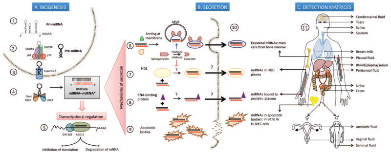
Background: The recent revolutionary advances made in genome-wide sequencing technology have transformed biology and molecular diagnostics, allowing new sRNA (small RNA) classes to be discovered as potential disease-specific biological indicators. Cell-free microRNAs (miRNAs) have been shown to exist stably in a wide spectrum of body fluids and their expression profiles have been shown to reflect an assortment of physiological conditions, underscoring the utility of this new class of molecules to function as noninvasive biomarkers of disease.

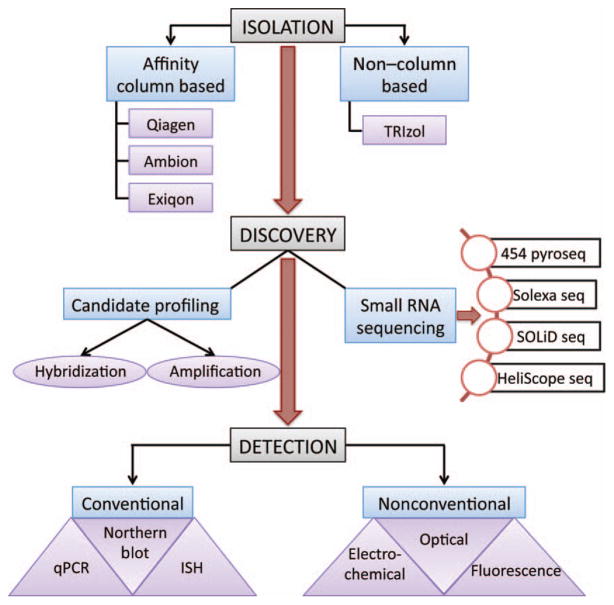
Content: We summarize information on the known mechanisms of miRNA protection and release into extracellular space and compile the current literature on extracellular miRNAs that have been investigated as biomarkers of 20 different cancers, 11 organ damage conditions and 10 diverse disease states. We also discuss the various strategies involved in the miRNA biomarker discovery workflow and provide a critical opinion on the impediments faced by this advancing field that need to be overcome in the laboratory.

Summary: The field of miRNA-centered diagnostics is still in its infancy, and basic questions with regard to the exact role of miRNAs in the pathophysiology of diseases, and the mechanisms of their release from affected cells into biological fluids are yet to be completely understood. Nevertheless, these noninvasive micromarkers have immense potential in translational medicine not only for use in monitoring the efficacy and safety of therapeutic regimens but also to guide the diagnosis of diseases, to determine the risk of developing diseases or conditions, and more importantly, to inform treatment options.


Conflict of interest statement

Authors’ Disclosures or Potential Conflicts of Interest: Upon manuscript submission, all authors completed the author disclosure form. Disclosures and/or potential conflicts of interest:

Employment or Leadership: V.S. Vaidya, Harvard Program in Therapeutic Sciences.

Consultant or Advisory Role: None declared.

Stock Ownership: None declared.

Honoraria: None declared.

Research Funding: V.S. Vaidya, National Institutes of Health/National Institutes of Environmental Health Sciences–Outstanding New Environmental Scientist Award (ES017543) and an Innovation in Regulatory Science Award from the Burroughs Wellcome Fund.

Expert Testimony: None declared.

Patents: None declared.

Figures

Fig. 1
Fig. 1. MicroRNA (miRNA) biogenesis and secretion into extracellular space
(A), Biogenesis. 1. Transcription of pri-miRNAs from genes in the nucleus by RNA polymerase II. 2. Processing of primary transcript into 70-nucleotide (nt)–long pre-miRNA by the Drosha-DGCR8 Microprocessor complex assisted by p68 (DDX5) and p72 (DDX17) DEAD-box RNA helicases that possibly act as scaffolding proteins to recruit cofactors. 3. Export into cytoplasm in a Ran-GTP– dependent manner through Exportin 5. 4. Cleavage of pre-miRNA by RNase III enzyme Dicer, into a 22-nt double-stranded RNA composed of the mature miRNA “guide” strand and the low-abundance miRNA* “passenger” strand; TRBP and protein activator of interferon-induced protein kinase PKR (PACT) are some of the molecules that regulate this step. 5. Incorporation of mature miRNA into the RISC, whose main components are AGO-2 and GW-182; partial complementarity of the seed region of miRNA to the 3′ untranslated region of target mRNAs causes translational repression while complete complementarity leads to degradation of the transcript. (B), Secretion of miRNAs into extracellular space by: 6. Packaging into multivesicular bodies (MVBs) that fuse with the plasma membrane and release as exosomes in a ceramide-dependent pathway positively regulated by nSMase2. 7. Encapsulation into HDL particles, a process which is repressed by nSMase2. 8. Binding to RNA-binding proteins, namely AGO-2 and nucleophosmin 1 (NPM1). 9. Incorporation into apoptotic bodies. 10. Pioneering studies describing exosomal miRNAs isolated from primary bone marrow derived mast cells; HDL-miRNA in human plasma; protein-bound miRNAs in human plasma (AGO-2) and human cell lines (NPM1 in HepG2 and A549); miR-126 from endothelial cell-derived apoptotic bodies in vitro in human umbilical vein endothelial cells (HUVEC). (C), Detection matrices. 11. Depiction of 13 different matrices where extracellular miRNAs have been discovered.
Fig. 2
Fig. 2
Tools and techniques used in miRNA bio-marker discovery in the RNA isolation, candidate discovery, and verification stages. Seq, sequencing.

References

    1. Lee R, Feinbaum R, Ambros V. The C. elegans heterochronic gene lin-4 encodes small RNAs with antisense complementarity to lin-14. Cell. 1993;75:843–54. - PubMed
    1. Reinhart B, Slack F, Basson M, Pasquinelli A, Bettinger J, Rougvie A, et al. The 21-nucleotide let-7 RNA regulates developmental timing in Caenorhabditis elegans. Nature. 2000;403:901–6. - PubMed
    1. Pasquinelli A, Reinhart B, Slack F, Martindale M, Kuroda M, Maller B, et al. Conservation of the sequence and temporal expression of let-7 heterochronic regulatory RNA. Nature. 2000;408:86–9. - PubMed
    1. Hsu P, Huang H-D, Hsu S-D, Lin L-Z, Tsou A-P, Tseng C-P, et al. MiRNAmap: genomic maps of microRNA genes and their target genes in mammalian genomes. Nucleic Acids Res. 2006;34:D135–9. - PMC - PubMed
    1. Winter J, Jung S, Keller S, Gregory RI, Diederichs S. Many roads to maturity: microRNA biogenesis pathways and their regulation. Nat Cell Biol. 2009;11:228–34. - PubMed

Publication types