. 2014 Apr;5(2):195-208.
doi: 10.1007/s13244-013-0308-y. Epub 2014 Jan 10.

Bowel wall thickening at CT: simplifying the diagnosis

Affiliations

Bowel wall thickening at CT: simplifying the diagnosis

Teresa Fernandes et al. Insights Imaging. 2014 Apr.

Abstract

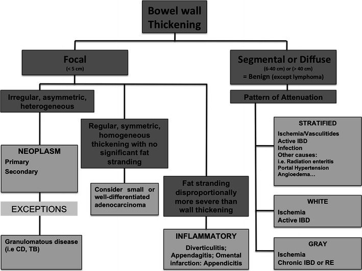
Objective: In this article we present a simplified algorithm-based approach to the thickening of the small and large bowel wall detected on routine computed tomography (CT) of the abdomen.

Background: Thickening of the small or large bowel wall may be caused by neoplastic, inflammatory, infectious, or ischaemic conditions. First, distinction should be made between focal and segmental or diffuse wall thickening. In cases of focal thickening further analysis of the wall symmetry and perienteric anomalies allows distinguishing between neoplasms and inflammatory conditions. In cases of segmental or diffuse thickening, the pattern of attenuation in light of clinical findings helps narrowing the differential diagnosis.

Conclusion: Focal bowel wall thickening may be caused by tumours or inflammatory conditions. Bowel tumours may appear as either regular and symmetric or irregular or asymmetric thickening. When fat stranding is disproportionately more severe than the degree of wall thickening, inflammatory conditions are more likely. With the exception of lymphoma, segmental or diffuse wall thickening is usually caused by benign conditions, such as ischaemic, infectious and inflammatory diseases.

Key points: • Thickening of the bowel wall may be focal (<5 cm) and segmental or diffuse (6-40 cm or >40 cm) in extension. • Focal, irregular and asymmetrical thickening of the bowel wall suggests a malignancy. • Perienteric fat stranding disproportionally more severe than the degree of wall thickening suggests an inflammatory condition. • Regular, symmetric and homogeneous wall thickening is more frequently due to benign conditions, but can also be caused by neoplasms such as well-differentiated adenocarcinoma and lymphoma. • Segmental or diffuse bowel wall thickening is usually caused by ischaemic, inflammatory or infectious conditions and the attenuation pattern is helpful in narrowing the differential diagnosis.


Figures

Fig. 1
Fig. 1
Algorithm approach to the bowel wall thickening. CD Crohn’s disease, TB tuberculosis, IBD inflammatory bowel disease, RE radiation enteritis. Adapted from the electronic poster “Bowel wall thickening—a complex subject made simple” DOI:10.5444/esgar2011/EE-063
Fig. 2
Fig. 2
Colon cancer. Axial contrast-enhanced CT scan shows focal asymmetric and irregular thickening of the ascending colon (arrow), a finding strongly suggestive of a neoplasm. Also note mild pericolonic fat stranding (asterisks), a frequent associated finding. Adapted from the electronic poster “Bowel wall thickening—a complex subject made simple” DOI:10.5444/esgar2011/EE-063
Fig. 3
Fig. 3
Intestinal tuberculosis. Axial (a) and reformatted coronal (b) contrast-enhanced CT scans show parietal irregular and asymmetric thickening of the caecum (large arrows), an appearance that mimics colon cancer. Also note low attenuation adenopathy (thin arrows), a usual finding in tuberculosis. Mild pericolonic fat stranding is also seen. Adapted from the electronic poster “Bowel wall thickening—a complex subject made simple” DOI:10.5444/esgar2011/EE-063
Fig. 4
Fig. 4
Crohn’s disease mimicking colon cancer. Reformatted coronal (a) and axial (b) unenhanced CT scans and axial contrast-enhanced CT scan (c) show spiculated irregular wall thickening of the caecal wall (arrow in ac) with heterogeneous contrast enhancement (c). Also note proliferation of the pericaecal fat (asterisk), a common finding in Crohn’s disease. Adapted from the electronic poster “Bowel wall thickening—a complex subject made simple” DOI:10.5444/esgar2011/EE-063
Fig. 5
Fig. 5
Well-differentiated adenocarcinoma of the descending colon. Axial contrast-enhanced CT scan showing focal concentric and regular thickening of the descending colon (arrows) with smooth contours and homogeneous enhancement. This proved to be a well-differentiated adenocarcinoma after biopsy. Adapted from the electronic poster “Bowel wall thickening—a complex subject made simple” DOI:10.5444/esgar2011/EE-063
Fig. 6
Fig. 6
Acute diverticulitis. Reformatted coronal contrast-enhanced CT scan showing sigmoid diverticuli (arrow), mild bowel wall thickening (arrowhead) and pericolonic disproportionate fat stranding. These findings are compatible with sigmoid diverticulitis
Fig. 7
Fig. 7
Epiploic appendagitis of the descending colon. Axial contrast-enhanced CT scan shows the inflamed epiploic appendage anterior to the colonic wall (asterisk) with adjacent disproportionate fat stranding and minimal wall thickening of the descending colon (arrow). Adapted from the electronic poster “Bowel wall thickening—a complex subject made simple” DOI:10.5444/esgar2011/EE-063
Fig. 8
Fig. 8
Omental infarction. Axial contrast-enhanced CT scan of a patient who presented with acute right upper quadrant pain shows an inhomogeneous fatty mass (large arrow) in the greater omentum, indicative of omental infarction. Note the mild wall thickening (arrow) of the adjacent colonic wall, which is clearly disproportionate relative to the fat stranding. Adapted from the electronic poster “Bowel wall thickening—a complex subject made simple” DOI:10.5444/esgar2011/EE-063
Fig. 9
Fig. 9
Acute appendicitis. Axial (a) and coronal (b) contrast-enhanced CT scan shows the retrocaecal enlarged fluid-filled appendix (arrows) associated with adjacent fat stranding and reactive wall thickening of the ascending colon (asterisk)
Fig. 10
Fig. 10
Small bowel lymphoma. Reformatted coronal contrast-enhanced CT scan shows concentric and regular segmental thickening of a small bowel loop (arrows) with homogeneous contrast enhancement. Adapted from the electronic poster “Bowel wall thickening—a complex subject made simple” DOI:10.5444/esgar2011/EE-063
Fig. 11
Fig. 11
Acute small bowel ischaemia. Axial contrast-enhanced CT scan shows diffuse thickening of the small bowel loops with a target appearance (arrow) due to submucosal oedema. Also note the engorgement of the mesenteric root vessels and ascites, common findings in cases of acute bowel ischaemia. Adapted from the electronic poster “Bowel wall thickening—a complex subject made simple” DOI:10.5444/esgar2011/EE-063
Fig. 12
Fig. 12
Bowel ischaemia secondary to systemic lupus erythaematosus (LES). Axial contrast-enhanced CT scan shows circumferential thickening of the small bowel loops (arrows) with a stratified appearance in a patient with LES presenting with bloody diarrhoea indicating bowel involvement by the vasculitis
Fig. 13
Fig. 13
Stratified appearance in Crohn’s disease. Axial contrast-enhanced CT scan of the abdomen shows concentric wall thickening of small bowel loops with a stratified appearance indicating active disease (arrows). Also note a fistula (arrowhead) connecting the bowel loops, a common finding in Crohn’s disease
Fig. 14
Fig. 14
Stratified attenuation pattern in pseudomembranous colitis. Axial contrast-enhanced CT scan shows significant wall thickening of the ascending and descending colon (arrows) due to submucosal oedema, resulting in the stratified appearance. The marked thickening of the bowel wall and the mild pericolonic fat stranding suggest pseudomembranous colitis
Fig. 15
Fig. 15
Radiation enteritis in a patient with cervical cancer. Axial contrast-enhanced CT scan shows concentric stratified wall thickening of low-lying small bowel loops (arrows). There are also some ascites. These findings in the context of radiation therapy are suggestive of radiation enteritis
Fig. 16
Fig. 16
Stratified attenuation pattern in the ascending colon of a cirrhotic patient. Reformatted coronal contrast-enhanced CT scan shows parietal regular and concentric thickening of the ascending colon (arrows) with a “target” appearance due to oedema of the submucosa. Note the irregular contours of the liver (arrowheads) consistent with hepatic cirrhosis. Adapted from the electronic poster “Bowel wall thickening—a complex subject made simple” DOI:10.5444/esgar2011/EE-063
Fig. 17
Fig. 17
Fat halo sign in Crohn’s disease. Axial contrast-enhanced CT image shows concentric wall thickening of the rectum (arrow) with fatty appearance of the submucosa in a patient with a long history of Crohn’s disease
Fig. 18
Fig. 18
Shock bowel in a patient with significant haemorrhage due to bleeding oesophageal varices. Axial contrast-enhanced CT scan shows thickened hyperattenuating small bowel loops (arrow) due to the increased vascular permeability in the context of severe hypovolaemia. Also note engorgement of the mesenteric vessels and small volume ascites
Fig. 19
Fig. 19
White attenuation pattern due to spontaneous intramural bowel wall haemorrhage in a patient with polyarteritis nodosa. Axial unenhanced (a) and contrast-enhanced (b) CT scans of the abdomen show concentric wall thickening of the third duodenal portion that showed spontaneous hyperenhancement of the bowel wall due to spontaneous haemorrhage. Adapted from the electronic poster “Bowel wall thickening—a complex subject made simple” DOI:10.5444/esgar2011/EE-063
Fig. 20
Fig. 20
White attenuation pattern in Crohn’s disease. Axial (a) and coronal (b) contrast-enhanced CT scans show homogeneous hyperenhancement (arrows) of a thickened and stenotic ileal loop indicating active disease. Note the proximal dilatation of the small bowel loops (asterisk) due to the obstruction
Fig. 21
Fig. 21
Grey attenuation pattern in small bowel ischaemia. Axial contrast-enhanced CT scans show distended fluid-filled small bowel loops (asterisks in a and b) with hypoenhancing thick walls (thin arrows in a and b) indicating ischaemia in a patient with partial occlusion of the superior mesenteric artery (large arrow in a). Adapted from the electronic poster “Bowel wall thickening—a complex subject made simple” DOI:10.5444/esgar2011/EE-063
Fig. 22
Fig. 22
Ischaemic colitis. Axial contrast-enhanced CT scans through the descending colon (a) and rectum (b) show the thickened hypoattenuating bowel wall due to ischaemia. The diagnosis was confirmed with colonoscopy and biopsy

Similar articles

Cited by

References

    1. Fernandes TC, Castro R, Pinto D, Oliveira MI, Carneiro A, Cunha R (2011) Bowel wall thickening – a complex subject made simple. Electronic Poster presented at 2011 ESGAR meeting doi:10.5444/esgar2011/EE-063#_blank
    1. Paulsen SR, Huprich JE, Fletcher JG, Booya F, Young BM, Fidler JL, Johnson CD, Barlow JM, Earnest F. CT enterography as a diagnostic tool in evaluating small bowel disorders: review of clinical experience with over 700 cases. Radiographics. 2006;26:641–657. doi: 10.1148/rg.263055162. - DOI - PubMed
    1. Macari M, Megibow AJ, Balthazar EJ. A pattern approach to the abnormal small bowel: observations at MDCT and CT enterography. AJR Am J Roentgenol. 2007;188:1344–1355. doi: 10.2214/AJR.06.0712. - DOI - PubMed
    1. Macari M, Balthazar EJ. CT of bowel wall thickening: significance and pitfalls of interpretation. AJR Am J Roentgenol. 2001;176:1105–1116. doi: 10.2214/ajr.176.5.1761105. - DOI - PubMed
    1. Wisner W, Khurana B, Ji H, Ros PR. CT of acute bowel ischemia. Radiology. 2003;226:635–650. doi: 10.1148/radiol.2263011540. - DOI - PubMed