

Detection accuracy of condylar defects in cone beam CT images scanned with different resolutions and units
- PMID: 24408818
- PMCID: PMC4064628
- DOI: 10.1259/dmfr.20130414
Detection accuracy of condylar defects in cone beam CT images scanned with different resolutions and units
Abstract
Objectives: To assess the impact of spatial resolution and cone beam CT (CBCT) unit on CBCT images for the detection accuracy of condylar defects.
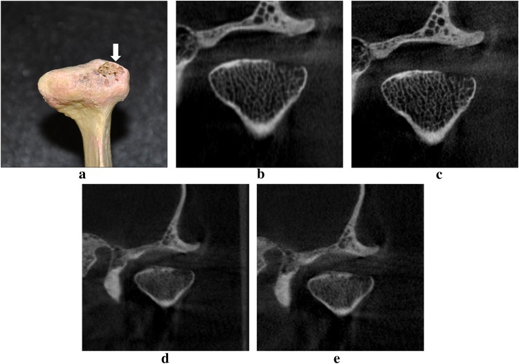
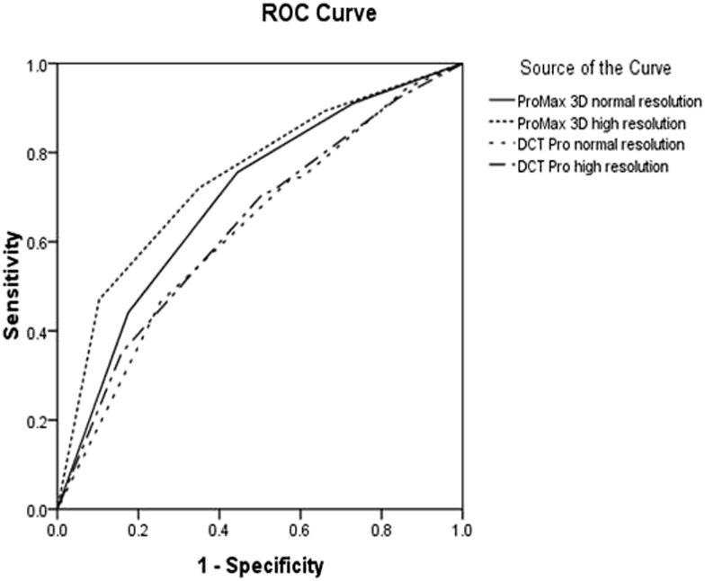
Methods: 42 temporomandibular joints were scanned, respectively, with the CBCT units ProMax® 3D (Planmeca Oy, Helsinki, Finland) and DCT PRO (Vatech, Co., Ltd., Yongin-Si, Republic of Korea) at normal and high resolutions. Seven dentists evaluated all the test images with respect to the presence or the absence of condylar defects. Receiver operating characteristic curve analysis was employed to define the detection accuracy. Two-way analysis of variance was used to analyse the values under the receiver operating characteristic curves for the differences among imaging groups and observers. Intraobserver variation was analysed using the Wilcoxon test.
Results: Macroscopic anatomy examination revealed that, of the 42 temporomandibular joint condylar surfaces, 18 were normal and 24 had defects on the surface of condyles. No significant differences were found between the images scanned with normal and high resolutions for both CBCT units ProMax 3D (p = 0.119) and DCT PRO (p = 0.740). Significant differences exist between image groups of DCT PRO and ProMax 3D (p < 0.05). Neither the inter- nor the intraobserver variability were significant.
Conclusions: The spatial resolution per se did not have an impact on the detection accuracy of condylar defects. The detection accuracy of condylar defects highly depends on the CBCT unit used for examination.
Keywords: cone-beam computed tomography; mandibular condyle; temporomandibular joint.
Figures
Similar articles
-
Temporomandibular Joint Anatomy Assessed by CBCT Images.Biomed Res Int. 2017;2017:2916953. doi: 10.1155/2017/2916953. Epub 2017 Feb 2. Biomed Res Int. 2017. PMID: 28261607 Free PMC article. Review.
-
Detection accuracy of condylar bony defects in Promax 3D cone beam CT images scanned with different protocols.Dentomaxillofac Radiol. 2013;42(5):20120241. doi: 10.1259/dmfr.20120241. Epub 2013 Feb 18. Dentomaxillofac Radiol. 2013. PMID: 23420852 Free PMC article.
-
Evaluation of cone-beam computed tomography in the diagnosis of simulated small osseous defects in the mandibular condyle.Am J Orthod Dentofacial Orthop. 2014 Feb;145(2):143-56. doi: 10.1016/j.ajodo.2013.10.014. Am J Orthod Dentofacial Orthop. 2014. PMID: 24485728
-
[Detection accuracy of occlusal caries by cone-beam computed tomography images scanned with different parameters].Beijing Da Xue Xue Bao Yi Xue Ban. 2012 Feb 18;44(1):70-4. Beijing Da Xue Xue Bao Yi Xue Ban. 2012. PMID: 22353904 Chinese.
-
Application of cone beam computed tomography for assessment of the temporomandibular joints.Aust Dent J. 2012 Mar;57 Suppl 1:109-18. doi: 10.1111/j.1834-7819.2011.01663.x. Aust Dent J. 2012. PMID: 22376103 Review.
Cited by
-
Prediction of detectability of the mandibular canal by quantitative image quality evaluation using cone beam CT.Dentomaxillofac Radiol. 2018 May;47(4):20170369. doi: 10.1259/dmfr.20170369. Epub 2018 Feb 13. Dentomaxillofac Radiol. 2018. PMID: 29376745 Free PMC article.
-
Temporomandibular joint diagnostics using CBCT.Dentomaxillofac Radiol. 2015;44(1):20140235. doi: 10.1259/dmfr.20140235. Dentomaxillofac Radiol. 2015. PMID: 25369205 Free PMC article. Review.
-
Temporomandibular Joint Anatomy Assessed by CBCT Images.Biomed Res Int. 2017;2017:2916953. doi: 10.1155/2017/2916953. Epub 2017 Feb 2. Biomed Res Int. 2017. PMID: 28261607 Free PMC article. Review.
-
In children and adolescents with temporomandibular disorder assembled with juvenile idiopathic arthritis - no association were found between pain and TMJ deformities using CBCT.BMC Oral Health. 2021 Oct 12;21(1):518. doi: 10.1186/s12903-021-01870-z. BMC Oral Health. 2021. PMID: 34641860 Free PMC article.
-
Comparative analysis of the structure of temporomandibular joint in human and rabbit.Acta Biomed. 2016 Jan 16;87(3):282-285. Acta Biomed. 2016. PMID: 28112695 Free PMC article.
References
-
- Scarfe WC. Farman AG cone-beam computed tomography. In: White SC, Pharoah MJ, eds. Oral radiology: principle and interpretation. 6th edn. Maryland Heights, MO: Mosby Inc.; 2009. pp. 225–43.
-
- Wenzel A, Haiter-Neto F, Frydenberg M, Kirkevang LL. Variable-resolution cone-beam computerized tomography with enhancement filtration compared with intraoral photostimulable phosphor radiography in detection of transverse root fractures in an in vitro model. Oral Surg Oral Med Oral Pathol Oral Radiol Endod 2009; 108: 939–45. - PubMed
MeSH terms
LinkOut - more resources
Full Text Sources
Other Literature Sources
