. 2014 Apr;25(4):693-705.
doi: 10.1681/ASN.2013050481. Epub 2014 Jan 9.

The regenerative potential of parietal epithelial cells in adult mice

Affiliations

The regenerative potential of parietal epithelial cells in adult mice

Katja Berger et al. J Am Soc Nephrol. 2014 Apr.

Abstract

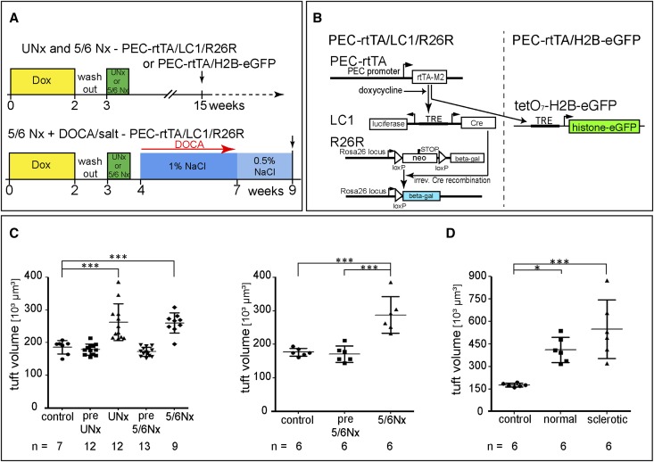
Previously, we showed that some podocytes in juvenile mice are recruited from cells lining Bowman's capsule, suggesting that parietal epithelial cells (PECs) are a progenitor cell population for podocytes. To investigate whether PECs also replenish podocytes in adult mice, PECs were genetically labeled in an irreversible fashion in 5-week-old mice. No significant increase in labeled podocytes was observed, even after 18 months. To accelerate a potential regenerative mechanism, progressive glomerular hypertrophy was induced by progressive partial nephrectomies. Again, no significant podocyte replenishment was observed. Rather, labeled PECs exclusively invaded segments of the tuft affected by glomerulosclerosis, consistent with our previous findings. We next reassessed PEC recruitment in juvenile mice using a different reporter mouse and confirmed significant recruitment of labeled PECs onto the glomerular tuft. Moreover, some labeled cells on Bowman's capsule expressed podocyte markers, and cells on Bowman's capsule were also directly labeled in juvenile podocyte-specific Pod-rtTA transgenic mice. In 6-week-old mice, however, cells on Bowman's capsule no longer expressed podocyte-specific markers. Similarly, in human kidneys, some cells on Bowman's capsule expressed the podocyte marker synaptopodin from 2 weeks to 2 years of age but not at 7 years of age. In summary, podocyte regeneration from PECs could not be detected in aging mice or models of glomerular hypertrophy. We propose that a small fraction of committed podocytes reside on Bowman's capsule close to the vascular stalk and are recruited onto the glomerular tuft during infancy to adolescence in mice and humans.


Figures

Figure 1.
Figure 1.
Absence of PEC recruitment in aging mice. (A) Timeline of the experiment. Mice were killed after 1.5, 3, 6, 12, and 18 months as indicated by the arrows (1.5–12 months, n=5; 18 months, n=3). Dox, doxycycline. (B–D) Representative histologic images of X-gal/eosin-stained kidney sections at different time points. At all time points, X-gal staining persisted in PECs (arrowheads). β-Gal–positive cells were rarely observed within the glomerular tuft and almost always localized to the vascular pole (arrows with tails). These cells are transitional cells, which are directly labeled by the PEC-rtTA mouse. Within the tubules, scattered tubular cells were labeled (arrows). (E) Statistical analysis of β-gal–positive cells in 100 glomerular cross-sections of X-gal–stained frozen sections in aging PEC-rtTA/LC1/R26R mice shows no significant changes over time. Glom., glomeruli. (F and G) Periodic acid–Schiff (PAS)-stained kidney of an 18-month-old PEC-rtTA mouse shows (F, arrow) protein casts, (G, arrow) mesangial expansion/sclerosis, and (G, arrowhead) a thickened irregular GBM. (H) Mesangial expansion and sclerosis were confirmed by transmission electron microscopy (arrow). In addition, the GBM was thickened with spikes/humps (arrowheads) and covered by podocytes with filopodial protrusions (open arrow).
Figure 2.
Figure 2.
Induction of progressive glomerular hypertrophy. (A) Experimental setups and timeline to induce glomerular hypertrophy using two alternative reporter mice: R26R and H2B-eGFP. All experiments included 14 days of doxycycline treatment and a 7-day washout before UNx or 5/6Nx. The 5/6Nx + DOCA salt model included 25 mg DOCA over 3 weeks combined with supplementation of 1% NaCl and 2 additional weeks with 0.5% NaCl by drinking water until euthanized. Arrow, time of euthanization. (B) Two alternative reporter genes were used in this study, which were both induced by the PEC-specific PEC-rtTA mouse. (Left) In the PEC-rtTA/LC1/R26R mouse, administration of doxycycline induces reversible expression of Cre recombinase from the LC1 transgene, which mediates irreversible activation of β-gal expression. (Right) Administration of doxycycline induces reversible expression of eGFP-tagged histone from the tetO7-H2B-eGFP transgene, which is deposited in the nucleus, where it persists because of a long-half life. (C) Glomerular hypertrophy after partial nephrectomy. Glomerular tuft volume was deduced from measured glomerular tuft areas of 80 glomerular cross-sections in kidneys of sham-operated (control) mice, resected kidneys (pre-UNx and pre-5/6Nx), and residual kidneys after 3 months (UNx and 5/6Nx) using the formula VG=(β/k)(Am)3/2. n, number of mice as indicated (one-way ANOVA followed by Bonferroni test). (D) PEC-rtTA/LC1/R26R mice were subjected to the 5/6Nx+DOCA/salt model to induce the most significant glomerular hypertrophy and glomerulosclerosis in a fraction of the glomeruli. Glomerular size was evaluated in histologically normal and sclerotic glomeruli individually. *P<0.05; *** P<0.001.
Figure 3.
Figure 3.
Tracing of labeled PECs after induction of glomerular hypertrophy in (A–D) PEC-rtTA/LC1/R26R or (F–H) rtTA/H2B-EGFP reporter mice. (A1–A3) X-gal–stained frozen sections of (A1) control (n=7) and (A2 and A3) experimental remnant kidneys (UNx, n=12; 5/6Nx, n=9). Staining quality is reduced, because no perfusion fixation was performed (for accurate kidney weights). Original magnification, ×400. (A4) β-Gal–positive cells close to the vascular pole (arrows). (B) Statistical analysis of 100 random glomerular cross-sections per animal including data from resected control kidneys (pre-UNx, n=12; pre-5/6Nx, n=13). Virtually no β-gal–positive cells were detected on the glomerular tuft (black bars, glomeruli with β-gal–positive cells within the tuft; error bars, SD). (C) Absolute numbers of β-gal–positive cells exclusively in those glomerular cross-sections showing β-gal positivity on the tuft. Error bars, SD. (D) Analysis of PEC to podocyte regeneration in aging PEC-rtTA/LC1/R26R mice after UNx shows no significant changes of time (1.5 and 3 months, n=5; 6 months, n=4; 12 months, n=3). (E) Costainings of synaptopodin or claudin-1 (red) and the reporter eGFP-histone (green) on paraffin sections of control and experimental remnant kidneys 3 months after 5/6Nx using PEC-rtTA/H2B-EGFP reporter mice. No significant numbers of eGFP-labeled cells were detected within the glomerular tuft in any of the experimental or control groups. Arrowheads mark eGFP-positive cells along the vascular stalk. Original magnification, ×400. (F) Statistical analysis of eGFP-positive cells on the glomerular tuft 3 months after 5/6Nx (control, sham operated; pre-5/6Nx, resected kidney; 5/6Nx, contralateral remnant kidney after 3 months; n=6 in each group). (G) The absolute number of eGFP-positive cells on the tuft in glomeruli, with eGFP positivity on the tuft ranging only between 1 and 1.5. Error bars, SD. (H) Absolute number of eGFP-positive PECs on glomerular cross-sections, with eGFP positivity on the capsule showing that GFP-histone labeling persisted in PECs. Error bars, SD.
Figure 4.
Figure 4.
Sclerotic lesions are populated by PECs in β-gal reporter mice. Serial cryosections of a glomerulus (A and B) stained with X-gal/eosin and immunostained for (A′) CD44/hemalaun or (B′) extracellular matrix derived from PECs (LKIV69). Although labeled PECs reside on Bowman’s capsule, activated PECs invade the glomerular tuft in several locations (arrows).
Figure 5.
Figure 5.
Tracking PECs in juvenile PEC-rtTA/H2B-eGFP mice. (A) Mice were induced with doxycycline 3 and 4 days after birth and analyzed 8 days or 6 weeks after birth (arrows). (B–C″) Immunofluorescent costainings for (B′ and C′) eGFP and (B and C) WT-1 of paraffin sections of 8-day-old PEC-rtTA/H2B-eGFP mice confirm labeling of PECs and absence of labeling in podocytes. At the vascular pole, WT-1–positive cells are also labeled (arrows). Original magnification, ×400. (D–E″) At 6 weeks, eGFP–labeled cells can be found along the hilus of the glomerular tuft and also, within the periphery of the glomeruli (arrows). Coexpression of WT-1 and location on the glomerular tuft suggests that these eGFP-positive cells are fully differentiated podocytes. Arrowheads mark labeled cells that have migrated onto the tuft but did not fully differentiate into podocytes (WT-1–negative and eGFP-positive). Nuclei counterstaining with Hoechst. Original magnification, ×400.
Figure 6.
Figure 6.
Direct labeling of synaptopodin-positive cells on Bowman’s capsule in juvenile PEC-rtTA/H2B-eGFP mice. (A–B″) When costaining for podocytes marker synaptopodin (red) and the reporter transgene eGFP-histone (green) 8 days after birth, weak expression of synaptopodin is noted in cells on Bowman’s capsule, mostly located close to the vascular pole (arrows). These synaptopodin-positive cells are also eGFP-labeled by the PEC-rtTA/H2B-eGFP mouse. *eGFP-labeled and synaptopodin-negative cells on Bowman’s capsule (classical PECs). Prominent synaptopodin expression was present in eGFP-positive cells at the vascular stalk (presumptive transitional cells with the round nucleus characteristic for podocytes; arrowheads). (C–D″) In 6-week-old mice, synaptopodin-positive cells were no longer present on Bowman’s capsule (arrows). Again, significant numbers of eGFP-histone–tagged cells were present within the glomerular capillary tuft and expressed synaptopodin, indicating that these cells represented differentiated podocytes recruited from Bowman’s capsule. (E) Total number of PECs and eGFP-labeled PECs in 60 random glomerular cross-sections at 8 days and 6 weeks of age as detected by Hoechst/eGFP double staining (n=4 mice per group; error bars, SD; no sign of differences between time points). (F) Recruitment of eGFP-labeled differentiated podocytes on the glomerular tuft was evaluated by eGFP/Hoechst/WT-1 or -/p57 triple staining of mice ages 8 days or 6 weeks (30 glomerular cross-sections in n=4 mice each; error bars, SD). ***P<0.0001. (G) The percentage of glomerular cross-sections with eGFP-positive transitional cells (as defined by their location at the vascular stalk) was about 20% in both age groups (n=4; 60 glomeruli per mouse).
Figure 7.
Figure 7.
The podocyte-specific Pod-rtTA/H2B-eGFP mouse directly labels cells on Bowman’s capsule in juvenile mice. (A) Labeling was induced on days 3 and 4 after birth in Pod-rtTA/H2B-eGFP mice. Kidneys were analyzed 8 days after birth (arrow). (B) Nuclear eGFP immunostaining (brown, PAS counterstaining) shows eGFP-histone–labeled cells on Bowman’s capsule (arrows). (C–E″) WT-1 (red) is expressed in podocytes on the glomerular tuft and to a lesser extent, the cell on the Bowman’s capsule. The reporter transgene eGFP-histone (green) is expressed in visceral podocytes and in addition, also by some WT-1–positive cells on Bowman’s capsule (arrows).
Figure 8.
Figure 8.
Synaptopodin expression is also in cells on Bowman’s capsule in juvenile human kidney. Staining of synaptopodin (green) and Hoechst (blue) on paraffin sections of kidneys derived from juvenile humans at different ages (A1 and A2, 2 weeks; B1 and B2, 5 months; C1–C3, 2 years; D1 and D2, 7 years of age). Arrowheads mark synaptopodin expression in cells on Bowman’s capsule. Visceral podocytes are densely packed on the glomerular tuft at younger ages (arrows). Scale bars, 50 µm. (E) Area covered by synaptopodin-positive cells on Bowman’s capsule in 20 random glomerular cross-sections for each time point shows a progressive decrease with increasing age. (F) Similarly, more area of Bowman’s (Bm’s) capsule was covered by synaptopodin-positive cells in smaller (and presumably, less mature) glomeruli. Tuft area was only measured in glomerular cross-sections, which also included the vascular stalk.
Figure 9.
Figure 9.
Proposed schematic of differentiation and subsequent migration during postnatal development in mouse and human. Induction of labeling in the PEC reporter mouse marks all cells on Bowman’s capsule: classic PECs as well as cells closer to the vascular pole destined to differentiate into podocytes. At this stage, these cells coexpressing markers were PECs as well as podocytes. (Left) They represent a reservoir of podocytes while there is not enough space on the relatively small glomerular tuft, which is closely packed by visceral podocytes (Figure 8, A1 and A2). During adolescence, glomeruli undergo progressive hypertrophy, and the densely packed podocytes need to cover an increasing filtration area. (Center panel) At this stage, genetically labeled (mouse) and synaptopodin-expressing (mouse and human) cells migrate from the Bowman’s capsule onto the tuft through the vascular stalk. (Right) Fully developed glomeruli show fully differentiated podocytes on the glomerular tuft and PECs on the Bowman’s capsule.

References

    1. Kriz W, Gretz N, Lemley KV: Progression of glomerular diseases: Is the podocyte the culprit? Kidney Int 54: 687–697, 1998 - PubMed
    1. Wharram BL, Goyal M, Wiggins JE, Sanden SK, Hussain S, Filipiak WE, Saunders TL, Dysko RC, Kohno K, Holzman LB, Wiggins RC: Podocyte depletion causes glomerulosclerosis: Diphtheria toxin-induced podocyte depletion in rats expressing human diphtheria toxin receptor transgene. J Am Soc Nephrol 16: 2941–2952, 2005 - PubMed
    1. Wiggins RC: The spectrum of podocytopathies: A unifying view of glomerular diseases. Kidney Int 71: 1205–1214, 2007 - PubMed
    1. Nagata M, Yamaguchi Y, Komatsu Y, Ito K: Mitosis and the presence of binucleate cells among glomerular podocytes in diseased human kidneys. Nephron 70: 68–71, 1995 - PubMed
    1. Pabst R, Sterzel RB: Cell renewal of glomerular cell types in normal rats. An autoradiographic analysis. Kidney Int 24: 626–631, 1983 - PubMed

Publication types

LinkOut - more resources