. 2014 Feb;55(2):248-55.
doi: 10.2967/jnumed.113.121442. Epub 2014 Jan 9.

Preserved coronary flow reserve effectively excludes high-risk coronary artery disease on angiography

Affiliations

Preserved coronary flow reserve effectively excludes high-risk coronary artery disease on angiography

Masanao Naya et al. J Nucl Med. 2014 Feb.

Abstract

Myocardial perfusion imaging has limited sensitivity for the detection of high-risk coronary artery disease (CAD). We tested the hypothesis that a normal coronary flow reserve (CFR) would be helpful for excluding the presence of high-risk CAD on angiography.

Methods: We studied 290 consecutive patients undergoing (82)Rb PET within 180 d of invasive coronary angiography. High-risk CAD on angiography was defined as 2-vessel disease (≥ 70% stenosis), including the proximal left anterior descending artery; 3-vessel disease; or left main CAD (≥ 50% stenosis). Patients with prior Q wave myocardial infarction, elevated troponin levels between studies, prior coronary artery bypass grafting, a left ventricular ejection fraction of less than 40%, or severe valvular heart disease were excluded.

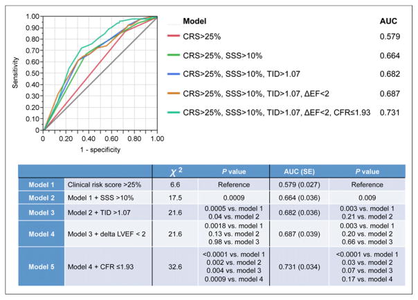
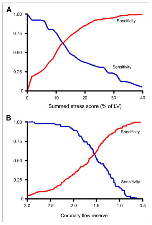
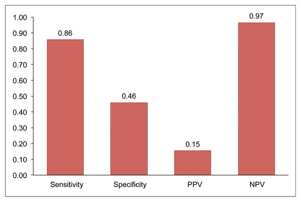
Results: Fifty-five patients (19%) had high-risk CAD on angiography. As expected, the trade-off between the sensitivity and the specificity of the CFR for identifying high-risk CAD varied substantially depending on the cutoff selected. In multivariable analysis, a binary CFR of less than or equal to 1.93 provided incremental diagnostic information for the identification of high-risk CAD beyond the model with the Duke clinical risk score (>25%), percentage of left ventricular ischemia (>10%), transient ischemic dilation index (>1.07), and change in the left ventricular ejection fraction during stress (<2) (P = 0.0009). In patients with normal or slightly to moderately abnormal results on perfusion scans (<10% of left ventricular mass) during stress (n = 136), a preserved CFR (>1.93) excluded high-risk CAD with a high sensitivity (86%) and a high negative predictive value (97%).

Conclusion: A normal CFR has a high negative predictive value for excluding high-risk CAD on angiography. Although an abnormal CFR increases the probability of significant obstructive CAD, it cannot reliably distinguish significant epicardial stenosis from nonobstructive, diffuse atherosclerosis or microvascular dysfunction.

Keywords: 82Rb PET; coronary artery disease; coronary flow reserve.


Figures

FIGURE 1
FIGURE 1
Multivariate ROC curves demonstrating ability of PET variables to detect high-risk CAD. Model starts with binary category of Duke clinical risk score (CRS) of greater than 25% (red line) and then adds %SSS (SSS) of greater than 10.2% (green line), binary of TID of greater than 1.07 (blue line), ΔLVEF (ΔEF) of less than 2 (orange line), and CFR of less than or equal to 1.93 (blue-green line). Table shows χ2 and area under curve (AUC) for each model as well as P values for comparisons of models.
FIGURE 2
FIGURE 2
Univariate ROC curves showing sensitivity (blue curve) and specificity (red curve) pairs for identification of patients with high-risk CAD (2-vessel disease, including proximal left anterior descending artery; 3-vessel disease; and left main CAD) by use of %SSS (A) and global CFR (B). This analysis demonstrated that %SSS of 10.2% and global CFR of 1.93 had best trade-off, with sensitivities of 0.75 and 0.89 and specificities of 0.51 and 0.36, respectively, for identification of high-risk CAD. Number of patients with high-risk CAD on angiography (truth reference) was denominator of sensitivity–specificity pair calculations illustrated by ROC curves. Results in A are consistent with notion that, like SPECT, semiquantitative myocardial perfusion imaging with PET often underestimates extent of CAD on angiography; this effect is likely related to the issue of balance flow reduction. In fact, finding that perfusion defect involving at least 10% of left ventricular (LV) mass was associated with sensitivity of approximately 60% for correctly identifying high-risk disease on angiography was nearly identical to that reported by Berman et al. using SPECT (56% sensitivity with same threshold) (20). ROC curves also showed trade-offs in sensitivity–specificity with changing semiquantitative thresholds of ischemia. For example, perfusion defect involving 40% of left ventricle would be expected to be associated with multivessel CAD (specificity and positive predictive value near 100%). However, such a threshold would miss large numbers of patients with smaller defects but high-risk CAD on angiography (low sensitivity). Conversely, the opposite would be true if smaller semiquantitative threshold for ischemia were used. Similar pattern can be observed with CFR in B.
FIGURE 3
FIGURE 3
Imaging results for 85-y-old woman who had history of hypertension and obesity and was referred for evaluation of atypical chest pain. (A and B) Selected coronary angiographic views of left (A) and right (B) coronary arteries. Images show extensive and severe CAD involving left main (LM), proximal left anterior descending, and left circumflex coronary (LCx) arteries. (C) Short-axis stress–rest myocardial perfusion images showing TID and medium but severe perfusion defect that involved lateral left ventricular wall but was completely reversible. Patient’s quantitative global CFR was 1.0.
FIGURE 4
FIGURE 4
Imaging results for 46-y-old woman who had history of diabetes mellitus, hypertension, high cholesterol, and smoking and was referred for evaluation of atypical chest pain and dyspnea. (A and B) Selected coronary angiographic views of left (A) and right (B) coronary arteries, without significant obstructive CAD. (C) Short-axis stress–rest myocardial perfusion images showing TID without significant regional perfusion abnormalities. Patient’s quantitative global CFR was 1.3.
FIGURE 5
FIGURE 5
Sensitivity, specificity, positive predictive value (PPV), and negative predictive value (NPV) of abnormal CFR (≤1.93) for detecting high-risk CAD in patients with normal or small to medium stress myocardial perfusion defects (%SSS of <10.2%) (n = 136).
FIGURE 6
FIGURE 6
Imaging results for 66-y-old woman who had history of high cholesterol and family history of CAD and was referred for evaluation of atypical chest pain. (A and B) Selected coronary angiographic views of left (A) and right (B) coronary arteries. Images show severe disease in first diagonal (Diag) branch, moderate stenosis in proximal right coronary artery (RCA), and diffuse disease in posterior descending coronary artery (PDA). (C) Short-axis stress–rest myocardial perfusion images showing small but severe perfusion defect that involved middle and apical anterior wall but was completely reversible. Patient’s quantitative global CFR was 2.03.

References

    1. Hendel RC, Berman DS, Di Carli MF, et al. ACCF/ASNC/ACR/AHA/ASE/SCCT/SCMR/SNM 2009 appropriate use criteria for cardiac radionuclide imaging: a report of the American College of Cardiology Foundation Appropriate Use Criteria Task Force, the American Society of Nuclear Cardiology, the American College of Radiology, the American Heart Association, the American Society of Echocardiography, the Society of Cardiovascular Computed Tomography, the Society for Cardiovascular Magnetic Resonance, and the Society of Nuclear Medicine. Circulation. 2009;119:e561–e587. - PubMed
    1. Shaw LJ, Hage FG, Berman DS, Hachamovitch R, Iskandrian A. Prognosis in the era of comparative effectiveness research: where is nuclear cardiology now and where should it be? J Nucl Cardiol. 2012;19:1026–1043. - PubMed
    1. Hachamovitch R, Hayes SW, Friedman JD, Cohen I, Berman DS. Comparison of the short-term survival benefit associated with revascularization compared with medical therapy in patients with no prior coronary artery disease undergoing stress myocardial perfusion single photon emission computed tomography. Circulation. 2003;107:2900–2907. - PubMed
    1. Fiechter M, Ghadri JR, Gebhard C, et al. Diagnostic value of 13N-ammonia myocardial perfusion PET: added value of myocardial flow reserve. J Nucl Med. 2012;53:1230–1234. - PubMed
    1. Ziadi MC, Dekemp RA, Williams K, et al. Does quantification of myocardial flow reserve using rubidium-82 positron emission tomography facilitate detection of multivessel coronary artery disease? J Nucl Cardiol. 2012;19:670–680. - PubMed

Publication types

MeSH terms

Substances