. 2014 Jan;6(1):120-40.
doi: 10.1002/emmm.201302890.

Activation of IL-27 signalling promotes development of postinfluenza pneumococcal pneumonia

Affiliations

Activation of IL-27 signalling promotes development of postinfluenza pneumococcal pneumonia

Ju Cao et al. EMBO Mol Med. 2014 Jan.

Abstract

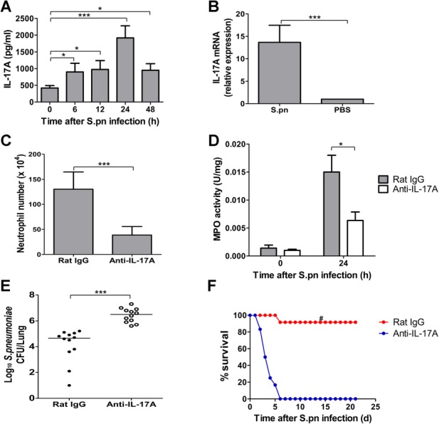
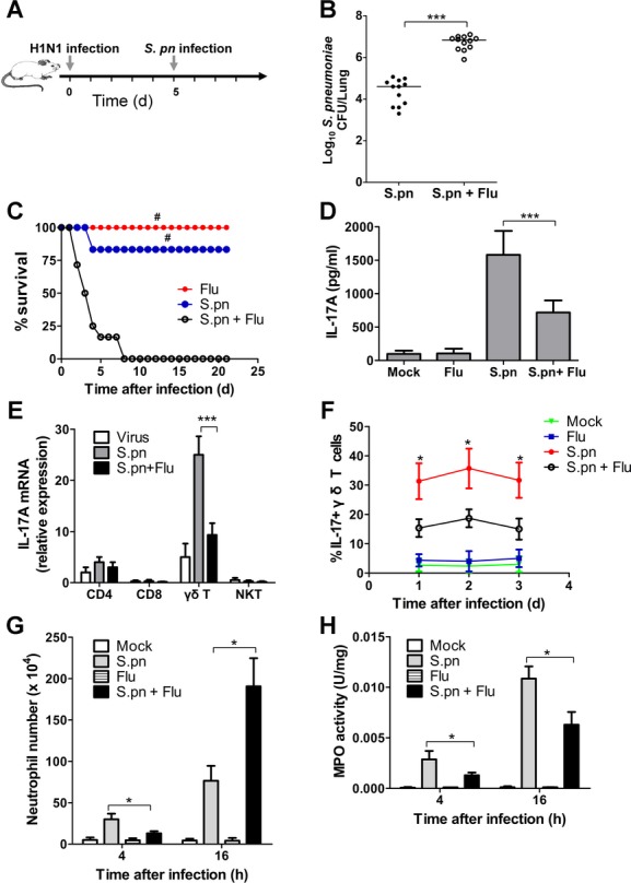
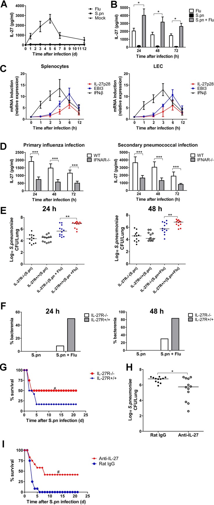
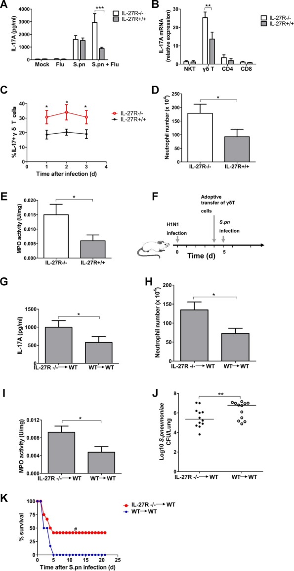
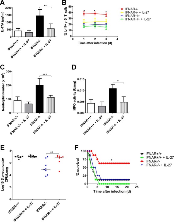
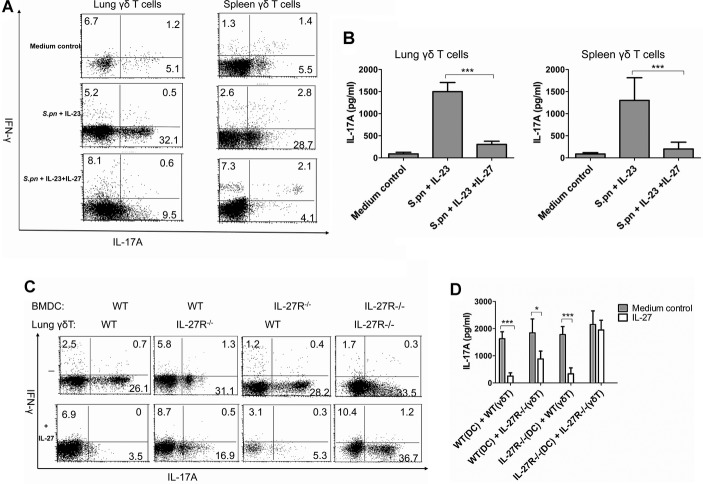
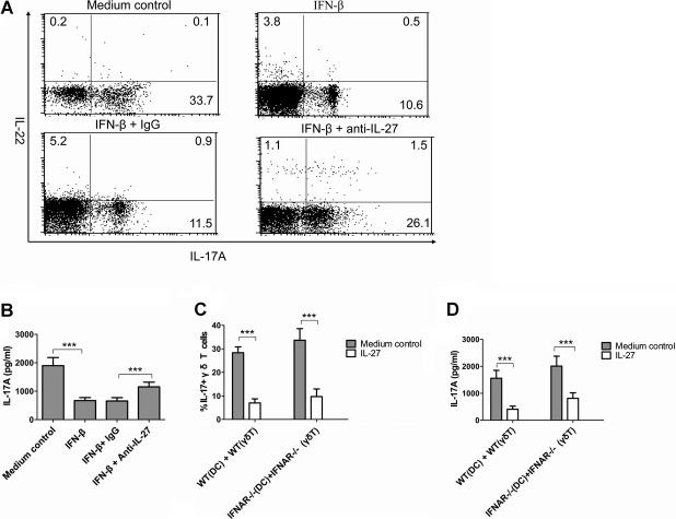
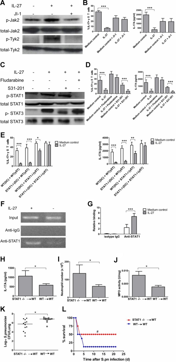
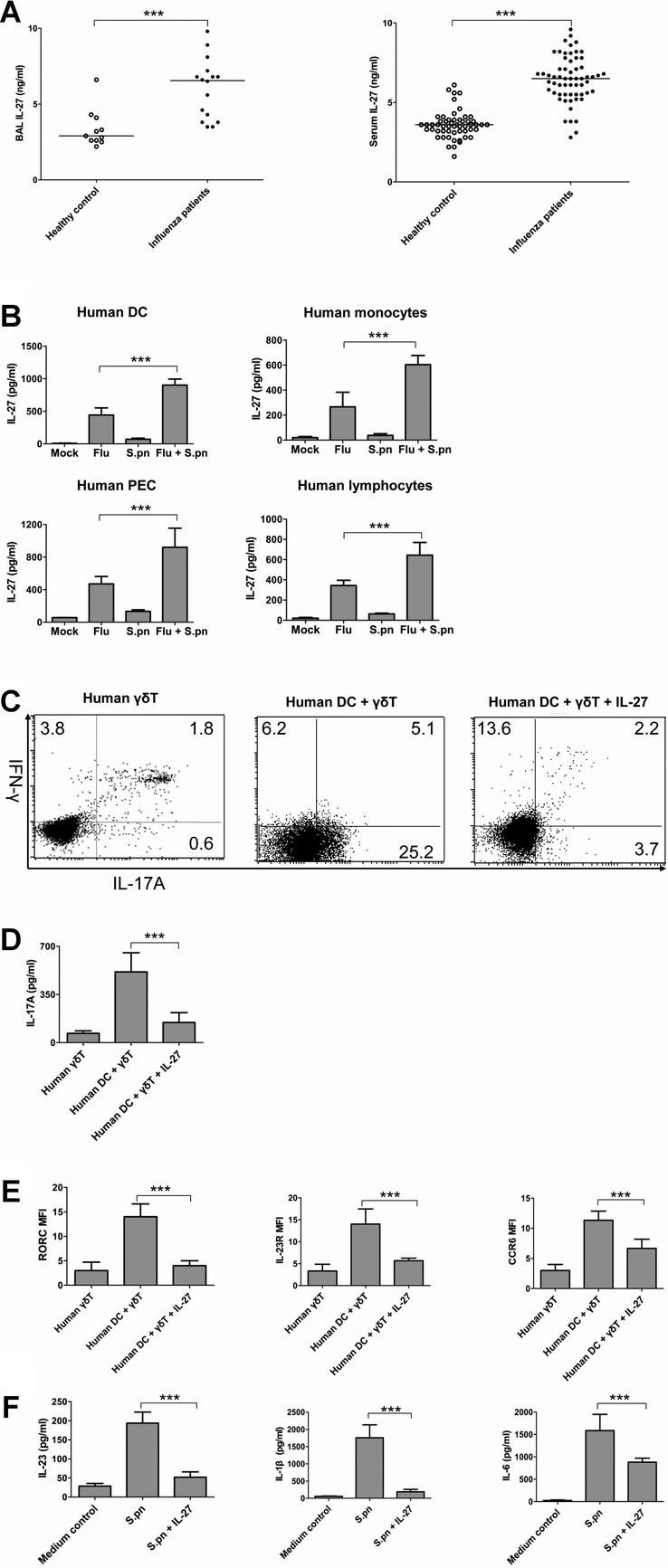
Postinfluenza pneumococcal pneumonia is a common cause of death in humans. However, the role of IL-27 in the pathogenesis of secondary pneumococcal pneumonia after influenza is unknown. We now report that influenza infection induced pulmonary IL-27 production in a type I IFN-α/β receptor (IFNAR) signalling-dependent manner, which sensitized mice to secondary pneumococcal infection downstream of IFNAR pathway. Mice deficient in IL-27 receptor were resistant to secondary pneumococcal infection and generated more IL-17A-producing γδ T cells but not αβ T cells, thereby leading to enhanced neutrophil response during the early phase of host defence. IL-27 treatment could suppress the development of IL-17A-producing γδ T cells activated by Streptococcus pneumoniae and dendritic cells. This suppressive activity of IL-27 on γδ T cells was dependent on transcription factor STAT1. Finally, neutralization of IL-27 or administration of IL-17A restored the role of γδ T cells in combating secondary pneumococcal infection. Our study defines what we believe to be a novel role of IL-27 in impairing host innate immunity against pneumococcal infection.


Figures

Figure 1
Figure 1
IL-17A was required for efficient clearance of S. pneumoniae in the lung. A  Dynamic changes of lung IL-17A levels in the mice upon intranasal challenge with Type 3 S. pneumoniae (n = 5). B  Lung IL-17A mRNA levels at 24 h after pneumococcal challenge (n = 5). C  Lung neutrophil numbers at 24 h after inhibition of IL-17A. Either anti-IL-17A neutralizing antibodies or control IgG were injected i.p. in mice, and mice were subsequently infected with S. pneumoniae intranasally (n = 5). D  Lung MPO activity at 24 h after inhibition of IL-17A (n = 5). E  Pulmonary pneumococcal burdens at 48 h after inhibition of IL-17A. The horizontal lines indicate the median CFU per lung (n = 12). F  Survival was examined for 21 days after pneumococcal challenge in the presence or absence of anti-IL-17A neutralizing antibodies (n = 12). *p < 0.05, ***p < 0.001 when compared between groups denoted by horizontal lines; #p < 0.05 when compared with mice treated with anti-IL-17A.
Figure 2
Figure 2
Influenza virus inhibited IL-17A production by γ´ T cells in postinfluenza pneumonia. A  Schematic representation model of postinfluenza pneumonia in C57BL/6 mice. B  Pulmonary pneumococcal burdens in the mice at 48 h after primary or secondary pneumococcal infection (n = 12). C  Survival for groups of mice after influenza infection, S. pneumoniae infection or secondary pneumococcal infection following influenza infection (n = 12). D  Lung IL-17A levels in the indicated groups of mice at 24 h after pneumococcal infection (n = 5). E  γδ T cells, CD4+ T cells, CD8+ T cells and NKT cells were purified by cell sorting. IL-17A gene expression was measured from different cell types. Results are presented relative to GAPDH (n = 5). F  Percentages of IL-17A producers among γδ T cells in the lungs of groups of mice at indicated times following pneumococcal challenge (n = 5). G  Lung neutrophil numbers in the mice at 4, 16 h following pneumococcal challenge (n = 5). H  Lung MPO activity in the mice at 4, 16 h following pneumococcal challenge (n = 5). *p < 0.05, ***p < 0.001 when compared between mice infected with S. pneumoniae alone and mice infected with influenza virus plus S. pneumoniae; #p < 0.05 when compared with mice of secondary pneumococcal infection.
Figure 3
Figure 3
Influenza-infected IL-27R-deficient mice were resistant to secondary pneumococcal pneumonia. A  Lungs from indicated groups of mice were harvested at the designated time points for assessment of IL-27 by ELISA (n = 5). B  Lung IL-27 production in the groups of mice at 24, 48, 72 h after influenza infection, S. pneumoniae infection or secondary pneumococcal infection following influenza infection (n = 5). C  WT splenocytes or LEC were stimulated with influenza virus (MOI = 1) for the indicated time points. IFN-β, EBI3, and IL-27p28 gene transcript level was detected by quantitative PCR (n = 3). D  IL-27 production in the lungs of IFNAR-deficient or WT mice after primary influenza infection or secondary pneumococcal infection (n = 5). E  Pulmonary pneumococcal burdens in IL-27R-deficient and WT mice at 24 or 48 h following primary pneumococcal infection or secondary pneumococcal infection (n = 12). F  Incidence of bacteraemia was measured in IL-27R-deficient and WT mice at 24 or 48 h following primary pneumococcal infection or secondary pneumococcal infection (n = 12). G  Survival for indicated groups of mice following secondary pneumococcal infection (n = 12). H  Pulmonary pneumococcal burdens in WT mice treated with anti-IL-27 blocking antibodies at 48 h after secondary pneumococcal infection (n = 12). I  Survival for indicated groups of mice treated with anti-IL-27 neutralizing antibodies or isotypical IgG after secondary pneumococcal infection (n = 12). *p < 0.05, **p < 0.01, ***p < 0.001 when compared between groups denoted by horizontal lines; #p < 0.05 when compared between indicated groups of mice.
Figure 4
Figure 4
IL-27 negatively regulated IL-17A production by γ´ T cell upon pneumococcal infection in mice. A  IL-17A production in the lungs from IL-27R-deficient and WT mice at 24 h after secondary pneumococcal challenge following influenza infection (n = 5). B  γδ T cells, CD4+ T cells, CD8+ T cells and NKT cells from IL-27R-deficient and WT mice were purified by cell sorting at 24 h after secondary pneumococcal infection. IL-17A gene expression was measured from different cell types (n = 5). C  Percentages of IL-17A producers among γδ T cells in the lungs from IL-27R-deficient and WT mice at indicated times following secondary pneumococcal challenge (n = 5). D  Lung neutrophil numbers in IL-27R-deficient and WT mice at 24 h following secondary pneumococcal challenge (n = 5). E  Lung MPO activity in IL-27R-deficient and WT mice at 24 h following secondary pneumococcal challenge (n = 5). F  Schematic representation model of γδ T cells adoptive transfer experiment. G  Lung IL-17A levels were determined at 24 h after secondary pneumococcal infection (n = 5). H  Lung neutrophil numbers in WT mice transferred with IL-27R-deficient or WT γδ T cells at 24 h after secondary pneumococcal infection (n = 5). I  Lung MPO activity at 24 h after secondary pneumococcal infection (n = 5). J  Pulmonary pneumococcal burdens at 48 h after secondary pneumococcal infection (n = 12). K  Survival for WT mice transferred with IL-27R-deficient or WT γδ T cells after secondary pneumococcal infection (n = 12). *p < 0.05, **p < 0.01, ***p < 0.001 when compared between groups denoted by horizontal lines; #p < 0.05 when compared with mice transferred with WT γδ T cells.
Figure 5
Figure 5
IL-27 mediated the inhibitory effects of IFNAR signalling on IL-17A production by γ´ T cells during secondary pneumococcal infection. Recombinant murine IL-27 was given i.t. into IFNAR-deficient or WT mice 24 h before intranasal pneumococcal administration, lungs were then collected for analysis at different time points. A  Lung IL-17A levels were determined at 24 h following secondary pneumococcal infection (n = 4). B  Percentages of IL-17A producers among γδ T cells in the lungs from groups of mice at indicated times following secondary pneumococcal challenge (n = 4). C  Lung neutrophil numbers at 24 h after secondary pneumococcal infection (n = 4). D  Lung MPO activity at 24 h after secondary pneumococcal infection (n = 4). E  Pulmonary pneumococcal burdens at 48 h after secondary pneumococcal infection (n = 8). F  Survival for IFNAR-deficient or WT mice treated with or without exogenous IL-27 after secondary pneumococcal infection (n = 8). *p < 0.05, **p < 0.01, ***p < 0.001 when compared between IFNAR-deficient mice treated with and without IL-27; #p < 0.05 when compared with mice treated with IL-27.
Figure 6
Figure 6
IL-27 inhibited IL-17A production by γ´ T cells in vitro. A  FACS analysis of IL-17A and IFN-γ expression in FACS-sorted lung or spleen γδ T cells cultured for 72 h under the stimulation of HkSp (1 × 10⁸ CFU/ml) and IL-23 (50 ng/ml) in the presence or absence of IL-27 (100 ng/ml). B  IL-17A concentrations in the supernatants of lung or spleen γδ T cells activated by HkSp and IL-23 in the presence or absence of IL-27 as determined by ELISA. C  FACS analysis of IL-17A and IFN-γ expression in FACS-sorted lung γδ T cells from IL-27R-deficient or WT mice, which were co-cultured with BMDC from IL-27R-deficient or WT mice activated by HkSp (1 × 10⁸ CFU/ml) in the presence or absence of IL-27 (100 ng/ml) for 72 h. Lung γδ T cells were gated for FACS analysis. D  IL-17A concentrations in the supernatants of lung γδ T cells from IL-27R-deficient or WT mice, which were co-cultured with BMDC from IL-27R-deficient or WT mice activated by HkSp in the presence or absence of IL-27 for 72 h. Results were from three independent experiments, and each was performed with cells isolated from three mice. *p < 0.05, ***p < 0.001 when compared between groups denoted by horizontal lines.
Figure 7
Figure 7
IL-27 directly contributed to IFN-b-mediated inhibition of IL-17A production in γ´ T cells. A, B  Spleen γδ T cells isolated from WT mice were restimulated with HkSp (1 × 10⁸ CFU/ml) and IL-23 (50 ng/ml) for 72 h in the presence or absence of supernatants from IFN-β-treated splenocytes plus anti-IL-27 neutralizing antibodies or control IgG. IL-17 levels were determined by (A) FACS analysis and (B) ELISA, respectively. C, D  WT or IFNAR-deficient spleen γδ T cells were co-cultured with WT or IFNAR-deficient BMDC activated by HkSp (1 × 10⁸ CFU/ml) in the presence or absence of IL-27 (100 ng/ml) for 72 h. (C) The percentage of IL-17A-producing γδ T cells was measured by FACS analysis, (D) while IL-17A secretion in the culture supernatants was detected by ELISA. Results were from three independent experiments, and each was performed with cells isolated from three mice. ***p < 0.001 when compared between groups denoted by horizontal lines.
Figure 8
Figure 8
STAT1 was critical for suppression of IL-17A in γ´ T cells by IL-27 in vitro and in vivo. A  Splenocytes were pretreated with JI-1 (1 nM) for 1 h, followed by stimulation with IL-27 (100 ng/ml) for a further 15 min. Phospho-Jak2 and phosphor-Tyk2 were detected by Western blot. B  FACS analysis of percentages of IL-17A producers among γδ T cells in FACS-sorted spleen γδ T cells co-cultured with BMDC infected with HkSp (1 × 10⁸ CFU/ml) in the presence or absence of IL-27 (100 ng/ml) and of JI-1 (1 nM) for 72 h, and IL-17A concentrations in the culture supernatants were also determined by ELISA. C  Splenocytes were pretreated with fludarabine (50 μM) or S31-201 (10 μM) for 1 h, followed by stimulation with IL-27 (100 ng/ml) for a further 15 min. Phospho-STAT1 (Tyr 701) and phosphor-STAT3 (Tyr 705) were detected by Western blot. D  FACS analysis of percentages of IL-17A producers among γδ T cells in FACS-sorted spleen γδ T cells co-cultured with BMDC infected with HkSp (1 × 10⁸ CFU/ml) in the presence or absence of IL-27 (100 ng/ml) and of fludarabine (50 μM) or S31-201 (10 μM) for 72 h, and IL-17A concentrations in the culture supernatants were also determined by ELISA. E  FACS analysis of IL-17A expression in FACS-sorted spleen γδ T cells from STAT1-deficient or WT mice, which were co-cultured with BMDC from STAT1-deficient or WT mice infected by HkSp in the presence or absence of IL-27 for 72 h. The percentages of IL-17A-producing γδ T cells were determined by FACS analysis, while IL-17A concentrations were determined by ELISA. F  FACS-sorted spleen γδ T cells were stimulated with HkSp (1 × 10⁸ CFU/ml) and IL-23 (50 ng/ml) in the presence or absence of IL-27 (100 ng/ml) for 72 h. Cell lysates were immunoprecipitated either with anti-STAT1 or isotype IgG. Bound DNA was analyzed by PCR with IL-17A promoter site-specific primers. G  Eluted DNA was quantitated by quantitative PCR with primers specific for the IL-17A promoter. H  Lung IL-17A concentrations in WT mice transferred with STAT1-deficient or WT γδ T cells at 24 h after secondary pneumococcal infection (n = 5). I  Lung neutrophil numbers in WT mice transferred with STAT1-deficient or WT γδ T cells at 24 h after secondary pneumococcal infection (n = 5). J  Lung MPO activity at 24 h after secondary pneumococcal infection (n = 5). K  Pulmonary pneumococcal burdens at 48 h after secondary pneumococcal infection (n = 12). L  Survival for WT mice transferred with STAT1-deficient or WT γδ T cells after secondary pneumococcal infection (n = 12). *p < 0.05, **p < 0.01, ***p < 0.001 when compared between groups denoted by horizontal lines; #p < 0.05 when compared with mice transferred with WT γδ T cells.
Figure 9
Figure 9
IL-27 inhibited IL-17A production in human Vγ9V´2 T cells. A  ELISA analysis for IL-27 levels in BAL and serum samples from healthy individuals and influenza-infected patients. B  ELISA analysis for IL-27 production in human cells. Human monocyte-derived DC, momocytes, pulmonary epithelial cells and lymphocytes were stimulated with influenza virus (MOI = 1) and HkSp (1 × 10⁸ CFU/ml). After 24 h, ELISA was performed to measure the IL-27 concentrations in the culture supernatants. C  FACS analysis for IL-17A and IFN-γ expression in human Vγ9Vδ2 T cells co-cultured with DC activated by HkSp (1 × 10⁸ CFU/ml) in the presence or absence of IL-27 (100 ng/ml) at 5 days. D  IL-17A concentrations in the supernatants of human Vγ9Vδ2 T cells co-cultured with DC activated by HkSp in the presence or absence of IL-27 at 5 days. E  The expression of RORγt, IL-23R and CCR6 in human Vγ9Vδ2 T cells co-cultured with DC infected with HkSp in the presence or absence of IL-27 F  ELISA analysis for cytokine production from human DC activated by HkSp in the presence or absence of IL-27. Results were from three independent experiments, and each was performed with cells isolated from three different donors. ***p < 0.001 when compared between groups denoted by horizontal lines.

Similar articles

Cited by

References

    1. Banchereau J, Pascual V, O'Garra A. From IL-2 to IL-37: the expanding spectrum of anti-inflammatory cytokines. Nat Immunol. 2012;13:925–931. - PMC - PubMed
    1. Brundage JF. Interactions between influenza and bacterial respiratory pathogens: implications for pandemic preparedness. Lancet Infect Dis. 2006;6:303–312. - PMC - PubMed
    1. Cao J, Gong Y, Dong S, Zhang L, Lai X, Zhang X, Yin Y. Pneumococcal ClpP modulates the maturation and activation of human dendritic cells: implications for pneumococcal infections. J Leukoc Biol. 2013;93:737–749. - PubMed
    1. Cao J, Gong Y, Yin Y. The role played by interleukin-10 in cytokine and chemokine dysregulation during secondary pneumococcal pneumonia after influenza. J Infect Dis. 2009;199:1555–1556. - PubMed
    1. Cao J, Wong CK, Yin Y, Lam CW. Activation of human bronchial epithelial cells by inflammatory cytokines IL-27 and TNF-alpha: implications for immunopathophysiology of airway inflammation. J Cell Physiol. 2010;223:788–797. - PubMed

Publication types

MeSH terms