. 2014 Feb 10;55(2):801-13.
doi: 10.1167/iovs.13-12812.

TWEAK/Fn14 pathway is a novel mediator of retinal neovascularization

Affiliations

TWEAK/Fn14 pathway is a novel mediator of retinal neovascularization

Hossein Ameri et al. Invest Ophthalmol Vis Sci. .

Abstract

Purpose: Retinal neovascularization (NV) is a major cause of vision loss in ischemia-induced retinopathy. Tumor necrosis factor (TNF)-like weak inducer of apoptosis (TWEAK) and its receptor, fibroblast growth factor inducible-14 (Fn14), have been implicated in angiogenesis, but their role in retinal diseases is unknown. The goal of this study was to investigate the role of TWEAK/Fn14 pathway in retinal NV.

Methods: Studies were performed in a mouse model of oxygen-induced retinopathy (OIR) and in primary human retinal microvascular endothelial cells (HRMECs). Hyperoxia treatment was initiated on postnatal day (P)14. Immunohistochemistry and quantitative PCR (qPCR) were used to assess retinal vascular changes in relation to expression of Fn14 and TWEAK.

Results: Fibroblast growth factor-inducible 14 mRNA was prominently increased from P13 to P17 in OIR retinas, whereas TWEAK level was slightly decreased. These alterations were normalized by hyperoxia treatment and were more striking in isolated retinal vessels. There was a discernible shift in the immunoreactivity of Fn14 and TWEAK from the neuronal layers in the healthy retina to the neovascular tufts in that of OIR. Blockade of TWEAK/Fn14 significantly prevented retinal NV while slightly accelerated revascularization. In contrast, activation of Fn14 positively regulated survival pathways in the B-cell lymphoma-2 (Bcl2) family and robustly enhanced HRMEC survival. Furthermore, gene analysis revealed the regulatory region of Fn14 gene contains several conserved hypoxia inducible factor (HIF)-1α binding sites. Overexpression of HIF-1α prominently induced Fn14 expression in HRMECs.

Conclusions: We found that the TNF-like weak inducer of apoptosis (TWEAK)/fibroblast growth factor inducible-14 (Fn14) pathway is involved in the development of pathologic retinal neovascularization. Hypoxia inducible factor-1α is likely implicated in the upregulation of Fn14.

Keywords: Fn14; TWEAK; ischemia-induced retinopathy; neovascularization; retina.


Figures

Figure 1
Figure 1
The expression of Fn14 and TWEAK is altered in retinas of OIR. Mice were subjected to OIR or maintained in RA as control. Retinas were collected at indicated time points (P12–P17), and Fn14 (A) or TWEAK mRNA (B) in the retinas was examined by qPCR and normalized to the age-matched RA control (n = 6). *P < 0.05 compared with relevant control.
Figure 2
Figure 2
Hyperoxia treatment reverses Fn14 and TWEAK expression in OIR mice. Oxygen-induced retinopathy mice were treated with hyperoxia (75% oxygen; HT) or maintained in RA (OIR) from P14 to P17. Mice maintained in RA from P1 to P17 are control. Fibroblast growth factor inducible–14 mRNA (A) and TWEAK mRNA (B) were determined by qPCR and normalized to RA control (n = 6). *P < 0.05 compared with RA. #P < 0.05 compared with OIR.
Figure 3
Figure 3
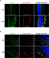
The expression of Fn14 and TWEAK is redistributed in the retinas of OIR. Retinal frozen sections from P17 OIR mice or RA mice were stained with isolectin B4 (red) to highlight the retinal vessels, DAPI (blue) for nuclei, and Fn14 antibody (green; [A]), or TWEAK antibody (green; [B]). The immunoreactivity of Fn14 and TWEAK shifted from the neuronal layers in healthy retinas to the neovascular tufts in OIR. Representative fluorescence microscopy images are shown (200×, n = 3). All images in OIR and RA were taken at the same setting except for isolectin B4 images that were adjusted individually in each photograph for better visibility of vessels. IPL, inner plexiform layer; OPL, outer plexiform layer; ONL, outer nuclear layer.
Figure 4
Figure 4
Fibroblast growth factor inducible–14 is highly expressed in the neovascular tufts in the retinas of OIR. Retinas isolated from P17 OIR mice or RA mice were immunostained with isolectin B4 (red) to highlight the retinal vessels and Fn14 antibody (green). The Fn14 protein was consistently highly expressed in neovascular tufts of central, intermediate, and peripheral retinal areas. Representative confocal images were taken at 200× magnification (n = 3).
Figure 5
Figure 5
The expression of Fn14 and TWEAK is altered in retinal vessels of OIR. Retinal vessels were isolated from P17 OIR or RA control mice, and RNA was extracted from pooled retinal vessels. Fibroblast growth factor inducible–14 mRNA (A) or TWEAK mRNA (B) in retinal vessels was quantified by qPCR and normalized to the RA control. n = 3 (each n represents a pool of six retinas). *P < 0.05 compared with RA.
Figure 6
Figure 6
The blockade of TWEAK/Fn14 results in decreased retinal NV in OIR. Oxygen-induced retinopathy mice were intravitreally treated with a soluble Fn14-Fc decoy receptor (2.5 μg/eye) or vehicle (PBS) at P12. Retinas were collected at P17 and stained with isolectin B4. Representative images of the retinal flat mounts are shown (40×; [A]). Neovascularization (B) and avascular areas (C) were quantified using ImageJ software (n = 8). *P < 0.05 compared with vehicle-treated mice.
Figure 7
Figure 7
The blockade of TWEAK/Fn14 does not influence VEGF expression in OIR. Oxygen-induced retinopathy mice were intravitreally treated with a soluble Fn14-Fc decoy receptor (2.5 μg/eye) or vehicle (PBS) at P12. Retinas were collected at P17 and VEGF mRNA level was measured by qPCR (n = 5). *P < 0.05 compared with vehicle-treated mice.
Figure 8
Figure 8
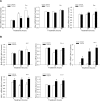
Tumor necrosis factor–like weak inducer of apoptosis treatment improves the survival of HRMECs. (A) Human retinal microvascular endothelial cells were treated with vehicle (EGM without serum) or TWEAK (200 ng/mL) for the time as indicated. Cell viability was determined by measuring the electrical cell-substrate impedance and normalized to that of cells treated with vehicle for 0 hour (n = 4). *P < 0.05 compared with cells treated with vehicle at the corresponding time. (B) After 4 days of treatment, cells in the electrode were stained with DAPI and representative images are shown (40×). Arrows show examples of cell nuclei. (C) Human retinal microvascular endothelial cells were treated with vehicle (EGM without serum) or TWEAK (200 ng/mL) for 4 days. Cell viability was determined by MTT assay (n = 3). *P < 0.05 compared with control.
Figure 9
Figure 9
Tumor necrosis factor–like weak inducer of apoptosis treatment regulates expression of Bcl2 family genes. Human retinal microvascular endothelial cells were treated with vehicle (EGM without serum) or TWEAK (200 ng/mL) for the times indicated. Ribonucleic acid was extracted and the expression of proapoptotic genes (Bim, Bax, and Bak; [A]) and antiapoptotic genes (Bcl2, Bcl-xl, Bfl-1, Mcl-1, and Bcl-W; [B]) were determined by qPCR analysis (n = 3). *P < 0.05 compared with 6-hours vehicle control. #P < 0.05 compared with relevant vehicle-treated values.
Figure 10
Figure 10
Identification of transcription factor binding sites in Fn14 gene region. Upper panel, rVISTA plot shows regions of homology between mouse and human in a 6-kb region of the Fn14 gene. The shaded area represents greater than 70% identity between two species. Lower panel, schematic diagram represents potential HIF-1α binding sites in the 6-kb region of Fn14 gene predicted by rVISTA2.0.
Figure 11
Figure 11
Hypoxia inducible factor–1α induces Fn14 expression in HRMECs. Human retinal microvascular endothelial cells were infected with adenovirus expressing HIF-1α (AdHIF-1α) or GFP (AdGFP) as control for 48 hours. Hypoxia inducible factor–1α protein was determined by Western blot and Tubulin was used as loading control (A). Fibroblast growth factor inducible–14 mRNA (B) and protein (C) in HRMECs were determined by qPCR (n = 5) and Western blot (n = 3). *P < 0.05 compared with control. (D) At 48 hours after infection, HRMECs were stimulated with VEGF (50 ng/mL), bFGF (20 ng/mL), or IL-1b (10 ng/mL) for 2 hours. Fibroblast growth factor inducible–14 mRNA was determined by qPCR and normalized to control cells that were infected with AdGFP and treated with vehicle (n = 3). *P < 0.05 compared with control.

Similar articles

Cited by

References

    1. Multicenter trial of cryotherapy for retinopathy of prematurity 3 1/2-year outcome–structure and function. Cryotherapy for Retinopathy of Prematurity Cooperative Group. Arch Ophthalmol. 1993; 111: 339–344 - PubMed
    1. Phelps DL. Retinopathy of prematurity. Pediatr Rev. 1995; 16: 50–56 - PubMed
    1. Tsilimbaris MK, Kontadakis GA, Tsika C, Papageorgiou D, Charoniti M. Effect of panretinal photocoagulation treatment on vision-related quality of life of patients with proliferative diabetic retinopathy. Retina. 2013; 33: 756–761 - PubMed
    1. Mintz-Hittner HA, Kennedy KA, Chuang AZ. Efficacy of intravitreal bevacizumab for stage 3+ retinopathy of prematurity. N Engl J Med. 2011; 364: 603–615 - PMC - PubMed
    1. Kurihara T, Westenskow PD, Bravo S, Aguilar E, Friedlander M. Targeted deletion of VEGF-A in adult mice induces vision loss. J Clin Invest. 2012; 122: 4213–4217 - PMC - PubMed

Publication types

MeSH terms