

OCT minimum intensity as a predictor of geographic atrophy enlargement
- PMID: 24408973
- PMCID: PMC3920825
- DOI: 10.1167/iovs.13-13199
OCT minimum intensity as a predictor of geographic atrophy enlargement
Abstract
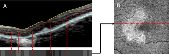
Purpose: We determined whether the minimum intensity (MI) of the optical coherence tomography (OCT) A-scans within the retina can predict locations of growth at the margin of geographic atrophy (GA) and the growth rate outside the margin.
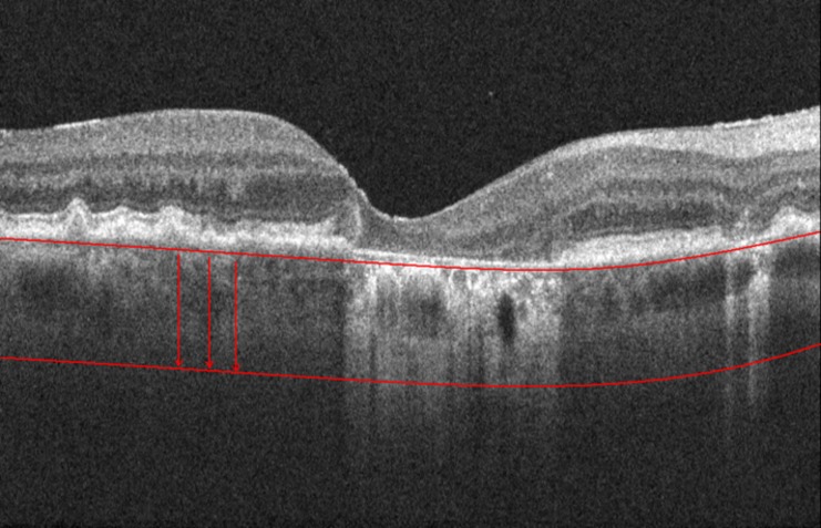
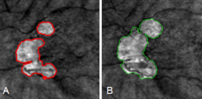
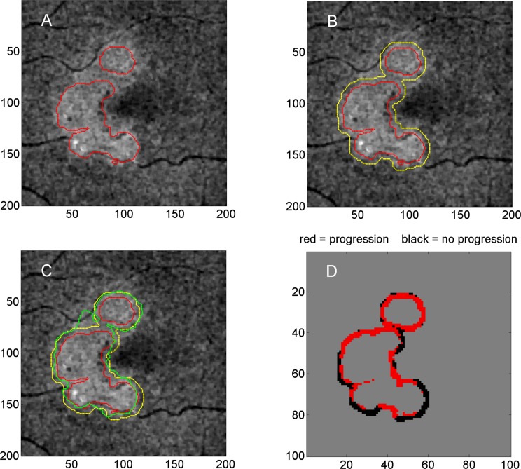
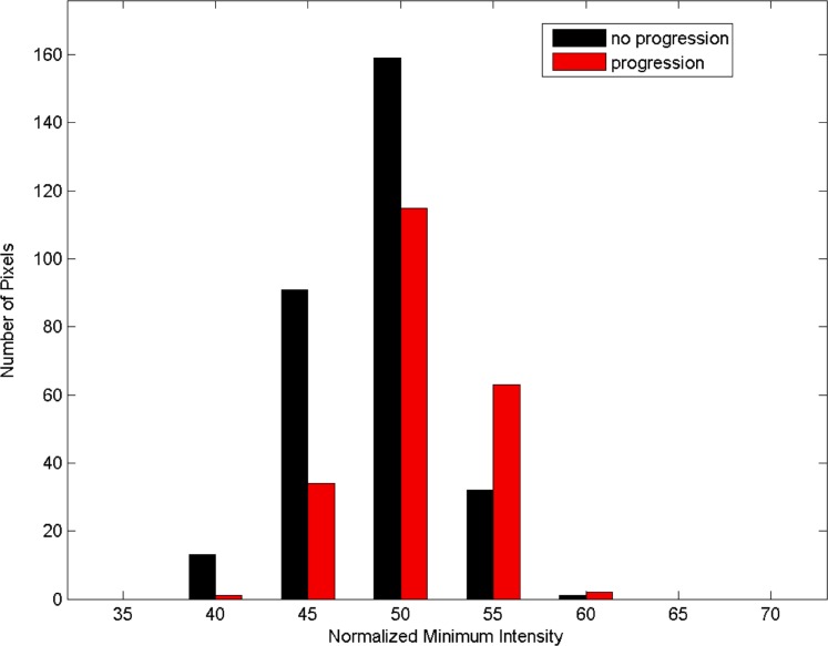
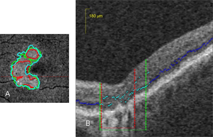
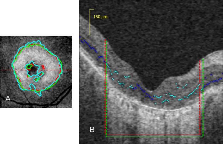
Methods: The OCT scans were analyzed at baseline and 52 weeks. Expert graders manually segmented OCT images of GA. The 52-week follow-up scans were registered to the baseline scan coordinates for comparison. The OCT MI values were studied within a 180-μm margin around the boundary of GA at baseline. Baseline MI values were compared in areas of progression and nonprogression of the GA, and sensitivity and specificity were assessed for prediction of growth at the margin. Average MI values in the margins were compared to overall growth rates to evaluate the prediction of growth outside the margins.
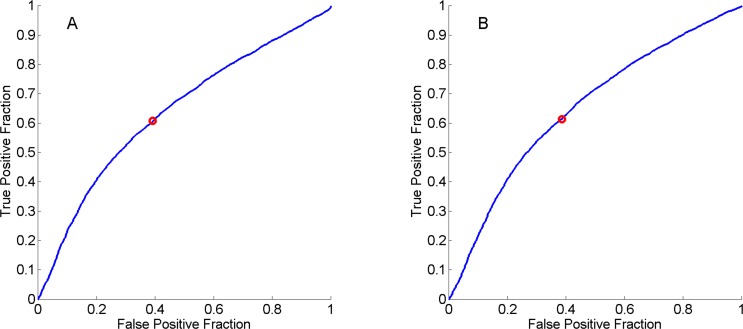
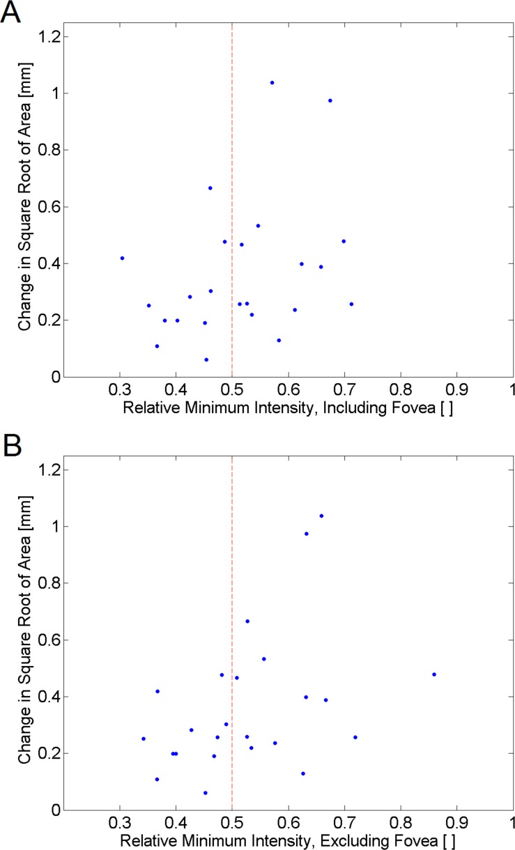
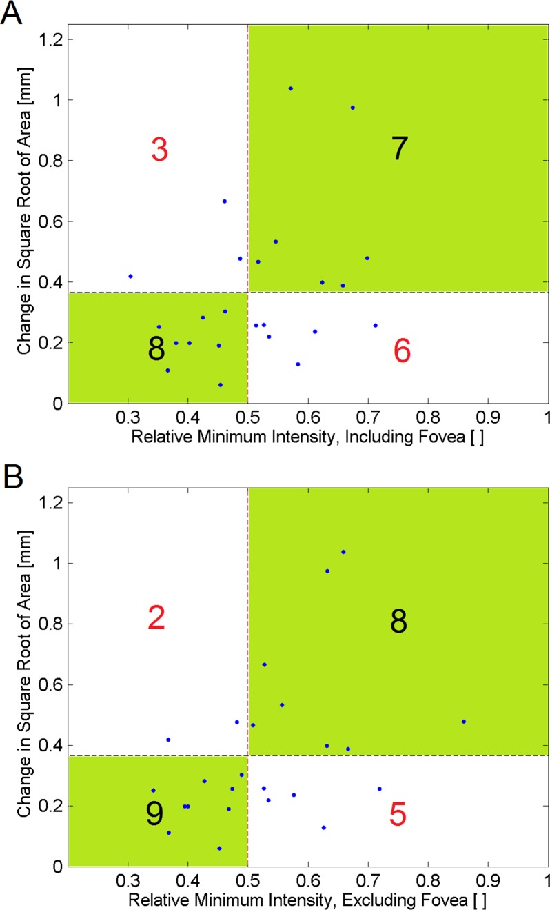
Results: A statistically significant increase in MI (P < 0.05) was seen in areas of growth in 21/24 cases (88%), and 22/24 cases (92%) when the foveal subfield was excluded. Locations of growth within the margins at 52 weeks were predicted with 61% sensitivity and 61% specificity. The MI values correlated significantly with overall growth rate, and high and low growth rate subjects were identified with 80% sensitivity and 64% specificity.
Conclusions: The MI may be increased at the margins of GA lesions before enlargement, which may indicate disruption or atrophy of the photoreceptors in these areas before GA becomes apparent. Increased MI may help predict areas of enlargement of GA, and may relate to overall growth rate and be a useful screening tool for GA. (ClinicalTrials.gov number, NCT00935883.).
Keywords: AMD; GA; geographic atrophy; image processing; macula.
Figures
References
-
- Sunness JS, Gonzalez-Baron J, Applegate CA, et al. Enlargement of atrophy and visual acuity loss in the geographic atrophy form of age-related macular degeneration. Ophthalmology. 1999; 106: 1768–1779 - PubMed
-
- Lujan BJ, Rosenfeld PJ, Gregori G, et al. Spectral domain optical coherence tomographic imaging of geographic atrophy. Ophthalmic Surg Lasers Imaging. 2008; 39 (suppl 4): S8–S14 - PubMed
-
- Helb HM, Charbel Issa P, Fleckenstein M, et al. Clinical evaluation of simultaneous confocal scanning laser ophthalmoscopy imaging combined with high-resolution, spectral-domain optical coherence tomography. Acta Ophthalmol. 2010; 88: 842–849 - PubMed
Publication types
MeSH terms
Associated data
Grants and funding
LinkOut - more resources
Full Text Sources
Other Literature Sources
Medical
