. 2014 Feb 4;55(2):688-95.
doi: 10.1167/iovs.13-12951.

Control of photoreceptor autophagy after retinal detachment: the switch from survival to death

Affiliations

Control of photoreceptor autophagy after retinal detachment: the switch from survival to death

Nicholas D Chinskey et al. Invest Ophthalmol Vis Sci. .

Abstract

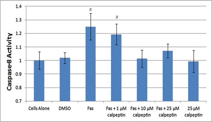
Purpose: To examine whether calpain inhibition following retinal detachment would prolong autophagy and result in reduced photoreceptor apoptosis.

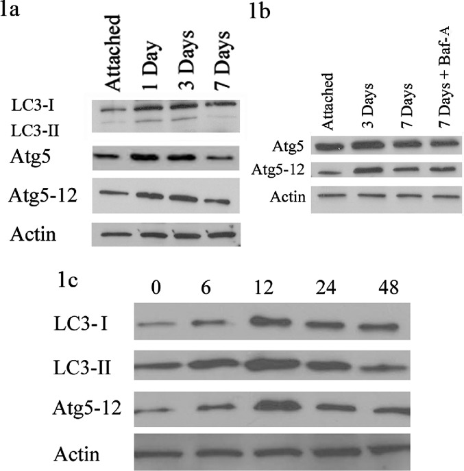
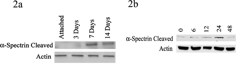
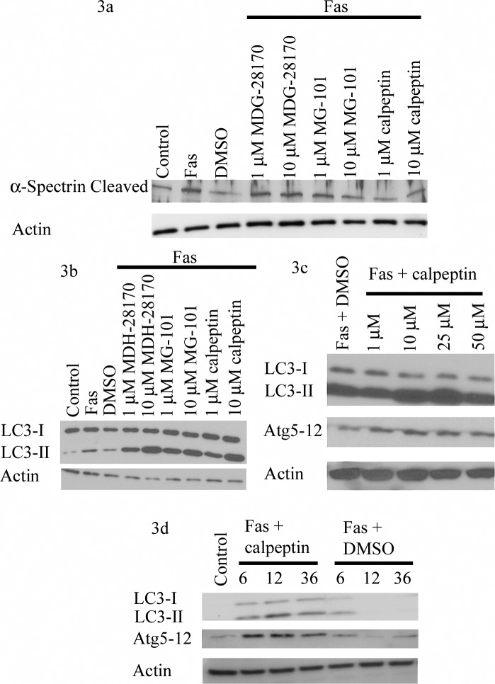
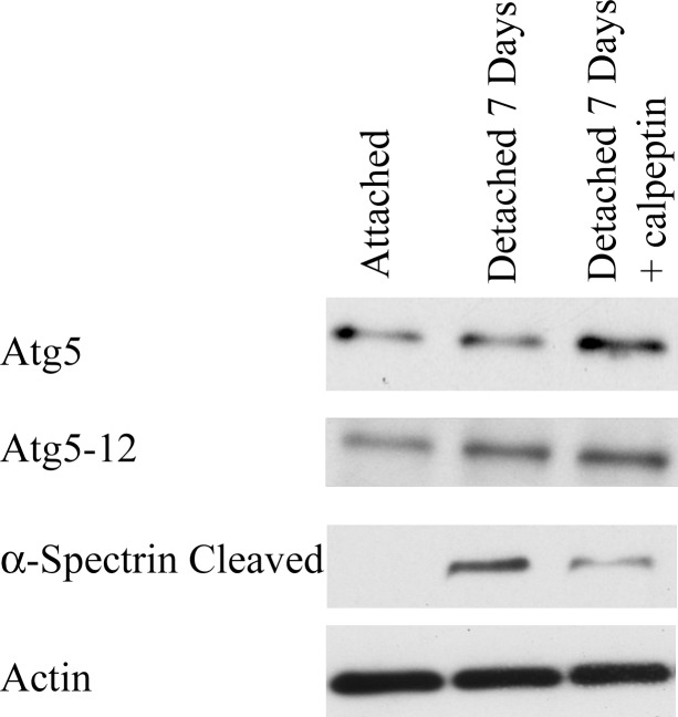
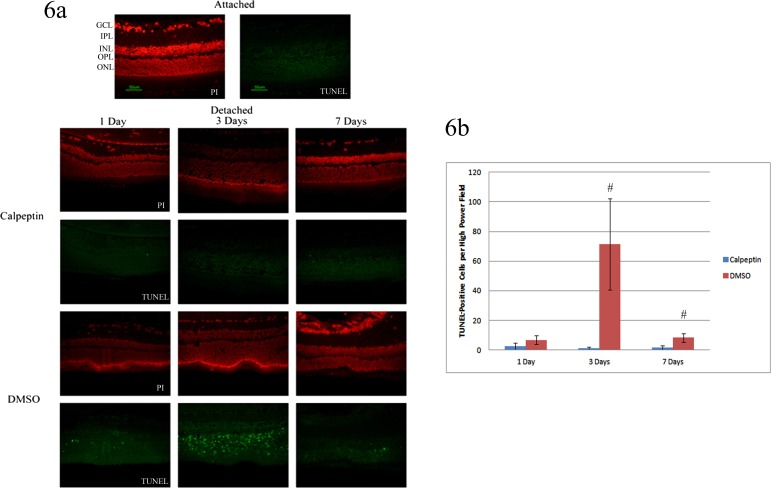
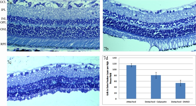
Methods: Retinal detachments were created in Brown-Norway rats by subretinal injection of 1% hyaluronic acid and simulated in vitro by Fas-receptor activation of 661W cells, a cone cell line. Protein levels of LC3 and autophagy-related gene 5 (Atg5), both of which are involved in the creation of the autophagosome, were assayed by Western blot. Calpain 1, the protease responsible for Atg5 cleavage and transitioning photoreceptors from autophagy to apoptosis, activity was monitored by α-spectrin cleavage. Various calpain inhibitors were added either to the subretinal space or cell culture media. Apoptosis was assessed in vitro by caspase-8 activity assays and in vivo via TUNEL assays. Cell counts were assessed in vivo at 2 months following detachment.

Results: Following retinal detachment or Fas-receptor activation of 661W cells, there was an increase in Atg5 and LC3-II that peaked at 3 days and decreased by 7-days postdetachment. Calpain 1 activity level peaked at 7 days and was associated with decreased autophagy. Calpain inhibition led to increased autophagy, a decrease in caspase-8 activation, reduced TUNEL-positive photoreceptors, and increased photoreceptor cell survival.

Conclusions: Our data suggest that calpain activation, which peaks at 7-days postdetachment, is a key step in triggering photoreceptors to shift from cell survival to death. Prolonging autophagy through calpain inhibition leads to significantly reduced photoreceptor apoptosis and increased cell survival.

Keywords: apoptosis; autophagy; retinal detachment.


Figures

Figure 1
Figure 1
Autophagy-related gene 5 activity following retinal detachment and Fas activation of 661W cells. (a) Protein from attached retinas and those detached for 1, 3, and 7 days were analyzed by Western blot. Actin was used as a protein-loading control. There was an increase in LC3-II, Atg5, and Atg5-12, peaking at the 1- and 3-day time points. (b) The attached retina, 3- and 7-day detached samples were again compared. Bafilomycin-A or equal volume of DMSO was injected into the subretinal space at the time of detachment. There was no significant difference in Atg5 or Atg5-12 at 7 days with Baf-A added. (c) 661W cells were treated with Fas activating antibody. Whole-cell lysates were collected at various time points, listed by number of hours following Fas activation. Both LC3-II and Atg5-12 peaked at 12 hours. Actin served as a loading control.
Figure 2
Figure 2
Calpain 1 activity following retinal detachment and Fas activation. (a) Whole retinas were harvested 3, 7, and 14 days after detachments were created. Attached retina served as controls. Cleaved α-spectrin levels were up sharply at 7 days compared with attached retinas and down by 14 days. Actin was used a loading control for all experiments. (b) 661W cells were treated with Fas activating antibody for 6 to 48 hours. There was an increase in total cleaved α-spectrin compared with untreated cells, which peaked at 24 hours.
Figure 3
Figure 3
Calpain inhibition in 661W cells with Fas activation. (a) Cells were treated for 24 hours with Fas activating antibody and various calpain inhibitors at two concentrations. Calpain inhibitors were added 1-hour prior to Fas activating antibody. All caused a drop in cleaved α-spectrin, with calpeptin causing the largest effect. Actin was used as a loading control for all experiments. (b) Similar samples were assayed for LC3-II. LC3-II was up at both concentrations at 24 hours after Fas activation. (c) Cells were treated at various concentrations of calpeptin for 24 hours. Both LC3-II and Atg5-12 complex peaked at the 25-μM levels. (d) 661W cells were treated with 25 μM of calpeptin for various lengths of time (in hours). There was an increase in both Atg5-12 and LC3-II following Fas activation. This signal lasted for an extended period of time with calpeptin when compared with vehicle (DMSO) alone.
Figure 4
Figure 4
Calpeptin inhibits calpain activity after retinal detachment. Retinas were harvested 7 days following detachment. Attached retinas served as controls. Either 0.2 μg of calpeptin or vehicle (DMSO) was injected into the subretinal space at the time of detachment. Protein was assayed by Western blot. Actin served as a loading control. There was significantly less cleaved α-spectrin with calpeptin. There was an increase in Atg5 and Atg5-12 with calpain inhibition when compared with detached retinas alone. Similar results were found at 3 days with and without calpeptin (data not shown).
Figure 5
Figure 5
Caspase-8 activity following Fas activation. 661W cells were treated with Fas activating antibody for 48 hours. Calpeptin was added 1-hour prior to treatment. Cells were then assayed for caspase-8 activity. Fas activation caused a significant elevation in caspase-8 activity. While 1 μM of calpeptin had little effect, both the 10- and 25-μM concentrations returned caspase-8 activity to baseline levels. Calpeptin alone had no effect on cells that were not treated with Fas activating antibody (P < 0.05; # compared with cells alone). Error bars indicate SD.
Figure 6
Figure 6
Photoreceptor apoptosis following retinal detachment with calpain inhibition. (a) Whole retinas were detached for 1 to 7 days. Calpeptin or DMSO alone was injected into the subretinal space at the time of detachment. Slides were analyzed via TUNEL staining at 1, 3, and 7 days following detachment. Propidium iodine (PI) was used to stain the nuclei of retinal cells. Attached retinas served as controls. (b) Three representative high power fields of three separate eyes were counted for number of TUNEL-positive cells in the ONL. The average of each eye was calculated and then the average of the three eyes for each condition was reported. There were significantly fewer TUNEL-positive cells at 3 and 7 days in retinas treated with calpeptin compared to vehicle alone (#, P < 0.05). Error bars indicate SD. GCL, ganglion cell layer; IPL, inner plexiform layer; INL, inner nuclear layer; OPL, outer plexiform layer; ONL, outer nuclear layer.
Figure 7
Figure 7
Cell survival with calpain inhibition. Whole-rat retinas were harvested 2 months following detachments. (a) Attached retinas were compared with retinas detached in the presence of (b) 0.2 μg calpeptin and (c) DMSO alone. (d) Three representative high power fields of three separate eyes were counted for number of cells in the ONL. The average of each eye was calculated and then the average of the three eyes for each condition was reported. The number of cells in the ONL was significantly greater in retinas detached in the presence of calpeptin compared with DMSO. Both detachment conditions had significantly fewer cells in the ONL compared with attached retinas. Error bars represent SD. All three conditions were significantly different with P < 0.05.

References

    1. Piccolino FC, de la Longrais RR, Ravera G, et al. The foveal photoreceptor layer and visual acuity loss in central serous chorioretinopathy. Am J Ophthalmol. 2005; 139: 87–99 - PubMed
    1. Burton TC. Recovery of visual acuity after retinal detachment involving the macula. Trans Am Ophthalmol Soc. 1982; 80: 475–497 - PMC - PubMed
    1. Zacks DN, Hänninen V, Pantcheva M, Ezra E, Grosskreutz C, Miller JW. Caspase activation in an experimental model of retinal detachment. Invest Ophthalmol Vis Sci. 2003; 44: 1262–1267 - PubMed
    1. Zacks DN, Zheng QD, Han Y, Bakhru R, Miller JW. FAS-mediated apoptosis and its relation to intrinsic pathway activation in an experimental model of retinal detachment. Invest Ophthalmol Vis Sci. 2004; 45: 4563–4569 - PubMed
    1. Zacks DN, Boehlke C, Richards AL, Zheng QD. Role of the Fas signaling pathway in photoreceptor neuroprotection. Arch Ophthalmol. 2007; 125: 1389–1395 - PubMed

Publication types

MeSH terms