

Pathophysiology of cerebral oedema in acute liver failure
- PMID: 24409052
- PMCID: PMC3882398
- DOI: 10.3748/wjg.v19.i48.9240
Pathophysiology of cerebral oedema in acute liver failure
Abstract
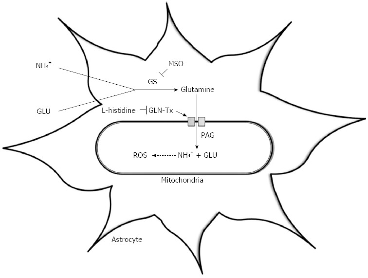
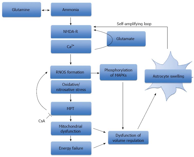
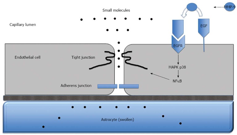
Cerebral oedema is a devastating consequence of acute liver failure (ALF) and may be associated with the development of intracranial hypertension and death. In ALF, some patients may develop cerebral oedema and increased intracranial pressure but progression to life-threatening intracranial hypertension is less frequent than previously described, complicating less than one third of cases who have proceeded to coma since the advent of improved clinical care. The rapid onset of encephalopathy may be dramatic with the development of asterixis, delirium, seizures and coma. Cytotoxic and vasogenic oedema mechanisms have been implicated with a preponderance of experimental data favouring a cytotoxic mechanism. Astrocyte swelling is the most consistent neuropathological finding in humans with ALF and ammonia plays a definitive role in the development of cytotoxic brain oedema. The mechanism(s) by which ammonia induces astrocyte swelling remains unclear but glutamine accumulation within astrocytes has led to the osmolyte hypothesis. Current evidence also supports an alternate 'Trojan horse' hypothesis, with glutamine as a carrier of ammonia into mitochondria, where its accumulation results in oxidative stress, energy failure and ultimately astrocyte swelling. Although a complete breakdown of the blood-brain barrier is not evident in human ALF, increased permeation to water and other small molecules such as ammonia has been demonstrated resulting from subtle alterations in the protein composition of paracellular tight junctions. At present, there is no fully efficacious therapy for cerebral oedema other than liver transplantation and this reflects our incomplete knowledge of the precise mechanisms underlying this process which remain largely unknown.
Keywords: Acute liver failure; Ammonia; Cerebral blood flow; Cerebral oedema; Hepatic encephalopathy; Intracranial hypertension; Intracranial pressure.
Figures
References
-
- Bernal W, Auzinger G, Dhawan A, Wendon J. Acute liver failure. Lancet. 2010;376:190–201. - PubMed
-
- Bower WA, Johns M, Margolis HS, Williams IT, Bell BP. Population-based surveillance for acute liver failure. Am J Gastroenterol. 2007;102:2459–2463. - PubMed
-
- Trey C, Davidson CS. The management of fulminant hepatic failure. Prog Liver Dis. 1970;3:282–298. - PubMed
-
- O’Grady JG, Schalm SW, Williams R. Acute liver failure: redefining the syndromes. Lancet. 1993;342:273–275. - PubMed
-
- Blei AT, Larsen FS. Pathophysiology of cerebral edema in fulminant hepatic failure. J Hepatol. 1999;31:771–776. - PubMed
Publication types
MeSH terms
Substances
Grants and funding
LinkOut - more resources
Full Text Sources
Other Literature Sources
