

Surgical outcomes of macular buckling techniques for macular retinoschisis in highly myopic eyes
- PMID: 24409086
- PMCID: PMC3885973
- DOI: 10.1016/j.sjopt.2013.08.001
Surgical outcomes of macular buckling techniques for macular retinoschisis in highly myopic eyes
Abstract
Purpose: To report the anatomic and visual results following macular buckling for patients with macular retinoschisis related to high myopia.
Methods: Thirty-nine highly myopic eyes (mean refractive error -16.7 D; range, -9 to -24 D) of 36 patients (mean age 59 years; range, 35-79 years) presenting with macular retinoschisis associated with a posterior staphyloma, who underwent combined vitrectomy and macular buckling were evaluated. Main outcome measures included best-corrected visual acuity (BCVA) and optical coherence tomography (OCT) findings. Three cases were excluded due to short follow-up (less than 3 months). The mean follow-up was 16 months.
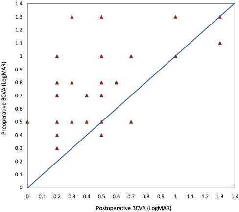
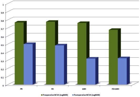
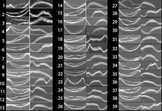
Results: The mean BCVA increased from 0.76 to 0.43 LogMAR (p = 0.001). Visual acuity improved in 30 eyes (83.3%), remained stable in three eyes (8.3%) and decreased in three eyes (8.3%). OCT showed resolution of foveoschisis with foveal reattachment in all eyes. None of the evaluated patients developed a macular hole during follow-up.
Conclusion: Macular buckling associated with vitrectomy results in good anatomic and visual outcomes in patients with myopic foveoschisis.
Keywords: High myopia; Lamellar macular hole; Macular buckling; Macular detachment; Myopic foveoschisis; Optical coherence tomography; Pars plana vitrectomy; Posterior staphyloma; Retinoschisis.
Figures

References
-
- Ward B. Degenerative myopia: myopic macular schisis and the posterior pole buckle. Retina. 2013;3(1):224–231. - PubMed
-
- Gass J.D.M. Myopic choroidal degeneration. In: Gass J.D.M., editor. Stereoscopic Atlas of Macular Diseases Diagnosis and Treatment. 4th ed. Mosby; St Louis: 1997. pp. 126–129.
-
- Ip M., Garza-Karren C., Duker J.S. Differentiation of degenerative retinoschisis from retinal detachment using optical coherence tomography. Ophthalmology. 1999;106:600–605. - PubMed
-
- Zhu Z., Xueying J., Zhang J., Ke G. Posterior scleral reinforcement in the treatment of macular retinoschisis in highly myopic patients. Clin Exp Ophthalmol. 2009;37:660–663. - PubMed
-
- Takano M., Kishi S. Foveal retinoschisis and retinal detachment in severely myopic eyes with posterior staphyloma. Am J Ophthamol. 1999;128:472–476. - PubMed
LinkOut - more resources
Full Text Sources
Other Literature Sources
Miscellaneous
