. 2014 Jan;12(1):e1001747.
doi: 10.1371/journal.pbio.1001747. Epub 2014 Jan 7.

Exocytosis of ATP from astrocytes modulates phasic and tonic inhibition in the neocortex

Affiliations

Exocytosis of ATP from astrocytes modulates phasic and tonic inhibition in the neocortex

Ulyana Lalo et al. PLoS Biol. 2014 Jan.

Erratum in

  • PLoS Biol. 2014 Apr;12(4):e1001857

Abstract

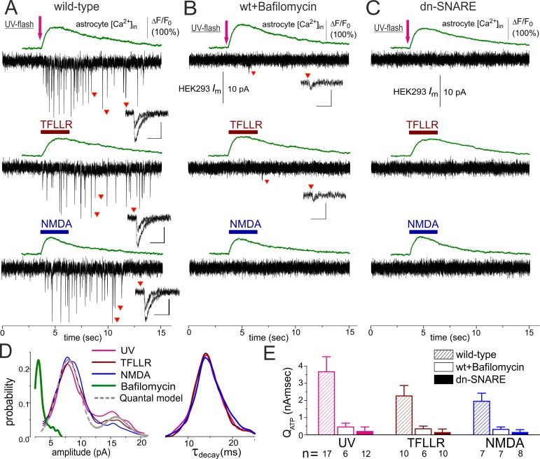
Communication between neuronal and glial cells is important for many brain functions. Astrocytes can modulate synaptic strength via Ca(2+)-stimulated release of various gliotransmitters, including glutamate and ATP. A physiological role of ATP release from astrocytes was suggested by its contribution to glial Ca(2+)-waves and purinergic modulation of neuronal activity and sleep homeostasis. The mechanisms underlying release of gliotransmitters remain uncertain, and exocytosis is the most intriguing and debated pathway. We investigated release of ATP from acutely dissociated cortical astrocytes using "sniff-cell" approach and demonstrated that release is vesicular in nature and can be triggered by elevation of intracellular Ca(2+) via metabotropic and ionotropic receptors or direct UV-uncaging. The exocytosis of ATP from neocortical astrocytes occurred in the millisecond time scale contrasting with much slower nonvesicular release of gliotransmitters via Best1 and TREK-1 channels, reported recently in hippocampus. Furthermore, we discovered that elevation of cytosolic Ca(2+) in cortical astrocytes triggered the release of ATP that directly activated quantal purinergic currents in the pyramidal neurons. The glia-driven burst of purinergic currents in neurons was followed by significant attenuation of both synaptic and tonic inhibition. The Ca(2+)-entry through the neuronal P2X purinoreceptors led to phosphorylation-dependent down-regulation of GABAA receptors. The negative purinergic modulation of postsynaptic GABA receptors was accompanied by small presynaptic enhancement of GABA release. Glia-driven purinergic modulation of inhibitory transmission was not observed in neurons when astrocytes expressed dn-SNARE to impair exocytosis. The astrocyte-driven purinergic currents and glia-driven modulation of GABA receptors were significantly reduced in the P2X4 KO mice. Our data provide a key evidence to support the physiological importance of exocytosis of ATP from astrocytes in the neocortex.


Conflict of interest statement

The authors have declared that no competing interests exist.

Figures

Figure 1
Figure 1. Detection of ATP released from cortical astrocytes with the aid of sniffer cells.
(A) Astrocytes acutely dissociated from the wild-type mouse neocortex have been loaded with UV-photoliable Ca2+-chelator NP-EGTA and Ca2+-indicator Fluo4-AM and placed over HEK293 cells expressing P2X2 receptors. The graphs below show the depolarization-activated currents and responses to application of 20 µM NMDA and 100 µM glutamate recorded in the astrocyte at a holding potential of −80 mV after an uncaging experiment; the response recorded under NBQX and D-AP5 was mediated by glutamate transporters, as evidenced by inhibition with specific blocker of glial glutamate transporters TFB-TBOA (300 nM). (B) Fluo-4 fluorescence was monitored in the astrocyte simultaneously with recording of transmembrane current in the HEK293-P2X2 cell voltage-clamped at −80 mV. UV uncaging of Ca2+ in the astrocyte was followed by the burst of phasic currents in the HEK293-P2X2 cell. Both the baseline and UV-elicited phasic currents were strongly inhibited after application of P2X receptor antagonist PPADS, confirming that they were mediated by ATP receptors. The diagram in the bottom panel shows the amplitude and frequency (mean ± SD for indicated numbers of HEK293-P2X2 cells) of phasic currents; statistical significance of the effect of PPADS was as indicated (*p<0.02 and **p<0.005, ANOVA). (C) Staining of cortical astrocyte with FM1-43 supports the existence of the mechanism of vesicular release. Acutely isolated cortical astrocytes were pre-incubated with 2.5 µM FM1-43 for 15 min and then washed out with extracellular medium for 15 min. The upper row shows the superposition of the DIC image of astrocyte and two-photon images of FM1-43 fluorescence (maximal intensity projections of Z-stack) recorded before (control) and after a 5-min-long application of the agonist of astroglial PAR-1 receptors TFLLR (10 µM) and glutamate; scale bar is 5 µm. Activation of astrocytes via PAR-1 and glutamate receptors led to FM1-43 destaining. To recover fluorescence, FM1-43 was applied for 5 min in the presence of glutamate (100 µM) and then washed out for 15 min. The bottom plot shows the changes in the FM1-43 fluorescent signal (mean ± SD for seven astrocytes) averaged over the whole-cell image.
Figure 2
Figure 2. Ca2+-dependent release of ATP can be evoked in the cortical astrocytes of the wild-type but not of the dn-SNARE mice.
Release of ATP from cortical astrocytes of wild-type (A, B) and dn-SNARE mice (C) was detected using the sniffer cells as described in Figure 1. (A) Elevation of cytosolic Ca2+ level was elicited in the astrocytes by UV uncaging and by rapid application of the agonist of PAR-1 metabotropic receptor TFLLR (10 µM) or the agonist of glutamate ionotropic receptor NMDA (20 µM). (B) Inhibition of vacuolar H-ATPase in the astrocytes with Bafilomycin A1 (1 µM for 2 h) dramatically decreased both the amplitude and frequency of phasic currents. (C) Elevation of the Ca2+ level in any of the dn-SNARE astrocytes did not lead to activation of phasic purinergic currents in the sniffer cell. Inlays in (A–C) show examples of individual phasic currents recorded at moments indicated; scale bars are 50 ms and 10 pA. (D) The amplitude and decay time distributions of purinergic currents recorded in the HEK293-P2X2 cells after stimulation of the astrocytes; data were pooled for number of experiments indicated in (E). The grey dotted line shows the best fit of quantal model to the distribution of UV-activated currents. (E) The pooled data (mean ± SD for indicated numbers of experiments) on net release of ATP were assessed as total charge transferred by spontaneous currents in the sniffer cell. The effects of bafylomicin and dn-SNARE expression on net charge transferred by purinergic currents were statistically significant at p = 0.005 (two-population t test).
Figure 3
Figure 3. Quantal release of ATP from astrocytes triggers fast purinergic currents in the cortical neurons in situ.
The spontaneous transmembrane currents were monitored in the layer 2/3 pyramidal neurons of neocortical slices of wild-type (A, D, G), dn-SNARE (B, E, H), and P2X4 KO mice (C, F, I). Expression of dn-SNARE in the astroglial cells of the neocortex is confirmed by EGFP-GFAP fluorescence (right column, scale bar 20 µm; see also Figure S15). Two distinct populations of spontaneous sEPSCs were recorded in the pyramidal neurons in the presence of TTX (1 µM), CNQX (50 µM), and D-APV (30 µM) at a holding potential of −80 mV: currents with larger amplitude and fast kinetics (orange dots) and currents with smaller amplitude and slow kinetics (green dots). (A, B, C) The representative transmembrane currents were recorded before and after activation of Ca2+ signaling in the astrocytes by selective agonist of astroglial PAR-1 receptor. The nonglutamatergic inward spontaneous currents (sEPSCs) were inhibited by P2 receptors antagonist PPADS (10 µM) and knocking out of P2X4 receptors. (D, E, F) Time course of changes in the frequency of sEPSCs currents during application of TFLLR (10 µM). Each dot represents the frequency calculated for a 1 min time window, and data show mean ± SD for number of neurons as follows: 17 (WT), 8 (dn-SNARE), and 12 (P2X4 KO). Activation of the PAR-1 receptor caused the appearance of a large number of spontaneous currents in the neurons of wild-type but not dn-SNARE mice; the frequency of sEPSCs was significantly lower in the P2X4 KO mice. (G, H, I) The amplitude and decay time distributions of purinergic sEPSCs recorded before and after application of TFLLR reveal the presence of a distinct population of spontaneous currents of smaller amplitude and slower kinetics in the wild-type but not in the dn-SNARE mice. Shaded areas show the amplitude distribution of background noise. Stimulation of astrocytes with TFLLR significantly increases the peaks corresponding to smaller and slower sEPSCs only in the neurons of the wild-type mice, whereas currents recorded in the neurons of dn-SNARE mice exhibit single-peaked amplitude and decay distributions. Deletion of P2X4 receptors attenuated amplitudes of both fast and slow inward currents and significantly reduced the proportion of slower currents evoked by TFLLR application.
Figure 4
Figure 4. Exocytosis of ATP from astrocytes in situ can be triggered by synaptic stimulation.
(A, B) Ca2+ signaling was monitored in astrocytes of somatosensory cortex layer 2/3 of wild-type (A) and dn-SNARE (B) mice simultaneously with voltage-clamp recordings of membrane currents in the pyramidal neurons. Spontaneous currents recorded in the pyramidal neurons at a holding potential of −80 mV in the presence of picrotoxin (100 µM) and CNQX (50 µM) were mediated by P2X receptors, as verified by inhibition with selective P2X antagonist NF279 (3 µM). The single episode of 100 Hz stimulation (HFS) triggered Ca2+ transients in the astrocytes of both wild-type and dn-SNARE mice; representatives are the Ca2+ transients and pseudo-color images (scale bar, 10 µm) recorded before (rest) and at the peak of response (stim). Ca2+ transients were followed by burst of spontaneous purinergic currents only in the neurons of wild-type mice (A), whereas neuronal spontaneous activity was not enhanced in the dn-SNARE mice (B). Inlays show the average waveforms (20 events) of spontaneous currents recorded before (rest) and 1 min after HFS (stim). (C, D). Each dot shows the average amplitude and frequency of spontaneous currents recorded in a 1 min time window in the pyramidal neurons of wild-type and dn-SNARE mice; data are presented as mean ± SD for six neurons. The asterisks (*) and (**) indicate the significant difference from the control values. The decrease in amplitude and significant increase in frequency of purinergic sEPSCs were observed in the wild-type but not in the dn-SNARE mice. (E, F) The amplitude and decay time distributions of purinergic sEPSCs recorded before, immediately after (0–30 s), and 1–3 min after HFS (pooled data for six neurons of each type) reveal the presence of a distinct population of spontaneous currents of smaller amplitude and slower kinetics in the wild-type but not in the dn-SNARE mice. Hence, these events are most likely activated by ATP released from astrocytes. The faster sEPSCs with larger amplitudes that underlie baseline activity both in wild-type and dn-SNARE mice are activated by ATP released from the nerve terminals. (G, H) The plot of decay time of purinergic sEPSCs against the amplitude demonstrates the presence of two populations of sEPSCs in the wild-type mice: slower currents of smaller amplitude (green area) and faster currents of higher amplitude (orange area). The corresponding waveforms (average of 20 traces) are shown in the inlay. HFS significantly increases the number of slower spontaneous currents. These currents were very rarely observed in the dn-SNARE mice, both in control and after HFS.
Figure 5
Figure 5. Intracellular perfusion of astrocyte with inhibitors of vesicular transport of ATP selectively affects slower purinergic EPSCs.
Whole-cell recordings of spontaneous EPSCs in the pyramidal neurons of somatosensory cortex of wild-type mice were carried out simultaneously with intracellular perfusion of astrocyte located in close proximity. Spontaneous currents recorded in the pyramidal neurons at a holding potential of −80 mV in the presence of picrotoxin (100 µM), CNQX (50 µM), and intracellular MK801 (10 µM) were mediated by P2X receptors, as demonstrated in Figures 3 and 4; the intracellular solution contained fluorescent dye Texas Red (30 µM). Whole-cell recording configuration was also established for neighboring astrocytes 15 min prior to the start of recording in the neuron, and the holding potential was −80 mV. Intracellular solution contained either fluorescent dye AlexaFluor 488 (30 µM) alone or AlexaFluor 488 and 1 mM of diadenosine-triphosphate (AP3A) or UTP. (A) A representative two-photon image of the recording outline and green and red fluorescence images (projection of Z-stack) are merged. (B) Two episodes of 100 Hz stimulation (HFS) of cortical afferents were delivered to trigger Ca2+ transients in the astrocytes, as demonstrated in Figure 4. Each dot shows the average amplitude and frequency of spontaneous currents recorded in a 1 min time window in the pyramidal neurons during perfusion of astrocyte with AlexaFluor 488 only (contro) and AlexaFluor and AP3A; data are presented as mean ± SD for five neurons. The asterisk (*) indicates the significant difference (p<0.05, unpaired t test) from the control values. The decrease in amplitude and significant increase in frequency of purinergic sEPSCs were attenuated by perfusion of astrocyte with AP3A, and the effect was more prominent after the second HFS episode. (C) The amplitude and decay time distributions of purinergic sEPSCs recorded before and 1–3 min after the second HFS (pooled data for five neurons in each case) reveal the decrease in number of sEPSCs of smaller amplitude and slower kinetics after perfusion of astrocyte with AP3A. (D) The representative average waveforms (20 events) of fast and slow spontaneous sEPSCs (separated as shown in Figures 4 and S3) recorded 1 min after the 2-s HFS episode. Note the decrease in the amplitude of slow sEPSCs recorded in the presence of AP3A and lack of changes in the fast sEPSC. (E) Diagrams show the amplitude and frequency of slow purinergic sEPCS averaged within a 3 min time window before (baseline) and after HFS episodes delivered in control and during perfusion of astrocytes with AP3A and UTP. Data are shown as mean ± SD for the five neurons. The statistical significance of the difference from the control values was indicated as (*) p<0.05 and (**) p<0.01. Note the decrease in the mean frequency and amplitude of slow sEPSCs.
Figure 6
Figure 6. Exocytosis of ATP from astrocytes leads to down-regulation of inhibitory synaptic currents in neocortex.
Astroglial exocytosis was elicited by application of PAR-1 receptor agonist as shown in Figures 2 and 3. (A) Plots show the time course of evoked IPSCs in the layer 2/3 pyramidal neurons of wild-type mice (left column) and dn-SNARE and P2X4 knockout mice (right column) during application of TFLLR (10 µM) and nonhydrolysable ATP-analog ATP-γS (10 µM). In the wild-type mice, TFLLR was also applied in the presence of P2 purinoreceptors antagonist PPADS (10 µM). Data points represent the mean amplitude and quantal content for six consecutive IPSCs evoked at −40 mV in the presence of CNQX (50 µM), D-APV (30 µM), and DPCPX (3 µM). Data show mean ± SD for the number of neurons as follows: 9 (WT, control), 7 (WT+PPADS), 6 (WT+ATP-γS), 8 (dn-SNARE), and 12 (P2X4-KO). The average waveforms (25 IPSCs) are shown below as indicated. (B) Time course of the amplitude of spontaneous mIPSCs recorded in the same conditions as above +1 µM TTX. Each point represents the average amplitude for a 1 min time window. Data points represent the mean amplitude and frequency of mIPSCs recorded with a 1 min time window. Data show mean ± SD for number of neurons as follows: 12 (WT, control), 5 (WT+PPADS), 5 (WT+ATP-γS), 8 (dn-SNARE), and 12 (P2X4-KO). The average waveforms (25 mIPSCs) are shown below. Note that the reduction in the inhibitory currents was strongly attenuated by the antagonist of ATP receptors and knocking out P2X4 receptors and there was no marked reduction in the IPSCs in the dn-SNARE mice. (C) Cumulative amplitude histograms of spontaneous mIPSCs (pooled for number of neurons indicated above) recorded in the baseline conditions (left) and after application of PAR-1 agonist (right). Histograms show the significant reduction in the mIPSCs amplitude caused by TFLLR. Amplitude histogram of mIPSCs recorded in dn-SNARE mice in control shows the marked right shift, indicating the up-regulation of baseline inhibitory transmission.
Figure 7
Figure 7. The presynaptic changes in the inhibitory synaptic currents in the neocortex evaluated by PPR.
The IPSCs were recorded in the pyramidal neuron in the same conditions as described in Figure 6A. (A–C) Graphs show the representative IPSCs (average of 25 traces) evoked with a 50 ms interval in control and during application of 10 µM TFLLR. To calculate the PPR, the amplitude of IPSC was evaluated by best fit with two curves of mono-exponential rise and decay, depicted by red and blue lines. (D) The pooled data (mean ± SD) on PPR of IPCS measured before and during application of 10 µM TFLLR and 10 µM ATP-γS in the wild-type, dn-SNARE, and P2X4 knockout mice for number of cells as indicated. Asterisks * and ** indicate statistical significance of difference from the control of the same mice strain (two population t test), and asterisks in parentheses (*) indicate statistical significance of difference in the effect of TFLLR from the wild-type mice. Note the increase in PPR under action of TFLLR and ATP-γS, which contrasts with the decrease in the IPSCs amplitudes (Figure 6).
Figure 8
Figure 8. Exocytosis of ATP from astrocytes down-regulates the tonic inhibitory synaptic signaling.
(A, B) Tonic GABA-mediated signaling was evaluated by upward shift in the whole-cell holding current (ΔIhold) caused by application of bicuculline (50 µM) to the layer 2/3 pyramidal neurons in brain slices of wild-type (A) and dn-SNARE (B) mice. Bicuculline was applied either in control (upper traces) or after activation of astrocytic signaling by 10 µM TFLLR (middle traces) or after application of TFLLR in the presence of ATP receptors antagonist PPADS (bottom trace). Currents were recorded in the presence of CNQX (50 µM) and D-APV (30 µM) at a holding potential of −80 mV. (C) The average amplitude of the tonic GABA-mediated current measured in different conditions in the wild-type, dn-SNARE, and P2X4 knockout mice for number of cells as indicated; statistical significance of difference in mean amplitude was as indicated (*) p<0.05 and (**) p<0.01. In the wild-type mice, activation of astroglial Ca2+ signaling significantly decreased the tonic GABA-mediate current; reduction in tonic current was attenuated by PPADS. In the dn-SNARE and P2X4 KO mice, tonic inhibitory current was significantly up-regulated, and activation of the astroglial Ca2+ had a much smaller effect on the tonic current than in the wild-type mice. These results suggest that exocytosis of ATP can lead to down-regulation of tonic inhibition in the neocortical neurons, most likely via P2X receptors.

Similar articles

Cited by

References

    1. Burnstock G (2007) Physiology and pathophysiology of purinergic neurotransmission. Physiol Rev 87: 659–797. - PubMed
    1. Abbracchio MP, Burnstock G, Verkhratsky A, Zimmermann H (2009) Purinergic signalling in the nervous system: an overview. Trends Neurosci 32: 19–29. - PubMed
    1. Guthrie PB, Knappenberger J, Segal M, Bennett MV, Charles AC, et al. (1999) ATP released from astrocytes mediates glial calcium waves. J Neurosci 19: 520–528. - PMC - PubMed
    1. Fields RD, Burnstock G (2006) Purinergic signalling in neuron-glia interactions. Nat Rev Neurosci 7: 423–436. - PMC - PubMed
    1. Lalo U, Pankratov Y, Parpura V, Verkhratsky A (2010) Ionotropic receptors in neuronal-astroglial signalling: what is the role of “excitable” molecules in non-excitable cells. Biochim Biophys Acta 1813: 992–1002. - PubMed

MeSH terms