

Projections of nucleus accumbens adenosine A2A receptor neurons in the mouse brain and their implications in mediating sleep-wake regulation
- PMID: 24409122
- PMCID: PMC3857888
- DOI: 10.3389/fnana.2013.00043
Projections of nucleus accumbens adenosine A2A receptor neurons in the mouse brain and their implications in mediating sleep-wake regulation
Abstract
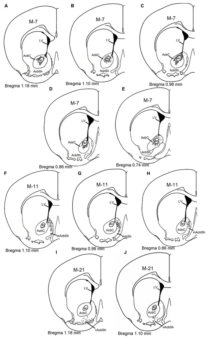
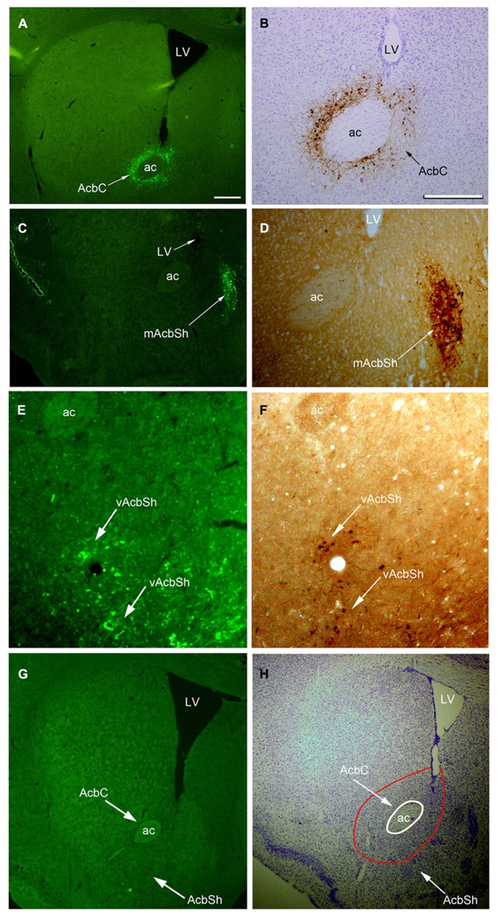
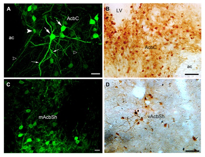
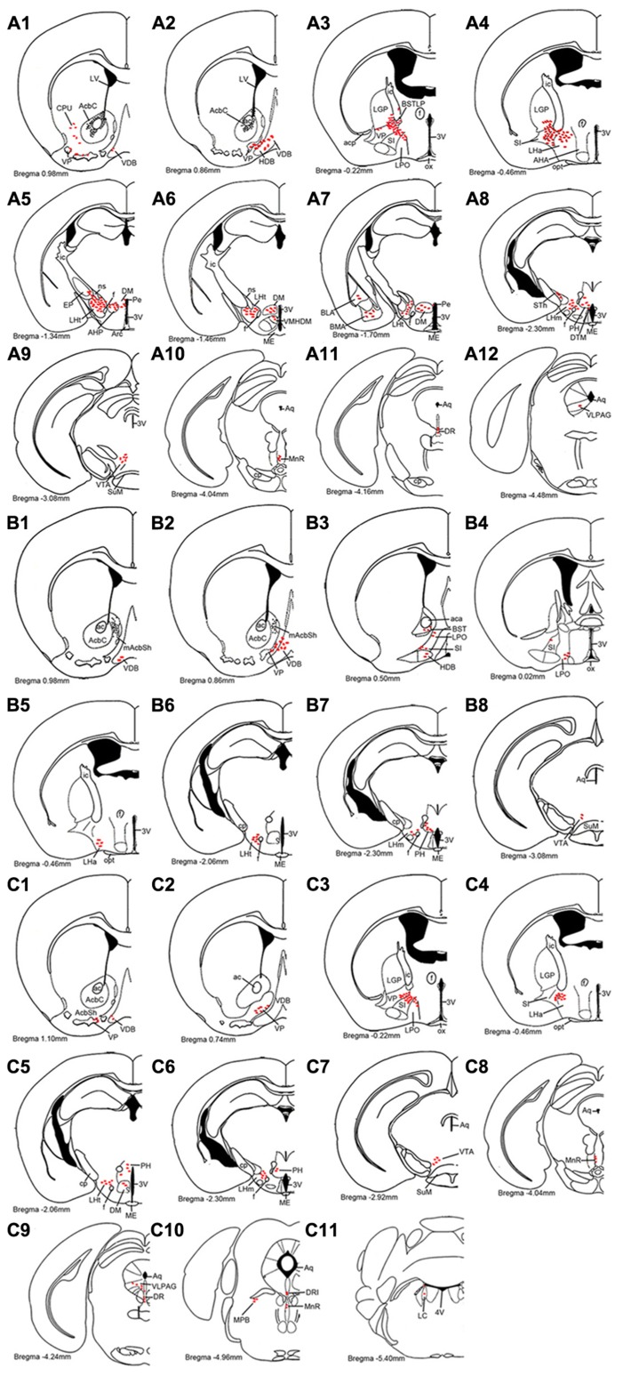
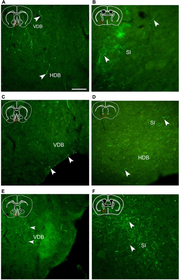
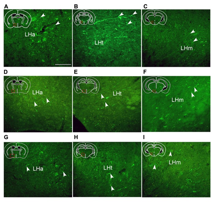
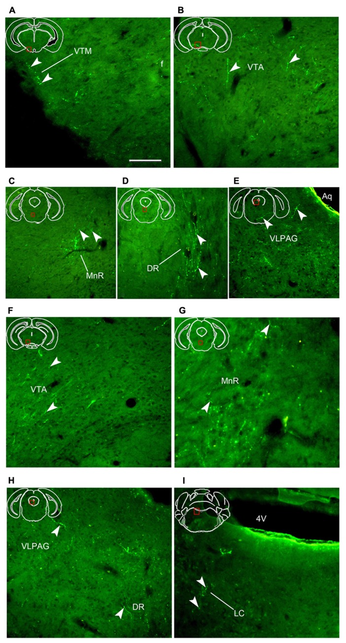
Adenosine A2A receptors (A2ARs) in the nucleus accumbens (Acb) have been demonstrated to play an important role in the arousal effect of adenosine receptor antagonist caffeine, and may be involved in physiological sleep. To better understand the functions of these receptors in sleep, projections of A2AR neurons were mapped utilizing adeno-associated virus (AAV) encoding humanized Renilla green fluorescent protein (hrGFP) as a tracer for long axonal pathways. The Cre-dependent AAV was injected into the core (AcbC) and shell (AcbSh) of the Acb in A2AR-Cre mice. Immunohistochemistry was then used to visualize hrGFP, highlighting the perikarya of the A2AR neurons in the injection sites, and their axons in projection regions. The data revealed that A2AR neurons exhibit medium-sized and either round or elliptic perikarya with their processes within the Acb. Moreover, the projections from the Acb distributed to nuclei in the forebrain, diencephalon, and brainstem. In the forebrain, A2AR neurons from all Acb sub-regions jointly projected to the ventral pallidum, the nucleus of the diagonal band, and the substantia innominata. Heavy projections from the AcbC and the ventral AcbSh, and weaker projections from the medial AcbSh, were observed in the lateral hypothalamus and lateral preoptic area. In the brainstem, the Acb projections were found in the ventral tegmental area, while AcbC and ventral AcbSh also projected to the median raphe nucleus, the dorsal raphe nucleus, and the ventrolateral periaqueductal gray. The results supply a solid base for understanding the roles of the A2AR and A2AR neurons in the Acb, especially in the regulation of sleep.
Keywords: Cre-Lox; adeno-associated virus; green fluorescent protein; mouse; nucleus accumbens; sleep.
Figures
Similar articles
-
Efferent projections of the nucleus accumbens in the rat with special reference to subdivision of the nucleus: biotinylated dextran amine study.Brain Res. 1998 Jun 22;797(1):73-93. doi: 10.1016/s0006-8993(98)00359-x. Brain Res. 1998. PMID: 9630528
-
Efferent projections of infralimbic and prelimbic areas of the medial prefrontal cortex in the Japanese monkey, Macaca fuscata.Brain Res. 2001 Jan 5;888(1):83-101. doi: 10.1016/s0006-8993(00)03013-4. Brain Res. 2001. PMID: 11146055
-
Identifying the efferent projections of leptin-responsive neurons in the dorsomedial hypothalamus using a novel conditional tracing approach.J Comp Neurol. 2010 Jun 1;518(11):2090-108. doi: 10.1002/cne.22323. J Comp Neurol. 2010. PMID: 20394060 Free PMC article.
-
[Neurochemical mechanisms of sleep regulation].Glas Srp Akad Nauka Med. 2009;(50):97-109. Glas Srp Akad Nauka Med. 2009. PMID: 20666118 Review. Serbian.
-
[Sleep-wake regulation by prostaglandin D2 and adenosine].Brain Nerve. 2012 Jun;64(6):621-8. Brain Nerve. 2012. PMID: 22647469 Review. Japanese.
Cited by
-
Enhanced functional connectivity in the reward circuitry in healthy adults with weekend catch-up sleep.Hum Brain Mapp. 2023 Oct 1;44(14):4927-4937. doi: 10.1002/hbm.26429. Epub 2023 Jul 19. Hum Brain Mapp. 2023. PMID: 37466297 Free PMC article.
-
Whole-Brain Neural Connectivity to Lateral Pontine Tegmentum GABAergic Neurons in Mice.Front Neurosci. 2019 Apr 24;13:375. doi: 10.3389/fnins.2019.00375. eCollection 2019. Front Neurosci. 2019. PMID: 31068780 Free PMC article.
-
Striatal adenosine A2A receptor neurons control active-period sleep via parvalbumin neurons in external globus pallidus.Elife. 2017 Oct 12;6:e29055. doi: 10.7554/eLife.29055. Elife. 2017. PMID: 29022877 Free PMC article.
-
The generation of knock-in mice expressing fluorescently tagged galanin receptors 1 and 2.Mol Cell Neurosci. 2015 Sep;68:258-71. doi: 10.1016/j.mcn.2015.08.006. Epub 2015 Aug 17. Mol Cell Neurosci. 2015. PMID: 26292267 Free PMC article.
-
Medial Parabrachial Nucleus Is Essential in Controlling Wakefulness in Rats.Front Neurosci. 2021 Mar 25;15:645877. doi: 10.3389/fnins.2021.645877. eCollection 2021. Front Neurosci. 2021. PMID: 33841086 Free PMC article.
References
LinkOut - more resources
Full Text Sources
Other Literature Sources
Molecular Biology Databases