. 2014 Jan 9:7:223.
doi: 10.3389/fnbeh.2013.00223. eCollection 2013.

Sex differences in stress-induced social withdrawal: role of brain derived neurotrophic factor in the bed nucleus of the stria terminalis

Affiliations

Sex differences in stress-induced social withdrawal: role of brain derived neurotrophic factor in the bed nucleus of the stria terminalis

Gian D Greenberg et al. Front Behav Neurosci. .

Abstract

Depression and anxiety disorders are more common in women than men, and little is known about the neurobiological mechanisms that contribute to this disparity. Recent data suggest that stress-induced changes in neurotrophins have opposing effects on behavior by acting in different brain networks. Social defeat has been an important approach for understanding neurotrophin action, but low female aggression levels in rats and mice have limited the application of these methods primarily to males. We examined the effects of social defeat in monogamous California mice (Peromyscus californicus), a species in which both males and females defend territories. We demonstrate that defeat stress increases mature brain-derived neurotrophic factor (BDNF) protein but not mRNA in the bed nucleus of the stria terminalis (BNST) in females but not males. Changes in BDNF protein were limited to anterior subregions of the BNST, and there were no changes in the adjacent nucleus accumbens (NAc). The effects of defeat on social withdrawal behavior and BDNF were reversed by chronic, low doses of the antidepressant sertraline. However, higher doses of sertraline restored social withdrawal and elevated BDNF levels. Acute treatment with a low dose of sertraline failed to reverse the effects of defeat. Infusions of the selective tyrosine-related kinase B receptor (TrkB) antagonist ANA-12 into the anterior BNST specifically increased social interaction in stressed females but had no effect on behavior in females naïve to defeat. These results suggest that stress-induced increases in BDNF in the anterior BNST contribute to the exaggerated social withdrawal phenotype observed in females.

Keywords: BDNF; BNST; SSRI; dose; sex; social defeat.


Figures

Figure 1
Figure 1
Experimental design and timeline. In Experiments 1 and 2 mice were tested in social interaction tests (day 17). In Experiment 3 mice were tested in social interaction tests (day 31), sucrose anhedonia (days 32–34), and forced swim (day 35). In Experiment 4, mice were tested in forced swim 1 h following social interaction (day 31). In Experiment 5 mice were tested in social interaction twice due to within-subjects design and were euthanized immediately following the second day of social interaction testing. Indicates injection(s) of sertraline. GDXIndicates gonadectomy. #Indicates cannula surgeries.
Figure 2
Figure 2
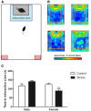
Exposure to social defeat stress decreased the amount of time female mice but not males spent in the interaction zone interacting with a novel target mouse. (A) The illustration describes the location of the “interaction zone” (highlighted in blue) and the “corners zone” (highlighted in red) within the social interaction arena. (B, clockwise from top left) Heat maps depicting space usage during the social interaction phase for control males, control females, stressed females, and stressed males. (C) Quantification of time spent in the interaction zone during social interaction phase. Data are shown as mean ± SE. (**p < 0.01 vs. control), n = 13–20 per group.
Figure 3
Figure 3
Social stress increased brain-derived neurotrophic factor (BDNF) protein in females but not males in the bed nucleus of the stria terminalis (BNST). (A) The anti-BDNF antibody is specific to BDNF protein as evaluated by Western blot using microdissections from California mouse brain bed nucleus of stria terminalis (BNST). The same blot was stained for β-Actin (top half) and BDNF (bottom half). A recombinant BDNF peptide was used as a positive control for the BDNF antibody and negative control for the β-Actin antibody. The Bio-Rad Precision Protein Plus Ladder (L) was run in the first lane. (B) BDNF protein in the BNST was significantly increased in stressed females. Western blot analysis of 2 mm-thick punch sample microdissections from BNST showed that females exposed to stress had significantly increased levels of BDNF protein in BNST, n = 12–19 per group. (C,D) Western blots from 500 micron-thick punches revealed increased BDNF expression specifically in anterior BNST of stressed female mice, n = 8–10 per group (representative image for panel C was cropped from full blot presented in panel A). Schematics diagramming tissue punch sites for BNST subdivisions are presented below blots. Illustrations are based on artwork from Paxinos and Franklin (2002), with permission from Academic Press. %BDNF was calculated from the ratio of BDNF:Actin normalized to control mice across blots. Data are shown as mean ± SE. (*p < 0.05, **p < 0.01 vs. control). Blots have been cropped for clarity to create representative gel pattern and analysis of BDNF levels in BNST.
Figure 4
Figure 4
Social defeat stress increased brain-derived neurotrophic factor (BDNF) immunoreactivity in females but not males in the anterior bed nucleus of the stria terminalis (BNST). (A) BDNF immunostaining from anterior-posterior serial sections in a representative stressed female, starting in nucleus accumbens (NAc) and continuing through to posterior BNST. Lower power photomicrographs, scale bar = 500 μm. Anterior commissure (ac), lateral ventricle (LV), island of Calleja major (ICjM), and fornix (f) labeled for reference. Bregma coordinates are based on cannula placements in this study and Campi et al. (2014). Puncta were observed in the anterior ventromedial BNST (BNSTVM), anteriorlateral BNST (BNSTAL) and anteriormedial BNST (BNSTAM) (B) Representative photomicrographs of BDNF immunostaining within BNSTVM from which BDNF immunoreactivity was quantified. Higher power photomicrographs, scale bar = 200 μm. (C) Quantification of immunoreactivity (BDNF-ir) revealed increased BDNF in BNSTVM (top graph) in stressed female mice but no differences in NAc core (bottom graph). Raw data are shown as %staining. Data are shown as mean ± SE. (**p < 0.01 vs. control), n = 3–5 per group.
Figure 5
Figure 5
(A) Nucleus accumbens (NAc) BDNF protein as evaluated by Western blot analyses. Social defeat had no effect on brain-derived neroutrophic factor (BDNF) in female or male mice. Blots have been cropped for clarity. %BDNF was calculated from the ratio of BDNF:Actin normalized to control mice across blots. Data are shown as mean ± SE. n = 5–8 per group. (B) There were no significant differences in Bdnf mRNA as evaluated by real-time PCR analyses using microdissections of bed nucleus of the stria terminalis (BNST) from intact and gonadectomized male and female California mice.
Figure 6
Figure 6
Measurements of time spent in (A) the interaction zone during the interaction phase of the social interaction test and (B) the center zone of an open field test (OFT) following chronic administration of a selective serotonin reuptake inhibitor (SSRI) antidepressant. The SSRI sertraline reversed avoidance behavior in stressed female mice when administered at a low dose (5 mg/kg). A moderate dose of sertraline (10 mg/kg) had an anxiolytic effect on time spent in the center of the OFT in male and female mice. (C) Low to moderate doses of sertraline reversed increases in brain-derived neurotrophic factor (BDNF) protein in the bed nucleus of the stria terminalis (BNST) in stressed females. 0 mg/kg female mice had significantly more BDNF in BNST than 0 mg/kg male mice, 5 mg/kg female mice and 10 mg/kg/day female mice. Blots have been cropped for clarity. %BDNF was calculated from the ratio of BDNF:Actin normalized to 0 mg/kg mice across blots. Data are shown as mean ± SE (p < 0.05, ††p < 0.01 sex difference, *p < 0.05, **p < 0.01 effects of sertraline), n = 5–7 per group.
Figure 7
Figure 7
(A,B,G) Effects of ANA-12 on behavior for stressed female mice in which needle tracks hit the anterior bed nucleus of the stria terminalis (BNST). ANA-12 increased time spent interacting with a novel mouse (A; interaction, **paired t-test p = 0.01) and decreased time spent in the corner zones opposite a novel mouse (B; interaction, nonsignificant). There was no effect of ANA-12 on behavior in the absence of a social stimulus (A,B; acclimation) or on behavior in an open field test (G). (C) Representative image of needle track hitting the anterior bed nucleus of the stria terminalis. Structures caudate-putamen (CPu), anterior commissure (ac), anterior-medial bed nucleus of the stria terminalis (BNSTam), ventromedial bed nucleus of the stria terminalis (BNSTmv) and lateral septum (LS) shown for reference. Photos captured at lower power, scale bar = 500 μm. (F) Representative image of needle track missing anerior BNST. (D,E,H) Effects of ANA-12 on behavior for stressed female mice in which needle tracks missed the anterior bed nucleus of the stria terminalis (BNST). ANA-12 did not affect time spent interacting with a novel mouse (D; interaction) or empty cage (D; acclimation). There was no effect of ANA-12 on time spent in the corner zones opposite a novel mouse or empty cage (E). There was no effect of ANA-12 on locomotor behavior during an open field test (H). Data are shown as mean ± SE, n = 4–6 per group. (I) Schematic representations of recorded hits (red dots) and misses (blue) from nissl-stained sections of stressed female mice with cannula guides implanted (based on artwork from Paxinos and Franklin (2002), with permission from Academic Press).

Similar articles

Cited by

References

    1. Aguilera G., Millan M. A., Hauger R. L., Catt K. J. (1987). Corticotropin-releasing factor receptors: distribution and regulation in brain, pituitary, and peripheral tissues. Ann. N.Y. Acad. Sci. 512, 48–66 10.1111/j.1749-6632.1987.tb24950.x - DOI - PubMed
    1. Ali A. E. A., Wilson Y. M., Murphy M. (2012). Identification of neurons specifically activated after recall of context fear conditioning. Neurobiol. Learn. Mem. 98, 139–147 10.1016/j.nlm.2012.07.004 - DOI - PubMed
    1. Anstrom K. K., Miczek K. A., Budygin E. A. (2009). Increased phasic dopamine signaling in the mesolimbic pathway during social defeat in rats. Neuroscience 161, 3–12 10.1016/j.neuroscience.2009.03.023 - DOI - PMC - PubMed
    1. Autry A. E., Adachi M., Nosyreva E., Na E. S., Los M. F., Cheng P.-F., et al. (2011). NMDA receptor blockade at rest triggers rapid behavioural antidepressant responses. Nature 475, 91–95 10.1038/nature10130 - DOI - PMC - PubMed
    1. Babb J. A., Masini C. V., Day H. E. W., Campeau S. (2013). Sex differences in activated corticotropin-releasing factor neurons within stress-related neurocircuitry and hypothalamic–pituitary–adrenocortical axis hormones following restraint in rats. Neuroscience 234, 40–52 10.1016/j.neuroscience.2012.12.051 - DOI - PMC - PubMed

LinkOut - more resources