. 2014 Jan 9:4:402.
doi: 10.3389/fphys.2013.00402. eCollection 2014.

CD13 promotes mesenchymal stem cell-mediated regeneration of ischemic muscle

Affiliations

CD13 promotes mesenchymal stem cell-mediated regeneration of ischemic muscle

M Mamunur Rahman et al. Front Physiol. .

Abstract

Mesenchymal stem cells (MSCs) are multipotent, tissue-resident cells that can facilitate tissue regeneration and thus, show great promise as potential therapeutic agents. Functional MSCs have been isolated and characterized from a wide array of adult tissues and are universally identified by the shared expression of a core panel of MSCs markers. One of these markers is the multifunctional cell surface peptidase CD13 that has been shown to be expressed on human and murine MSCs from many tissues. To investigate whether this universal expression indicates a functional role for CD13 in MSC biology we isolated, expanded and characterized MSCs from bone marrow of wild type (WT) and CD13(KO) mice. Characterization of these cells demonstrated that both WT and CD13(KO) MSCs expressed the full complement of MSC markers (CD29, CD44, CD49e, CD105, Sca1), showed comparable proliferation rates and were capable of differentiating toward the adipogenic and osteogenic lineages. However, MSCs lacking CD13 were unable to differentiate into vascular cells, consistent with our previous characterization of CD13 as an angiogenic regulator. Compared to WT MSCs, adhesion and migration on various extracellular matrices of CD13(KO) MSCs were significantly impaired, which correlated with decreased phospho-FAK levels and cytoskeletal alterations. Crosslinking human MSCs with activating CD13 antibodies increased cell adhesion to endothelial monolayers and induced FAK activation in a time dependent manner. In agreement with these in vitro data, intramuscular injection of CD13(KO) MSCs in a model of severe ischemic limb injury resulted in significantly poorer perfusion, decreased ambulation, increased necrosis and impaired vascularization compared to those receiving WT MSCs. This study suggests that CD13 regulates FAK activation to promote MSC adhesion and migration, thus, contributing to MSC-mediated tissue repair. CD13 may present a viable target to enhance the efficacy of mesenchymal stem cell therapies.

Keywords: CD13; adhesion; cell transplantation; hindlimb ischemia; mesenchymal stem cells.


Figures

Figure 1
Figure 1
Mesenchymal stem cell culture and characterization. (A) Phase contrast image of BM derived MSCs at Day 0 and Day 21(Bar = 100 μm). (B) CD13 expression in WT-MSCs by fluorescence immunostaining (Bar = 20 μm) and protein expression of CD13 in WT-MSC. (C) RT PCR analysis of stem cell expression profiles. (D) Flow cytometric analysis of MSCs. Cells were characteristically positive for CD29, CD49e and negative for CD19, CD31, and CD34. Unstained;WT-MSC; KO-MSC–. (E) Both WT-MSC and KO-MSC expressed transcription factor OCT3/4 (Bar = 20 μm). (F) Confluent MSCs were transferred to adipogenic and osteogenic medium for 3 weeks. Adipocytes were detected by oil red O staining and osteoblasts by alizarin red staining (Bar = 200 μm). (G) 1 × 10⁵ cells were seeded on Matrigel coated 6-well plates and incubated for 12 h. cells isolated from CD13KO mice are unable to form capillary networks and form fewer branches (Bar = 200 μm).
Figure 2
Figure 2
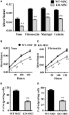
Lack of CD13 impairs MSC adhesion, proliferation, migration, and invasion. (A) Adhesion assay: Cells (1 × 10⁴) were seeded in 96 well plates coated separately with fibronectin, Matrigel, or gelatin and allowed to adhere for 1 h at 37°C. After PBS wash, adherent cells were detected by MTT assay. n = 6, **P < 0.01. (B,C) Proliferation assay: Cells (0.5 × 10⁴) were seeded in 96 well plate and cell proliferation detected by MTT assay at the indicated time points. n = 6, *P < 0.05, **P < 0.01. (D) Migration assay: 1 × 10⁴ cells were seeded in FluoroBlok chambers. After 4 h. incubation the cells were stained with DAPI and counted. n = 4, **P < 0.01. (E) Invasion assay: 1 × 10⁵ cells were seeded on Matrigel (1:5 dilution) coated FluoroBlok chambers. After 6 h. incubation the cells were counted. n = 4, **P < 0.01.
Figure 3
Figure 3
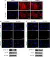
Mesenchymal stem cell defects in CD13KO mice are cell intrinsic. (A) phalloidin-stained MSC cells isolated from injured muscles of CD13KO mice showed remarkable cytoskeletal disruption compared to cells from WT mice; Objective 40X (Bar = 100 μm). (B,C) Immunofluorescent detection of FAK phosphorylation at tyrosine residues 397 (B) and 925 (C). Protein lysates of MSC were probed for phospho-FAK (Y397) and phospho-FAK (Y925) with β-actin as the loading control. CD13KO MSCs expressed lower levels of phospho-FAK protein (Bar = 20 μm).
Figure 4
Figure 4
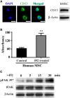
CD13 activation increases monolayer adhesion and FAK phosphorylation in human MSCs. (A) Human mesenchymal stem cells also express CD13 by immunofluorescence (green); Objective 63X (Bar = 20 μm) and immunoblot of human cell lysates. (B) Colorimetric quantification of adhesion of human MSCs treated with the CD13 activating mAb 452 to HUVEC monolayers. Data represents the mean ± s.e.m. n = 3 from two independent experiments (**P < 0.01). (C) CD13 crosslinking with activating mAb 452 temporally induces FAK tyrosine phosphorylation in human MSCs.
Figure 5
Figure 5
Lack of CD13 on exogenously administered MSCs impairs perfusion recovery in an in vivo hindlimb ischemia model. (A) Laser Doppler flow imaging of perfusion in mice at the indicated time points. Representative color-coded images of three groups (PBS, WT-MSC, and KO-MSC) of mice on day 0, 3, 7, 14, and 21 after surgery and cell transplantation assessed by laser Doppler imaging. Red indicates highest perfusion velocity, green intermediate, and blue, lowest velocity. (B) Cumulative results for PBS (n = 7), WT-MSC (n = 6), and KO-MSC (n = 6) injected mice are shown graphically as ratios of blood flow in ischemic limb (I) to that in the non-ischemic limb (NI) at each time point. Functional assessment of ischemic muscles. Cumulative results are shown graphically as (C) the ambulatory impairment score; (D) ischemic tissue damage score as described in Methods. (E) Images of Non-ischemic and PBS, WT-MSC, and KO-MSC injected ischemic paw. Black nail indicates necrosis. #P < 0.05 compare to PBS and *P < 0.05 compare to KO-MSC; (score assessment criteria in Methods).
Figure 6
Figure 6
Muscle regeneration and capillary formation are impaired in mice injected with CD13KO MSC following ischemic injury. (A) Hematoxylin and eosin (H&E) staining of gastrocnemius muscle regeneration was confirmed by the presence of multiple, centrally located myocyte nuclei, 20X objective; Bar = 100 μm. (B) Capillaries were visualized by immunofluorescent staining with CD31 (red) and nuclei with DAPI (blue); Objective, 40X objective; Bar = 50 μm. (C) The area of the injured tissue was compared among PBS, WT-MSC, and KO-MSC groups. A significant increase in muscle regeneration (average) was observed in WT-MSC group compared with PBS group at day 21. (D) The ratio of capillary density per fiber was measured in ischemic gastrocnemius muscles. Capillary density was significantly increased in WT-MSC compared with other groups, PBS and KO-MSC. All data were quantified by ImagePro Plus. Values are shown as mean ± s.e.m. (*P < 0.05) (PBS n = 7; WT-MSC n = 6; KO-MSC n = 6).
Figure 7
Figure 7
Engraftment of MSCs in vivo. Representative images of localized PKH26 (red) labeled WT-MSCs (A) and PKH67 (green) labeled KO-MSCs (B) at 7 days after cell injection. (C) Quantification of engrafted MSCs. Dye positive cells was quantified. *P < 0.05 WT-MSC vs. KO-MSC. 5X objective, Bar = 500 μm; 63X objective, Bar = 50 μm.

Similar articles

Cited by

References

    1. Armulik A., Genove G., Betsholtz C. (2011). Pericytes: developmental, physiological, and pathological perspectives, problems, and promises. Dev. Cell 21, 193–215 10.1016/j.devcel.2011.07.001 - DOI - PubMed
    1. Aust L., Devlin B., Foster S. J., Halvorsen Y. D., Hicok K., du Laney T., et al. (2004). Yield of human adipose-derived adult stem cells from liposuction aspirates. Cytotherapy 6, 7–14 10.1080/14653240310004539 - DOI - PubMed
    1. Bhagwat S. V., Lahdenranta J., Giordano R., Arap W., Pasqualini R., Shapiro L. H. (2001). CD13/APN is activated by angiogenic signals and is essential for capillary tube formation. Blood 97, 652–659 10.1182/blood.V97.3.652 - DOI - PMC - PubMed
    1. Bhagwat S. V., Petrovic N., Okamoto Y., Shapiro L. H. (2003). The angiogenic regulator CD13/APN is a transcriptional target of ras signaling pathways in endothelial morphogenesis. Blood 101, 1818–1826 10.1182/blood-2002-05-1422 - DOI - PubMed
    1. Covas D. T., Piccinato C. E., Orellana M. D., Siufi J. L., Silva W. A., Jr., Proto-Siqueira R., et al. (2005). Mesenchymal stem cells can be obtained from the human saphena vein. Exp. Cell Res. 309, 340–344 10.1016/j.yexcr.2005.06.005 - DOI - PubMed