Review
. 2014 Jan 8:4:404.
doi: 10.3389/fphys.2013.00404.

Phosphoinositide regulation of inward rectifier potassium (Kir) channels

Affiliations
Review

Phosphoinositide regulation of inward rectifier potassium (Kir) channels

Oliver Fürst et al. Front Physiol. .

Abstract

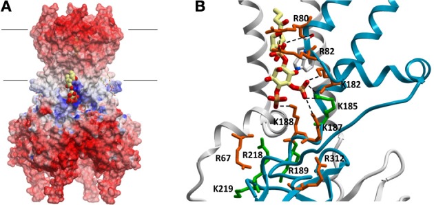
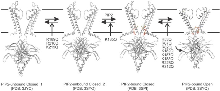
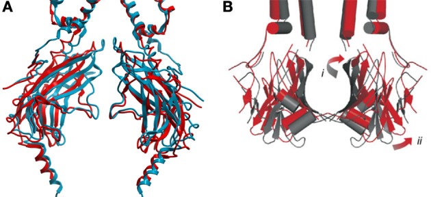
Inward rectifier potassium (Kir) channels are integral membrane proteins charged with a key role in establishing the resting membrane potential of excitable cells through selective control of the permeation of K(+) ions across cell membranes. In conjunction with secondary anionic phospholipids, members of this family are directly regulated by phosphoinositides (PIPs) in the absence of other proteins or downstream signaling pathways. Different Kir isoforms display distinct specificities for the activating PIPs but all eukaryotic Kir channels are activated by PI(4,5)P2. On the other hand, the bacterial KirBac1.1 channel is inhibited by PIPs. Recent crystal structures of eukaryotic Kir channels in apo and lipid bound forms reveal one specific binding site per subunit, formed at the interface of N- and C-terminal domains, just beyond the transmembrane segments and clearly involving some of the key residues previously identified as controlling PI(4,5)P2 sensitivity. Computational, biochemical, and biophysical approaches have attempted to address the energetic determinants of PIP binding and selectivity among Kir channel isoforms, as well as the conformational changes that trigger channel gating. Here we review our current understanding of the molecular determinants of PIP regulation of Kir channel activity, including in context with other lipid modulators, and provide further discussion on the key questions that remain to be answered.

Keywords: Kir channel; inward rectifier potassium channels; ion channel; ion channel gating; ligand-gated ion channels; lipid protein interactions; phosphoinositides.


Figures

Figure 1
Figure 1
(A) Electrostatic potential map of chicken Kir2.2 channels (PDB:3SPI). Large regions of negative potentials are present at the extracellular and intracellular surfaces, which are important for ion and inhibitor binding. A band of positively charged residues lie just below the lipid bilayer which (i) form critical interactions necessary to establish a three dimensional pocket for PIP lipids, and (ii) in the case of some of these residues, help to co-ordinate the ligands in the pocket. PI(4,5)P2 molecules are shown in space-filling and stick representations in this binding pocket. (B) PI(4,5)P2 (and likely all other PIPs) is coordinated by residues at the interface of 2 subunits. Postively charged residues thought to contribute to PIP2 sensitivity are highlighted using Kir2.1 channel numbering. Despite their proximity and in some cases their involvement in co-ordinating the lipid, not all residues contribute to the energy of PIP2 binding. Residues in green contribute to the energetics of binding, while residues in red appear to primarily affect gating transitions [adapted from Hansen et al. (2011)].
Figure 2
Figure 2
A proposed model for the predominant pathway of PIP2-dependent channel activation. Kir channels may undergo a conformational change whereby the cytoplasmic domain moves from an extended 3JYC-type closed conformation toward the plasma membrane and interacts through a hydrogen bond network with the slide helix. This leads to a compact PI(4,5)P2 unbound structure (similar to what was observed for the Kir3.2 apo structure PDB: 3SYO). Kir2.1 mutations in R189, R218, and K219 appears to disrupt this equilibrium thereby leading to reduced binding of PIP ligands. However, once the transition occurs, this state enables PI(4,5)P2 to bind by generating a three-dimensional pocket that can co-ordinate the ligand (PDB: 3SPI). Ligand binding within the pocket appears to be disrupted only by a K185Q mutation in Kir2.1 channels. PI(4,5)P2 binding in a particular conformation may then trigger rotation of the S6, and conformational changes in the cytoplasmic domain that lead to channel opening (PDB: 3SYQ). Most mutants that affect PI(4,5)P2 sensitivity seem to alter activity through this final transition step.
Figure 3
Figure 3
(A) Comparison of GIRK2 in the closed PIP2 bound conformation (PDB: 3SYA, blue) and in the maximally open (R201A) + PIP2 bound conformation (PDB: 3SYQ, red). (B) Comparison of the KirBac1.1 channel cytoplasmic domain (subunits A and C) in the closed (PDB: 1P7B, gray) and predicted “open” (red) conformations. Opening requires outward twisting and tilting of the major βI sheet (i), and outward motion of the minor βII sheet (ii) [adapted from Wang et al. (2012)]. Qualitatively, the movements involved in GIRK2 channel opening appear similar to those predicted for the prokaryotic KirBac1.1 channel.

References

    1. Baukrowitz T., Schulte U., Oliver D., Herlitze S., Krauter T., Tucker S. J., et al. (1998). PIP2 and PIP as determinants for ATP inhibition of KATP channels. Science 282, 1141–1144 10.1126/science.282.5391.1141 - DOI - PubMed
    1. Bavro V. N., De Zorzi R., Schmidt M. R., Muniz J. R., Zubcevic L., Sansom M. S., et al. (2012). Structure of a KirBac potassium channel with an open bundle crossing indicates a mechanism of channel gating. Nat. Struct. Mol. Biol. 19, 158–163 10.1038/nsmb.2208 - DOI - PMC - PubMed
    1. Beeler G. W., Jr., Reuter H. (1970). Voltage clamp experiments on ventricular myocarial fibres. J. Physiol. 207, 165–190 - PMC - PubMed
    1. Cheng W. W., D'Avanzo N., Doyle D. A., Nichols C. G. (2011). Dual-Mode phospholipid regulation of human inward rectifying potassium channels. Biophys. J. 100, 620–628 10.1016/j.bpj.2010.12.3724 - DOI - PMC - PubMed
    1. Cheng W. W., Enkvetchakul D., Nichols C. G. (2009). KirBac1.1: it's an inward rectifying potassium channel. J. Gen. Physiol. 133, 295–305 10.1085/jgp.200810125 - DOI - PMC - PubMed