. 2014 Jan 8:4:505.
doi: 10.3389/fimmu.2013.00505. eCollection 2014.

Nuclear Factor κB is Required for Tumor Growth Inhibition Mediated by Enavatuzumab (PDL192), a Humanized Monoclonal Antibody to TweakR

Affiliations

Nuclear Factor κB is Required for Tumor Growth Inhibition Mediated by Enavatuzumab (PDL192), a Humanized Monoclonal Antibody to TweakR

James W Purcell et al. Front Immunol. .

Abstract

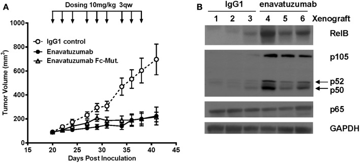
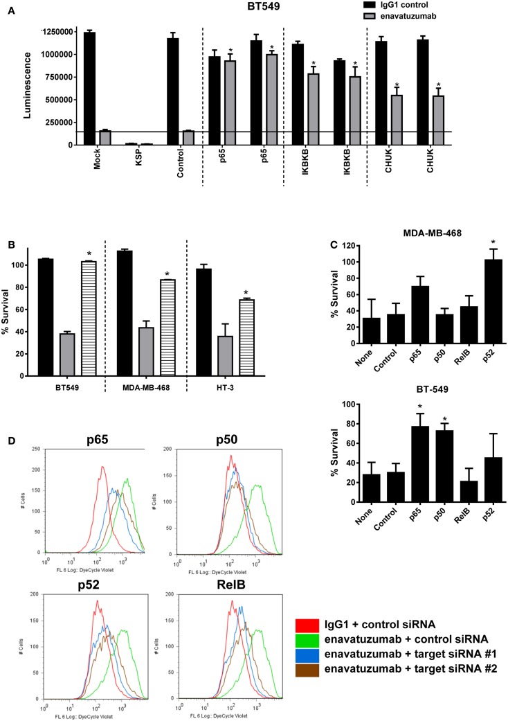
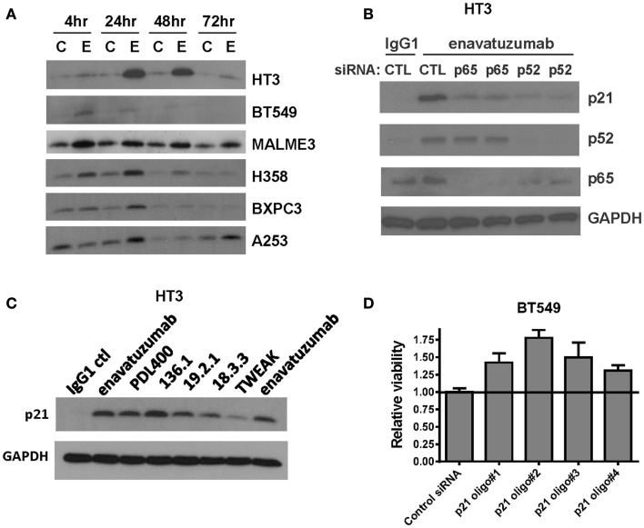
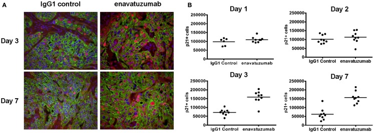
TweakR is a TNF receptor family member, whose natural ligand is the multifunctional cytokine TWEAK. The growth inhibitory activity observed following TweakR stimulation in certain cancer cell lines and the overexpression of TweakR in many solid tumor types led to the development of enavatuzumab (PDL192), a humanized IgG1 monoclonal antibody to TweakR. The purpose of this study was to determine the mechanism of action of enavatuzumab's tumor growth inhibition and to provide insight into the biology behind TweakR as a cancer therapeutic target. A panel of 105 cancer lines was treated with enavatuzumab in vitro; and 29 cell lines of varying solid tumor backgrounds had >25% growth inhibition in response to the antibody. Treatment of sensitive cell lines with enavatuzumab resulted in the in vitro and in vivo (xenograft) activation of both classical (p50, p65) and non-classical (p52, RelB) NFκB pathways. Using NFκB DNA binding functional ELISAs and microarray analysis, we observed increased activation of NFκB subunits and NFκB-regulated genes in sensitive cells over that observed in resistant cell lines. Inhibiting NFκB subunits (p50, p65, RelB, p52) and upstream kinases (IKK1, IKK2) with siRNA and chemical inhibitors consistently blocked enavatuzumab's activity. Furthermore, enavatuzumab treatment resulted in NFκB-dependent reduction in cell division as seen by the activation of the cell cycle inhibitor p21 both in vitro and in vivo. The finding that NFκB drives the growth inhibitory activity of enavatuzumab suggests that targeting TweakR with enavatuzumab may represent a novel cancer treatment strategy.

Keywords: Fn14; NFκB; TweakR; enavatuzumab; monoclonal antibody; p21.


Figures

Figure 1
Figure 1
Enavatuzumab has growth inhibitory activity in a broad range of cancer cell lines in vitro. One hundred and five cell lines representing multiple solid tumor types were tested for sensitivity to enavatuzumab in the presence of anti-human crosslinking antibody. The 38 cell lines that had ≥20% growth inhibition after 5-day treatment with enavatuzumab are shown.
Figure 2
Figure 2
The NFκB pathway is activated in response to enavatuzumab treatment in vitro. (A) A NFκB luciferase reporter construct was transfected into BT549 cells, then treated with antibody/crosslinker. Luciferase expression was measured 24 h post antibody treatment. (B) NFκB activation was examined by Western blotting for p-IκBα expression. Cell lysates were prepared at 4 and 24 h post antibody treatment. C, samples treated with control antibody/crosslinker; E, enavatuzumab/crosslinker-treated samples. (C,D) Enavatuzumab-induced the NFκB pathway in HT3 cells. HT3 cells were treated with enavatuzumab (E) or IgG1 control antibody (C) in the presence of crosslinking antibody for the times indicated. Cell lysates were prepared and activation of NFκB pathway members was analyzed by Western blot.
Figure 3
Figure 3
Differential NFκB pathway activation in sensitive compared to resistant cell lines. (A) NFκB functional DNA binding ELISAs were performed on sensitive (black bars) and resistant (gray bars) cell lines. The relative level of subunit binding (y-axis) was calculated by subtracting the OD450 of the control samples from that of the enavatuzumab-treated samples. (B) Enavatuzumab-induced changes in gene expression were measured 6–72 h after antibody treatment by gene expression profiling. The average of duplicate samples is shown for each time point treated with enavatuzumab or IgG1 control in the presence of a crosslinking antibody. Changes in NFκB responsive genes are shown for a sensitive (BT549) and resistant (T47D) cell line.
Figure 4
Figure 4
In vivo efficacy and NFκB activation in H358 xenografts in response to enavatuzumab. (A) H358 xenograft tumors at ~100 mm3 in ICR SCID mice were treated i.p. with enavatuzumab, enavatuzumab-Fc mutant, or IgG1 control at 10 mg/kg q3w as indicated. (B) H358 xenografts were treated on days 0 and 3 with enavatuzumab or control antibody. Tumors from three mice from each treatment group were excised on day 4, after which cell lysates were prepared and analyzed by Western blot. Activation of NFκB was examined by measuring RelB, p105/p50, p52, and p65 levels in the tumors.
Figure 5
Figure 5
Inhibition of NFκB activation prevents the growth inhibitory activity of enavatuzumab. (A) BT549 cells were transfected with two different siRNAs against p65 or upstream kinases IKK1 (IκBKβ), IKK2 (CHUK). Transfection controls were non-targeting control (negative) or KSP (positive control for transfection efficiency). After 48 h, cells were treated with enavatuzumab or IgG1 control at 10 μg/mL for an additional 5 days in the presence of anti-human crosslinking antibody (3.5 μg/mL), and the cell viability was determined. siRNA significantly reduced growth inhibition by enavatuzumab, compared to mock or control siRNA transfected cells (horizontal line) (*p-value < 0.05). (B) Enavatuzumab sensitive lines were treated with IKK inhibitor IKK16 at 160 nM (black bars), enavatuzumab/crosslinker (gray bars), or IKK16 plus enavatuzumab/crosslinker (striped bars) and % survival was measured after 5 days. IKK16 significantly blocked growth inhibition by enavatuzumab compared to enavatuzumab alone (*p-value < 0.05). (C) Targeting individual NFκB subunits (p50, p65, RelB, p52) by siRNA reduced enavatuzumab activity in sensitive cell lines (MDA-MB-468 and BT549). Cells were transfected with pooled targeting siRNA or control siRNA for 48 h, prior to treatment with enavatuzumab or IgG1 control for 5 days in the presence of a crosslinking antibody. Percent survival was calculated from the relative viability of cells treated with enavatuzumab versus control-treated cells (*p-value < 0.05). (D) The effect of NFκB induced by enavatuzumab on cell division was investigated by pre-labeling HT3 cells with the Cell Trace™ reagent. Cells treated with enavatuzumab/crosslinker (green line) are compared to the IgG1 control-treated cells (red line). Cells transfected with two different siRNAs to p65, p50, p52, or RelB and treated with enavatuzumab/crosslinker (blue and brown lines) are also displayed.
Figure 6
Figure 6
Enavatuzumab inhibits cell division and upregulates p21 in vitro and in vivo in an NFκB-dependent manner. (A) Sensitive cell lines HT3, BT549, MALME-3M, H358, BXPC3, and A253 were treated with IgG1 control antibody (C) or enavatuzumab (E) for 4–72 h in the presence of crosslinking antibody, after which p21 expression was assessed by Western blot. (B) The role of NFκB in enavatuzumab-induced p21 expression was examined by knocking down the expression of p65 or p52 with two different siRNAs to each. Samples were transfected with siRNA for 24 h, then treated with IgG1 control or enavatuzumab for an additional 24 h in the presence of crosslinking antibody. Cell lysates were analyzed by Western blot for p21, p52, and p65 expression. (C) p21 is activated by multiple antibodies targeting TweakR targeting and TWEAK ligand. HT3 cells were treated (10 μg/mL) with antibodies that bind and activate TweakR including enavatuzumab, PDL400, 136.1, 19.2.1, 18.3.3, or TWEAK ligand (300 ng/mL) for 24 h in the presence of crosslinking antibody. p21 expression was then assessed by Western blot. (D) siRNA inhibition of p21 reduces the relative growth inhibition caused by enavatuzumab treatment. BT549 cells were transfected with four different siRNA oligos targeting p21. Two days post transfection, cells were treated with enavatuzumab for 5 days in the presence of crosslinking antibody and cell viability was determined. Relative viability of 1.0 represents the viability of cells transfected with the non-targeting control siRNA and treated with enavatuzumab. An increase above 1.0 in cells transfected with p21 siRNAs indicates an increase in cell viability relative to the control siRNA transfected cells (p < 0.05). Control transfected cells treated with the IgG1 control antibody exhibited a relative viability of 3.75.
Figure 7
Figure 7
Enavatuzumab treatment of H358 xenograft tumors results in p21 upregulation in vivo. (A) H358 xenografts were grown in ICR SCID mice to ~100 mm3 and mice were then treated with a single i.p. injection of IgG1 control or enavatuzumab at 10 mg/kg. Tumors were excised and the levels of p21 were determined by multi-color IHC. A representative image with p21 expression in red, cytokeratin in green, and nuclei in blue is shown (40× magnification). (B) Quantification of p21 expression from H358 xenografts excised on days 1, 2, 3, and 7 post treatment. Data points represent a single 40× magnification field with three fields for three individual xenografts analyzed per time point for each treatment (p < 0.001 IgG1 control versus enavatuzumab days 3 and 7). The y-axis represents quantification of p21+ cells in arbitrary units.

Similar articles

Cited by

References

    1. Hu S, Tamada K, Ni J, Vincenz C, Chen L. Characterization of TNFRSF19, a novel member of the tumor necrosis factor receptor superfamily. Genomics (1999) 62:103–710.1006/geno.1999.5979 - DOI - PubMed
    1. Brown SA, Richards CM, Hanscom HN, Feng SL, Winkles JA. The Fn14 cytoplasmic tail binds tumour-necrosis-factor-receptor-associated factors 1, 2, 3 and 5 and mediates nuclear factor-kappaB activation. Biochem J (2003) 371:395–40310.1042/BJ20021730 - DOI - PMC - PubMed
    1. Winkles JA. The TWEAK-Fn14 cytokine-receptor axis: discovery, biology and therapeutic targeting. Nat Rev Drug Discov (2008) 7:411–2510.1038/nrd2488 - DOI - PMC - PubMed
    1. Chicheportiche Y, Bourdon PR, Xu H, Hsu YM, Scott H, Hession C, et al. TWEAK, a new secreted ligand in the tumor necrosis factor family that weakly induces apoptosis. J Biol Chem (1997) 272:32401–1010.1074/jbc.272.51.32401 - DOI - PubMed
    1. Jakubowski A, Browning B, Lukashev M, Sizing I, Thompson JS, Benjamin CD, et al. Dual role for TWEAK in angiogenic regulation. J Cell Sci (2002) 115:267–74 - PubMed