Review
. 2014 Jan;7(1):33-51.
doi: 10.1177/1756285613493469.

Targeting heat shock proteins to modulate α-synuclein toxicity

Affiliations
Review

Targeting heat shock proteins to modulate α-synuclein toxicity

Daryl Rhys Jones et al. Ther Adv Neurol Disord. 2014 Jan.

Abstract

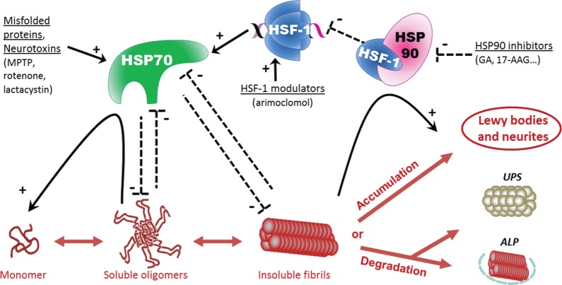
Parkinson's disease is a slowly progressive neurodegenerative disorder typically characterized by the loss of dopaminergic neurons within the substantia nigra pars compacta, and the intraneuronal deposition of insoluble protein aggregates chiefly comprised of α-synuclein. Patients experience debilitating symptoms including bradykinesia, rigidity and postural instability. No curative treatment currently exists and therapeutic strategies are restricted to symptomatic treatment only. Over the past decade a class of molecular chaperones called the heat shock proteins has emerged as a potentially promising therapeutic target. Heat shock proteins aid in the folding and refolding of proteins, and target denatured proteins to degradation systems. By targeting heat shock proteins through various means including overexpression and pharmacological enhancement, researchers have shown that α-synuclein aggregation and its associated cytotoxicity can be therapeutically modulated in an array of cell and animal models. This review highlights the relevant progress in this field and discusses the relevance of heat shock proteins as therapeutic modulators of α-synuclein toxicity to the rapidly evolving understanding of Parkinson's disease pathogenesis.

Keywords: heat shock protein; molecular chaperones; parkinsonism; α-synuclein.


Conflict of interest statement

Conflict of interest statement: The authors declare no conflict of interest in preparing this article.

Figures

Figure 1.
Figure 1.
Dopaminergic synapse and dopamine metabolism. (a,b) In the presynaptic terminal of dopaminergic neurons, tyrosine is transformed into l-DOPA by the action of tyrosine hydroxylase. l-DOPA is subsequently transformed to the neurotransmitter dopamine (DA) by action of the DOPA decarboxylase. (b) DA is then transferred in vesicles by the vesicular monoamine transporter 2 (VMAT-2). After exocytosis of the DA vesicles, DA binds to DA receptors on the postsynaptic membrane, leading to the transduction of the signal in the postsynaptic neuron. (a,b) DA is then recycled by reuptake via the DA transporter, or catabolized by the action of monoamine oxidase (MAO), cathecol-O-methyl transferase (COMT) and aldehyde dehydrogenase (AD) enzymes. The dopaminergic synapse is the principal site of action of current PD treatments. By increasing dopamine metabolism (l-DOPA treatment, ➊) or by inhibiting dopamine catabolism (COMT or MAO inhibition, ➋), or by directly activating postsynaptic dopamine receptors ➌, these treatments boost the activity of the dopaminergic synapses.
Figure 2.
Figure 2.
α-Synuclein structures and the pathogenic process of polymerization (a) Schematic illustration of the primary structure of endogenous α-synuclein protein. The 140 amino acid chain is divided into three major regions; the amphipathic N-terminus, the NAC region (which contains the stretch of hydrophobic 12 amino acid residues believed to be essential for filament assembly) and the acidic C-terminus. (b) A three-dimensional representation of the secondary structure of α-synuclein [Ulmer et al. 2005]. The N-terminus forms two α-helical regions whereas the rest of the protein is natively unfolded. α-Synuclein monomers are believed to adopt different secondary structures. (b) α-Synuclein is physiologically an unstructured soluble monomer but is also found bound to membranes with a secondary structure comprised of two α-helices. Under pathological conditions the hydrophobic regions of α-synuclein may acquire a β-sheet configuration, and gain the propensity to dimerize and subsequently polymerize into oligomers and fibrillar species. The exact cytotoxic species is disputable; however, the large insoluble species are now believed to be less toxic than the prefibrillar oligomeric species.
Figure 3.
Figure 3.
Activation of heat shock transcription factor-1 by Hsp90-inhibition and modulation of α-synuclein aggregation by heat shock proteins. In PD, α-synuclein adopts anomalous misfolded structures ranging from small soluble oligomers to large insoluble aggregates called Lewy bodies and Lewy neurites (depicted in red). Heat shock proteins (HSPs) are strongly implicated in the correct folding of proteins. Whereas HSP90 seems to promote fibril formation and growth, HSP70 was shown to stabilize the monomeric form and to block oligomerization, aggregation and fibril growth of α-synuclein. Moreover, HSP70 promotes α-synuclein degradation through the HSP-mediated autophagy–lysosomal pathway (ALP) and by directing α-synuclein to the ubiquitin–proteasome system (UPS). Conversely, α-synuclein oligomers have been shown to inhibit the UPS and HSP70 chaperone function [Snyder et al. 2003; Hinault et al. 2010]. HSP90 regulates Hsp70 by sequestering heat shock transcription factor 1 (HSF-1). Consequently, HSP90 inhibition induces the release of HSF-1 and subsequently upregulates HSP70. 17-AAG, 17-(allylamino)-17-demethoxygeldanamycin; GA, geldanamycin; MPTP, 1-methyl-4-phenyl-1,2-3,6-tetrahydropyridine.

References

    1. Abeliovich A., Schmitz Y., Farinas I., Choi-Lundberg D., Ho W., Castillo P., et al. (2000) Mice lacking alpha-synuclein display functional deficits in the nigrostriatal dopamine system. Neuron 25: 239–252 - PubMed
    1. Adachi H., Waza M., Tokui K., Katsuno M., Minamiyama M., Tanaka F., et al. (2007) CHIP overexpression reduces mutant androgen receptor protein and ameliorates phenotypes of the spinal and bulbar muscular atrophy transgenic mouse model. J Neurosci 27: 5115–5126 - PMC - PubMed
    1. Alcain F., Villalba J. (2009) Sirtuin activators. Expert Opin Ther Pat 19: 403–414 - PubMed
    1. Al-Ramahi I., Lam Y., Chen H., De Gouyon B., Zhang M., Perez A., et al. (2006) CHIP protects from the neurotoxicity of expanded and wild-type ataxin-1 and promotes their ubiquitination and degradation. J Biol Chem 281: 26714–26724 - PubMed
    1. Anfinsen C. (1973) Principles that govern the folding of protein chains. Science 181: 223–230 - PubMed