. 2014 Jan;7(1):60-70.
doi: 10.1177/1756285613496861.

Treatment of complex neurovascular lesions: an interdisciplinary angio suite approach

Affiliations

Treatment of complex neurovascular lesions: an interdisciplinary angio suite approach

Philipp Dammann et al. Ther Adv Neurol Disord. 2014 Jan.

Abstract

Objective: The objective of this study was to analyse our initial experience using an interdisciplinary angio suite approach to neurosurgical treatment of complex neurovascular lesions and expound technical feasibility and possible applications.

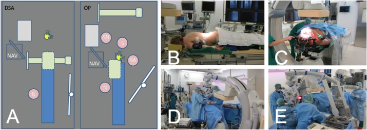
Subjects: Six out of 451 patients with cranial or spinal neurovascular lesions were surgically treated in the angio suite (biplane angiographic system) during a 28-month observation period. Clinical baseline data, radiological and intraoperative findings as well as clinical and radiological outcome were assessed.

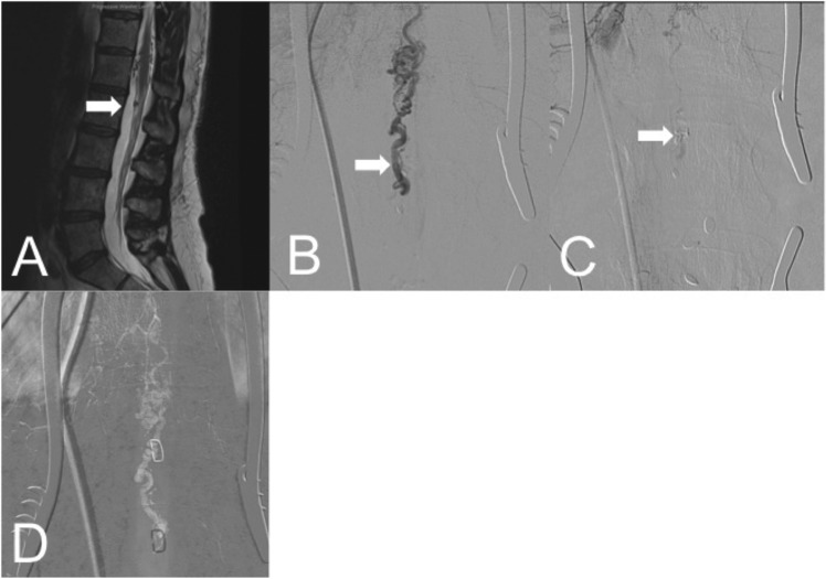
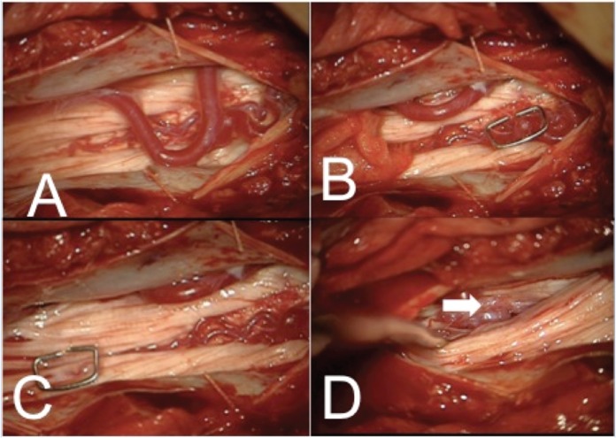
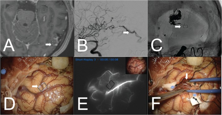
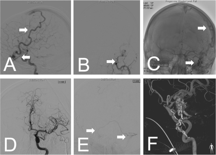
Results: A ventral spinal perimedullary arteriovenous malformation, a ventral spinal perimedullary fistula, two diffuse frontal dural arteriovenous fistulas, a multifocal temporal arteriovenous malformation and a partially embolized fronto-temporo-basal dural arteriovenous fistula were successfully treated with angiographically confirmed complete occlusion and unimpaired neurological condition of the patients at the 12-month follow up.

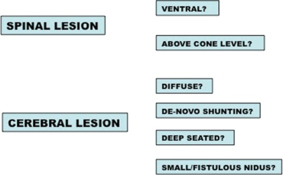
Conclusion: This study demonstrates the feasibility of this approach and points out possible indications, namely ventrally located spinal lesions and diffuse, deep seated cranial lesions.

Keywords: arteriovenous malformation; dural arteriovenous fistula; intraoperative angiography; neurovascular; surgical treatment.


Conflict of interest statement

Conflict of interest statement: The authors declare that there are no conflicts of interest.

Figures

Figure 1.
Figure 1.
(A) A blueprint of the angio suite setting during initial angiography (DSA), respective cranial surgery (OP). S, surgeon; SA, surgical assistant; SN, surgical nurse; N, neuroradiologist; NAV, neuro-navigation system; green, X-ray arms/detector systems, ceiling mounted arm retracted during surgery; yellow, operative microscope; blue, patient table; white, DSA monitor; grey, surgical table. (B), (D) Images of positioning and the intraoperative situation in the spinal set up with the femoral catheter already in position. (C), (E) Images of positioning and the intraoperative situation in the cranial set up using a radiolucent Mayfield© clamp. In both cases the C-arm is retracted from the situs.
Figure 2.
Figure 2.
Exemplary images of a ventral dural arteriovenous fistula (dAVF; case f) with the arteriovenous (AV) shunt located at level L1/2. (A) A sagittal T2-weighted MRI scan with a white arrow indicating the suggested AV shunt. After dorsal approach via laminotomy, anterior posterior intraoperative angiography (IOA; catheter placed in left segment artery Th11) localized the exact fistula point prior to occlusion (B) and confirmed complete occlusion after clipping of the feeding artery (C). (D) A roadmapping image with different metal clamp positions that were used to correlate the fistula point in situ (see also Figure 3).
Figure 3.
Figure 3.
Intradural situation after laminotomy L1 and L2 (case f) and careful dissection of filum tissue to approach the fistula. (A) may display the unclear vascular situation. Initially a vessel convolute located more cranially was suspected and marked with a metal clamp (B). Intraoperative angiography (IOA) (see Figure 2D), however, located the arteriovenous (AV) shunt more caudally (C) where it could be approached (white arrow (D)) and occluded.
Figure 4.
Figure 4.
Initial T1-weighted contrast enhanced MRI scan of the ‘remnant’ fistulous arteriovenous malformation (AVM) left temporo-occipital (A) and initial intraoperative angiography (IOA) displaying early venous drainage (B). Correlation of the deep-seated fistula point via roadmapping using a metal clamp (C), (D). (E) Indocyanine-green angiography (ICG) angiography prior to tissue dissection could not reveal the arteriovenous (AV) shunt located in eloquent temporodorsal area (F).
Figure 5.
Figure 5.
Exemplary images (case (b)) showing a diffuse frontal dural arteriovenous fistula (dAVF). The main feeder is a dilated ophthalmic artery draining via a dilated superficial vein into the superior sagittal sinus (A). Additional multiple feeders come from ethmoidal branches of the external carotid artery (B). Initial embolization was incomplete leaving the main feeder and some small ethmoidal feeders untouched, still showing arteriovenous (AV) shunting (C). During the procedure recruitment of new AV shunts feeding the fistula was observed. Therefore. surgery in the angio suite was planned to assure localization of all feeders and control complete obliteration. After obliteration of the main AV shunts, a new AV shunt draining via a superficial temporal vein into the sigmoid sinus was observed by angiographic control (E). Finally, the fistula was completely occluded. (F) The three-dimensional reconstruction of the control by rotational angiography.
Figure 6.
Figure 6.
The key features of the neurovascular lesions that were considered for angio suite treatment in this series.

Similar articles

Cited by

References

    1. Barrow D., Boyer K., Joseph G. (1992) Intraoperative angiography in the management of neurovascular disorders. Neurosurgery 30: 153–159 - PubMed
    1. Benes L., Wakat J., Sure U., Bien S., Bertalanffy H. (2003) Intraoperative spinal digital subtraction angiography: technique and results. Neurosurgery 52: 603-609; discussion 608–609. - PubMed
    1. Chalouhi N., Theofanis T., Jabbour P., Dumont A., Gonzalez L., Starke R., et al. (2012) Safety and efficacy of intra-operative angiography in craniotomies for cerebral aneurysms and arteriovenous malformations: a review of 1089 cases. Neurosurgery 71(2): E550–E551 - PubMed
    1. Dehdashti A., Thines L., Da Costa L., terBrugge K., Willinsky R., Wallace M., et al. (2009) Intraoperative biplanar rotational angiography during neurovascular surgery. Technical note. J Neurosurg 111: 188–192 - PubMed
    1. Enzmann D., Brody W., Djang W., Riederer S., Keyes G., Collins W., et al. (1983) Intraarterial digital subtraction spinal angiography. AJNR Am J Neuroradiol 4: 25–26 - PMC - PubMed

LinkOut - more resources