

A staging scheme for the development of the moth midge Clogmia albipunctata
- PMID: 24409296
- PMCID: PMC3883683
- DOI: 10.1371/journal.pone.0084422
A staging scheme for the development of the moth midge Clogmia albipunctata
Abstract
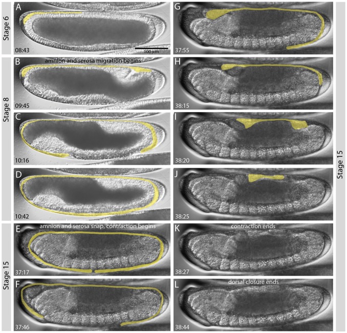
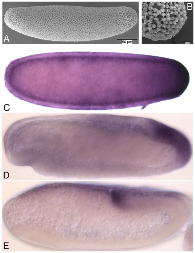
Model organisms, such as Drosophila melanogaster, allow us to address a wide range of biological questions with experimental rigour. However, studies in model species need to be complemented by comparative studies if we are to fully understand the functional properties and evolutionary history of developmental processes. The establishment of new model organisms is crucial for this purpose. One of the first essential steps to establish a species as an experimental model is to carefully describe its life cycle and development. The resulting staging scheme serves as a framework for molecular studies, and allows us to homologise developmental processes between species. In this paper, we have characterised the life cycle and development of an emerging non-drosophilid dipteran model system: the moth midge Clogmia albipunctata. In particular, we focus on early embryogenesis (cleavage and blastoderm cycles before gastrulation), on formation and retraction of extraembryonic tissues, and on formation of the germ line. Considering the large evolutionary distance between the two species (approximately 250 million years), we find that the development of C. albipunctata is remarkably conserved compared to D. melanogaster. On the other hand, we detect significant differences in morphology and timing affecting the development of extraembryonic tissues and the germ line. Moreover, C. albipunctata shows several heterochronic shifts, and lacks head involution and associated processes during late stages of development.
Conflict of interest statement
Figures
Similar articles
-
Clogmia albipunctata (Nematocera; Psychodidae) as the Etiologic Agent of Myiasis: True or False?Diagnostics (Basel). 2022 Sep 1;12(9):2129. doi: 10.3390/diagnostics12092129. Diagnostics (Basel). 2022. PMID: 36140530 Free PMC article. Review.
-
A staging scheme for the development of the scuttle fly Megaselia abdita.PLoS One. 2014 Jan 7;9(1):e84421. doi: 10.1371/journal.pone.0084421. eCollection 2014. PLoS One. 2014. PMID: 24409295 Free PMC article.
-
Evolution of early development in dipterans: reverse-engineering the gap gene network in the moth midge Clogmia albipunctata (Psychodidae).Biosystems. 2014 Sep;123:74-85. doi: 10.1016/j.biosystems.2014.06.003. Epub 2014 Jun 6. Biosystems. 2014. PMID: 24911671
-
A systematic analysis of the gap gene system in the moth midge Clogmia albipunctata.Dev Biol. 2010 Aug 1;344(1):306-18. doi: 10.1016/j.ydbio.2010.04.019. Epub 2010 Apr 28. Dev Biol. 2010. PMID: 20433825
-
What's eating you? Io moth (Automeris io).Cutis. 2008 Jul;82(1):21-4. Cutis. 2008. PMID: 18712020 Review. No abstract available.
Cited by
-
Twenty-seven ZAD-ZNF genes of Drosophila melanogaster are orthologous to the embryo polarity determining mosquito gene cucoid.PLoS One. 2023 Jan 3;18(1):e0274716. doi: 10.1371/journal.pone.0274716. eCollection 2023. PLoS One. 2023. PMID: 36595500 Free PMC article.
-
Stagewise resolution of temperature-dependent embryonic and postembryonic development in the cowpea seed beetle Callosobruchus maculatus (F.).BMC Ecol. 2020 Sep 11;20(1):50. doi: 10.1186/s12898-020-00318-2. BMC Ecol. 2020. PMID: 32917176 Free PMC article.
-
Clogmia albipunctata (Nematocera; Psychodidae) as the Etiologic Agent of Myiasis: True or False?Diagnostics (Basel). 2022 Sep 1;12(9):2129. doi: 10.3390/diagnostics12092129. Diagnostics (Basel). 2022. PMID: 36140530 Free PMC article. Review.
-
Quantitative system drift compensates for altered maternal inputs to the gap gene network of the scuttle fly Megaselia abdita.Elife. 2015 Jan 5;4:e04785. doi: 10.7554/eLife.04785. Elife. 2015. PMID: 25560971 Free PMC article.
-
A two-level staging system for the embryonic morphogenesis of the Mediterranean fruit fly (medfly) Ceratitis capitata.PLoS One. 2024 Dec 30;19(12):e0316391. doi: 10.1371/journal.pone.0316391. eCollection 2024. PLoS One. 2024. PMID: 39774542 Free PMC article.
References
-
- Abzhanov A, Extavour CG, Groover A, Hodges SA, Hoekstra HE, et al. (2008) Are we there yet? Tracking the development of new model systems. Trends Genet 24: 353–60. - PubMed
-
- Yeates DK, Wiegmann BM (1999) Congruence and Controversy: Toward a Higher-Level Phylogeny of Diptera. Ann Rev Entomol 44: 397–428. - PubMed
Publication types
MeSH terms
LinkOut - more resources
Full Text Sources
Other Literature Sources
