. 2014 Jan 7;9(1):e84472.
doi: 10.1371/journal.pone.0084472. eCollection 2014.

PTK 7 is a transforming gene and prognostic marker for breast cancer and nodal metastasis involvement

Affiliations

PTK 7 is a transforming gene and prognostic marker for breast cancer and nodal metastasis involvement

Silvia Gärtner et al. PLoS One. .

Abstract

Protein Tyrosin Kinase 7 (PTK7) is upregulated in several human cancers; however, its clinical implication in breast cancer (BC) and lymph node (LN) is still unclear. In order to investigate the function of PTK7 in mediating BC cell motility and invasivity, PTK7 expression in BC cell lines was determined. PTK7 signaling in highly invasive breast cancer cells was inhibited by a dominant-negative PTK7 mutant, an antibody against the extracellular domain of PTK7, and siRNA knockdown of PTK7. This resulted in decreased motility and invasivity of BC cells. We further examined PTK7 expression in BC and LN tissue of 128 BC patients by RT-PCR and its correlation with BC related genes like HER2, HER3, PAI1, MMP1, K19, and CD44. Expression profiling in BC cell lines and primary tumors showed association of PTK7 with ER/PR/HER2-negative (TNBC-triple negative BC) cancer. Oncomine data analysis confirmed this observation and classified PTK7 in a cluster with genes associated with agressive behavior of primary BC. Furthermore PTK7 expression was significantly different with respect to tumor size (ANOVA, p = 0.033) in BC and nodal involvement (ANOVA, p = 0.007) in LN. PTK7 expression in metastatic LN was related to shorter DFS (Cox Regression, p = 0.041). Our observations confirmed the transforming potential of PTK7, as well as its involvement in motility and invasivity of BC cells. PTK7 is highly expressed in TNBC cell lines. It represents a novel prognostic marker for BC patients and has potential therapeutic significance.


Conflict of interest statement

Competing Interests: The authors have read the journal's policy and have the following conflicts: S. Gärtner, T. Knyazeva, P. Knyazev and A. Ullrich are listed as inventors on the European Patent Application No. 10075169.2 with the title "PTK-7 protein involved in breast cancer". This does not alter the authors' adherence to all the PLOS ONE policies on sharing data and materials.

Figures

Figure 1
Figure 1. PTK7 is able to confer oncogenic potential to NIH3T3 cells.
NIH3T3 cells were infected with PTK7wt, PTK7DN, empty vector or v-src as a positive control and then seeded for Focus Formation Assay (A) or Colony Formation Assay in Soft Agar (B).
Figure 2
Figure 2. Analysis of PTK7 Expression in BC cell lines and primary tumors.
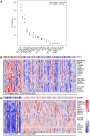
(A) Classification of BC cell lines by PTK7 expression: Cell lines Hs 578T, MDA-MB-157, BT-20, MDA-MB-468, MDA-MB-231, MDA-MB-435S, MDA-MB-436, BT-549, MCF10A1, SUM-149PT classified as basal like, MDA-MB-453, Sk-BR-3, BT-474, T-74D, ZR-75-1, MDA-MB-175VII, MDA-MB 361, BT-483, ZR-75-30, MCF7, and MDA-MB415 classified as luminal cell lines were analysed by RT-PCR. Higher PTK7 expression in BC cell lines which lack expression of ER and are grouped as basal-like. (B) Heat map of genes co-expressed with PTK7 in primary breast carcinomas (van de Vijver, Oncomine), which were grouped by ER status. The colors relate to expression units which are z-normalized to depict relative values within rows. (C) Heat map of genes co-expressed with oncogene HER3 in primary breast carcinomas (van de Vijver, Oncomine).
Figure 3
Figure 3. Expression of full length PTK7 correlates with motility and invasivity of breast cancer cells.
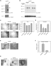
(A) Expression of full length and dominant negative PTK7 protein in wild type or PTK7DN overexpressing Hs578T cells which were subsequently used in cellular assays. Whole cell lysates were used to detect the protein level of PTK7 by immunoblot analysis (top) with α-Tubulin as loading control (bottom). (B) Expression of PTK7 protein 48 and 72 hrs after siRNA transfection. Whole cell lysates were used to detect the protein level of PTK7 by immunoblot analysis (top) with α-Tubulin as loading control (bottom). (C) The motility of cells was analysed by Oris™ cell migration assay. Cells were allowed to migrate into the wound area for 32 hrs. Cell migration was visualized at 4× magnification, representative micrographs are shown (scale bar  = 500 µm). Error bars indicate SEM (n = 3). (D) In vitro migration assay. Cells were allowed to migrate through transwell inserts for 4.5 hours. Error bars indicate SEM (n = 3). (E) The invasivity of cells was analysed by Matrigel outgrowth assay. Cells were seeded on the surface of Matrigel. Colony outgrowth was visualized at 10× magnification. (F) Matrigel Invasion Chamber Assay. Cells were allowed to invade through the Matrigel matrix and membrane pores for 20 hours. Error bars indicate SEM. (G) Effect of an anti-PTK7 polyclonal antibody on the invasivity of Hs578T cells in Matrigel outgrowth assay. Cells were seeded on the surface of Matrigel and treated with anti-PTK7 antibody or control antibody for 3 days. Colony outgrowth was visualized at 10× magnification.
Figure 4
Figure 4. Co-expression of Breast Cancer Related Genes and PTK7.
PTK7 and BC-related genes (Her2, Her3, Pai1, MMP1, CK19 and CD44) expression was assessed by RT-PCR and compared in LN metastasis (LNmet) and tumorfree LN (LNtfr) of 128 patients. All genes (PTK7, Her2, Her3, Pai1, MMP1, CK19) showed an overexpression in LN metastasis compared to tumorfree LN.
Figure 5
Figure 5. PTK7 serves as a prognostic marker in lymph nodes involvement.
Kaplan-Meier curve showing disease-free survival (DFS) in breast cancer patients with lymph node metastasis classified into PTK7 low and high groups according to RT-PCR expression. PTK7 expression is associated with DFS among breast cancer patients with lymph node metastasis expressing high PTK7. P-values were calculated by the log-rank test.

References

    1. Ferlay SH, Bray F, Forman D, Mathers C, Parkin DM (2010) GLOBOCAN 2008 v1.2, Cancer Incidence and Mortality Worldwide: IARC CancerBase No. 10 [Internet]. Lyon, France: International Agency for Research on Cancer; 2010. Available: http://globocan.iarc.fr. Accessed 2013 Apr 17.
    1. Bundred NJ (2001) Prognostic and predictive factors in breast cancer. Cancer Treat Rev 27: 137–142. - PubMed
    1. Gobble RM, Qin LX, Brill ER, Angeles CV, Ugras S, et al. (2011) Expression profiling of liposarcoma yields a multigene predictor of patient outcome and identifies genes that contribute to liposarcomagenesis. Cancer research 71: 2697–2705. - PMC - PubMed
    1. Mossie K, Jallal B, Alves F, Sures I, Plowman GD, et al. (1995) Colon carcinoma kinase-4 defines a new subclass of the receptor tyrosine kinase family. Oncogene 11: 2179–2184. - PubMed
    1. Park SK, Lee HS, Lee ST (1996) Characterization of the human full-length PTK7 cDNA encoding a receptor protein tyrosine kinase-like molecule closely related to chick KLG. Journal of biochemistry 119: 235–239. - PubMed

Publication types

MeSH terms