. 2014 Jan 7;9(1):e84767.
doi: 10.1371/journal.pone.0084767. eCollection 2014.

Inhibition of HIF prolyl-4-hydroxylases by FG-4497 reduces brain tissue injury and edema formation during ischemic stroke

Affiliations

Inhibition of HIF prolyl-4-hydroxylases by FG-4497 reduces brain tissue injury and edema formation during ischemic stroke

Stefan Reischl et al. PLoS One. .

Abstract

Ischemic stroke results in disruption of the blood-brain barrier (BBB), edema formation and neuronal cell loss. Some neuroprotective factors such as vascular endothelial growth factor (VEGF) favor edema formation, while others such as erythropoietin (Epo) can mitigate it. Both factors are controlled by hypoxia inducible transcription factors (HIF) and the activity of prolyl hydroxylase domain proteins (PHD). We hypothesize that activation of the adaptive hypoxic response by inhibition of PHD results in neuroprotection and prevention of vascular leakage. Mice, subjected to cerebral ischemia, were pre- or post-treated with the novel PHD inhibitor FG-4497. Inhibition of PHD activity resulted in HIF-1α stabilization, increased expression of VEGF and Epo, improved outcome from ischemic stroke and reduced edema formation by maintaining BBB integrity. Additional in vitro studies using brain endothelial cells and primary astrocytes confirmed that FG-4497 induces the HIF signaling pathway, leading to increased VEGF and Epo expression. In an in vitro ischemia model, using combined oxygen and glucose deprivation, FG-4497 promoted the survival of neurons. Furthermore, FG-4497 prevented the ischemia-induced rearrangement and gap formation of the tight junction proteins zonula occludens 1 and occludin, both in cultured endothelial cells and in infarcted brain tissue in vivo. These results indicate that FG-4497 has the potential to prevent cerebral ischemic damage by neuroprotection and prevention of vascular leakage.


Conflict of interest statement

Competing Interests: The authors have read the journal’s policy and have the following conflicts. Gail Walkinshaw and Lee Flippin are employees of and own equity in FibroGen Inc., which owns the commercial rights to FG-4497. This does not alter the authors’ adherence to all the PLOS ONE policies on sharing data and materials.

Figures

Figure 1
Figure 1. FG-4497 increases HIF-α and PHD3 protein abundance in endothelial cells and astrocytes.
Mouse cerebrovascular bEnd.3 cells (A) and primary murine astrocytes (B) were treated with either 1 mM DMOG or FG-4497 (5, 20 and 50 µM) for 6 hours. As DMOG and FG-4497 were dissolved in DMSO, 0.2% DMSO was used as a control. Then, cellular proteins were isolated and HIF-1α, HIF-2α and PHD3 abundance was quantified by Western blot. Values are normalized to β-actin and expressed as fold change to CTRL (control = non-treated cells). Significant differences determined by one-way ANOVA combined with Bonferroni post-test are indicated with *(p<0.05), **(p<0.01) or ***(p<0.001). N = 3.
Figure 2
Figure 2. FG-4497 induces HIF target gene expression in endothelial cells and astrocytes.
Mouse cerebrovascular bEnd.3 cells (A) and primary murine astrocytes (B) were treated with either 1 mM DMOG or FG-4497 (5, 20 and 50 µM) or were exposed to hypoxia (1% O2) for 12 hours. Subsequently, RNA was isolated and gene expression was analyzed by real-time PCR. Values are normalized to Rps12 and expressed as fold change to CTRL. Significant differences determined by one-way ANOVA combined with Bonferroni post-test are indicated with *(p<0.05), **(p<0.01) or ***(p<0.001). N = 3.
Figure 3
Figure 3. FG-4497 prevents subcellular delocalization of ZO-1 in OGD-stressed endothelial cells.
Mouse cerebrovascular bEnd.3 cells were exposed to OGD in the absence or presence of 20 µM FG-4497 for 6 and 24 hours or were treated with 20 µM FG-4497 for 6 hours under basal conditions. Immunofluorescent staining was used to visualize the subcellular localization of ZO-1. Cell membrane regions not immunoreactive for ZO-1 (gaps; denoted by white arrowheads) were quantified in five different randomly chosen microscopic fields for each condition per experiment. Significant differences determined by one-way ANOVA combined with Bonferroni post-test are indicated with *(p<0.05) or ***(p<0.001). Scale bar = 20 µm. N = 4.
Figure 4
Figure 4. Effect of FG-4497 on the VEGF expression in endothelial cells exposed to ischemic conditions.
Mouse cerebrovascular bEnd.3 cells were treated with 20 µM FG-4497 for 6 hours under OGD or basal conditions. Subsequently, RNA was isolated and gene expression was analyzed by real-time PCR. Values are normalized to Rps12 and expressed as fold change to untreated cells cultured under non-ischemic conditions. Significant differences determined by one-way ANOVA combined with Bonferroni post-test are indicated with **(p<0.01). N = 3.
Figure 5
Figure 5. FG-4497 attenuates neuronal cell death under ischemic conditions.
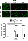
Mouse hippocampal HT-22 neuronal cells were exposed to OGD conditions in the absence or presence of 20 µM FG-4497 for 6, 24 and 48 hours. Cell death/survival was determined by using the Live/Dead assay. Dead (red) and living (green) cells were quantified in three randomly chosen microscopic fields for each condition per experiment, and the ratio dead/living cells was calculated. Significant differences determined by one-way ANOVA combined with Bonferroni post-test are indicated with *(p<0.05), **(p<0.01) or ***(p<0.001). Scale bar = 100 µm. N = 6.
Figure 6
Figure 6. FG-4497 promotes the HIF signaling pathway in cerebral tissue in vivo.
100/kg FG-4497 or an equal volume of 0.9% NaCl was applied intraperitoneally to adult C57BL/6 mice. After 6 hours brains were removed, nuclear proteins and RNA were prepared from cerebral tissue and Western blot (A) and real-time PCR (B) was performed, respectively. Values are normalized to TBP (protein) or Rps12 (RNA) and expressed as fold change to vehicle treated animals. Significant differences determined by unpaired two-tailed t-test are indicated with **(p<0.01). N = 5–8 (per group).
Figure 7
Figure 7. FG-4497 pre-treatment decreases infarct size and the formation of vasogenic edema upon transient cerebral ischemia.
100/kg FG-4497 or an equal volume of 0.9% NaCl was applied intraperitoneally to adult C57BL/6 mice. After 6 hours mice underwent 60 min of MCAO followed by 24 hours reperfusion. Subsequently, brains were removed and from each brain, 24 coronal cryosections (10 µm thick each; 0.4 mm apart) were prepared and submitted to cresyl violet staining for quantification of the infarct and edema size. Significant differences determined by unpaired two-tailed t-test are indicated with *(p<0.05). N = 8–9 (per group).
Figure 8
Figure 8. FG-4497 sustains subcellular localization of occludin and ZO-1 in cerebral blood capillaries upon ischemic stroke.
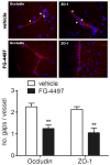
100/kg FG-4497 or an equal volume of 0.9% NaCl was applied intraperitoneally to adult C57BL/6 mice. After 6 hours mice underwent 60 min of MCAO followed by 24 hours reperfusion. Subsequently, brains were removed, coronal cryosections were prepared and immunofluorescent detection of occludin and ZO-1 was performed. Disruptions (gaps; denoted by white arrowheads) of the regular occludin and ZO-1 localization pattern, respectively were counted in at least three randomly chosen blood vessels localized within the peri-infarct region per animal, and the number of gaps per vessel was calculated. Significant differences determined by unpaired two-tailed t-test are indicated with **(p<0.01). N = 4 (per group).
Figure 9
Figure 9. FG-4497 increases VEGF and Epo expression in infarcted brain tissue.
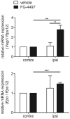
100/kg FG-4497 or an equal volume of 0.9% NaCl was applied intraperitoneally to adult C57BL/6 mice. After 6 hours mice underwent 60 min of MCAO followed by 24 hours reperfusion. Then, brains were removed, RNA was prepared from non-ischemic contralateral and ischemic ipsilateral hemisphere and real-time PCR was performed. Values are normalized to Rps12 and expressed as fold change to vehicle treated animals. Significant differences determined by unpaired two-tailed t-test are indicated with *(p<0.05), **(p<0.01) or ***(p<0.001). N = 3–4 (per group).
Figure 10
Figure 10. FG-4497 post-treatment reduces infarct size upon permanent cerebral ischemia.
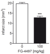
Adult C57BL/6 mice underwent permanent MCAO followed by intraperitoneal application of 100 mg/kg FG-4497 or an equal volume of 0.9% NaCl 1 hour later. 7 days after induction of permanent cerebral ischemia, brains were removed and from each brain, 24 coronal cryosections (10 µm thick each; 0.4 mm apart) were prepared and submitted to cresyl violet staining for quantification of the infarct size. Significant differences determined by unpaired two-tailed t-test are indicated with ***(p<0.001). N = 6 (per group).

Similar articles

Cited by

References

    1. Zhao Y, Rempe DA (2011) Prophylactic neuroprotection against stroke: low-dose, prolonged treatment with deferoxamine or deferasirox establishes prolonged neuroprotection independent of HIF-1 function. J Cereb Blood Flow Metab 31: 1412–1423. - PMC - PubMed
    1. Dirnagl U, Iadecola C, Moskowitz MA (1999) Pathobiology of ischaemic stroke: an integrated view. Trends Neurosci 22: 391–397. - PubMed
    1. Gelderblom M, Leypoldt F, Steinbach K, Behrens D, Choe CU, et al. (2009) Temporal and spatial dynamics of cerebral immune cell accumulation in stroke. Stroke 40: 1849–1857. - PubMed
    1. Sandoval KE, Witt KA (2008) Blood-brain barrier tight junction permeability and ischemic stroke. Neurobiol Dis 32: 200–219. - PubMed
    1. Siddiq A, Aminova LR, Ratan RR (2007) Hypoxia inducible factor prolyl 4-hydroxylase enzymes: center stage in the battle against hypoxia, metabolic compromise and oxidative stress. Neurochem Res 32: 931–946. - PMC - PubMed

Publication types

MeSH terms

Substances