. 2014 Jan 7;9(1):e84941.
doi: 10.1371/journal.pone.0084941. eCollection 2014.

Ovarian cancer spheroid cells with stem cell-like properties contribute to tumor generation, metastasis and chemotherapy resistance through hypoxia-resistant metabolism

Affiliations

Ovarian cancer spheroid cells with stem cell-like properties contribute to tumor generation, metastasis and chemotherapy resistance through hypoxia-resistant metabolism

Jianqun Liao et al. PLoS One. .

Abstract

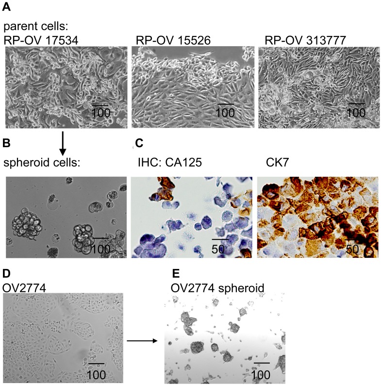
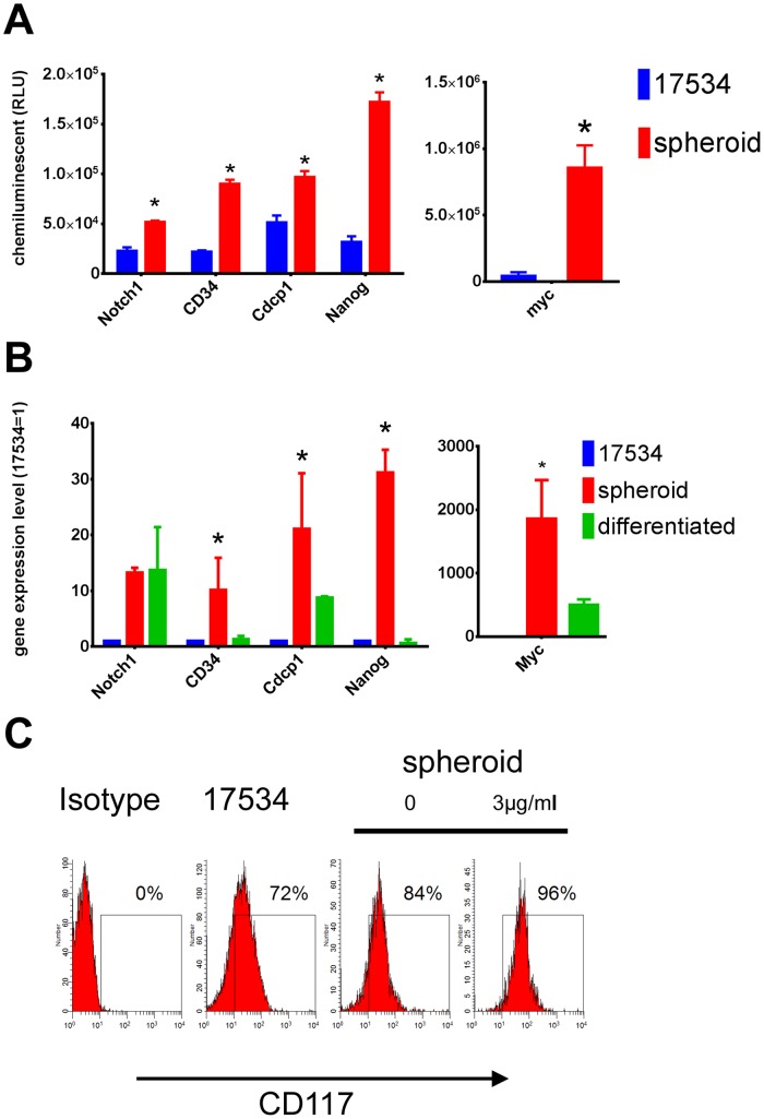
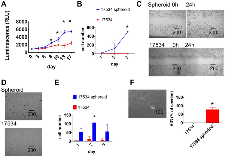
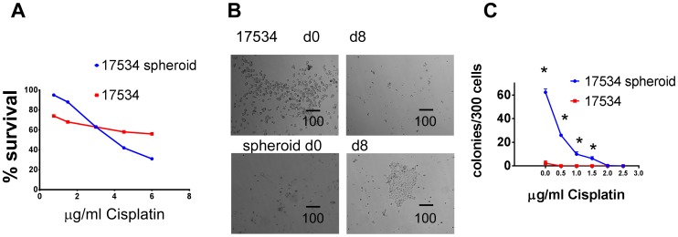
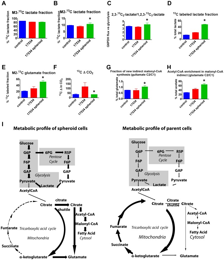
Cells with sphere forming capacity, spheroid cells, are present in the malignant ascites of patients with epithelial ovarian cancer (EOC) and represent a significant impediment to efficacious treatment due to their putative role in progression, metastasis and chemotherapy resistance. The exact mechanisms that underlie EOC metastasis and drug resistance are not clear. Understanding the biology of sphere forming cells may contribute to the identification of novel therapeutic opportunities for metastatic EOC. Here we generated spheroid cells from human ovarian cancer cell lines and primary ovarian cancer. Xenoengraftment of as few as 2000 dissociated spheroid cells into immune-deficient mice allowed full recapitulation of the original tumor, whereas >10(5) parent tumor cells remained non-tumorigenic. The spheroid cells were found to be enriched for cells with cancer stem cell-like characteristics such as upregulation of stem cell genes, self-renewal, high proliferative and differentiation potential, and high aldehyde dehydrogenase (ALDH) activity. Furthermore, spheroid cells were more aggressive in growth, migration, invasion, scratch recovery, clonogenic survival, anchorage-independent growth, and more resistant to chemotherapy in vitro. (13)C-glucose metabolic studies revealed that spheroid cells route glucose predominantly to anaerobic glycolysis and pentose cycle to the detriment of re-routing glucose for anabolic purposes. These metabolic properties of sphere forming cells appear to confer increased resistance to apoptosis and contribute to more aggressive tumor growth. Collectively, we demonstrated that spheroid cells with cancer stem cell-like characteristics contributed to tumor generation, progression and chemotherapy resistance. This study provides insight into the relationship between tumor dissemination and metabolic attributes of human cancer stem cells and has clinical implications for cancer therapy.


Conflict of interest statement

Competing Interests: The authors have declared that no competing interests exist.

Figures

Figure 1
Figure 1. Spheroid cells were generated from primary and established EOC cells.
(A) Three primary cell lines generated from EOC patients show epithelial cells morphology. (B) One of primary EOC cell line generated 3-D independent, self-renewing spheroid cells under stem-cell selection media. (C) Primary cell lines express ovarian carcinoma marker CA-125 and epithelial marker CK-7, as shown by IHC for RP-OV17534. (D) EOC cell line OV2774 is able to generate OV2774 spheroid cells (E). All scale bar unit in this figure and following figures are in µm.
Figure 2
Figure 2. RP-OV17534 Spheroid cells generated tumors in immune-deficient mice.
(A) Different amount of spheroid cells s.c. injected into SCID mice formed tumors. (B) Spheroid cells i.p. injected into SCID mice formed bloody ascites and tumors in different organs as indicated by arrows. (C) Representative H&E staining sections (upper panel) shows RP-OV17534 primary tumor, subcutaneous graft tumor from spheroids, intraperitoneal graft tumor from spheroids, and serial intraperitoneal graft tumor from SCID mouse ascites. Histological analysis (lower panel) shows frequent metastasis of tumor cells into various organs of spheroid cells recipients.
Figure 3
Figure 3. Spheroid cells overexpressed stem cell genes.
(A) Total RNA isolated from spheroid cells and parent cells were examined for their gene expression by a human stem cell markers cDNA array and stem cell-genes showed higher expression levels in spheroid cells to non-spheroid cells. (B) The overexpression levels of Notch1, Nanog, Cdcp1, CD34, and Myc in spheroid cells compared to non-spheroid cells were confirmed by quantitative real-time PCR. These stem cell-genes’ expression were lost or down-regulated when spheroid cells were differentiated by culturing in CM without growth factors for 14 days. Expression levels are represented as fold changes compared to these of non-spheroid cells. (C) FACS examination of CD117 expression on spheroid cells and parent cells showing high CD117 expression in spheroid cells. Some spheroid cells were treated with Cisplatin for 24 hrs before surface staining by CD117 Abs. Error bars: SD, N = 3. *: p<0.05.
Figure 4
Figure 4. Increased ALDH activity in spheroid cells.
(A) The ALDEFLOUR kit labels the population with a high ALDH enzymatic activity in spheroid cells. An aliquot of each sample of cells was treated with ALDH inhibitor DEAB as negative control for setting FACS gate. (B) The BioVision ALDH colorimetric assay detected enhanced ALDH activity in spheroid cell lysate. Error bars: SD, N = 2. *: p<0.05.
Figure 5
Figure 5. Spheroid cells show a more aggressive growth pattern.
(A) Spheroid cells had higher proliferation potential than their parent cells. Both spheroid cells and parental cells were cultured in CM for 24 h, then 5000/well cells were cultured in CM in 96 well plates. Half of media was changed every 3 days. Cell proliferations were detected from day 0 to day 17 by CellTiter-Glo Luminescent Cell Viability Assay. (B) Spheroid cells had higher migration ability than parent cells. Parental and spheroid EOC cells were treated with RPMI only medium for 24 hrs. In a transwell plate, 2×10⁵/well cells were put into insert with 0.2%BSA-RPMI medium, the lower chamber contains no cells except RPMI medium with 2.5% FBS. After culture for 1, 2, and 3 days, the numbers of cells that passed through membrane as well as cells fall into chamber were calculated under microscope. (C) Spheroid cells recovered ‘scratch’ made by 200-µL pipette tip after 24 hours more efficient than their parent cells. Experiments were repeated three times with similar results. (D) More spheroid cells invaded into matrigel than parent cells as shown by microscopy. Spheroid cells and parental cells were treated with serum-free medium RPMI for 24 h. In a transwell plate, 40000/well of cells were put into insert coated with matrigel with 0.2% BSA, while the chamber had only media with 5% FBS, after culture for 1, 2, and 3 days, The number of cells invading into matrigel were counted after 0.1% crystal blue staining. (E) Invasion tests were done in duplicate and average and SD were shown. *,p<0.01. (F) Spheroid cells formed colonies in top agar detected in anchorage-independent growth assay (AIG) as shown under microscopy. 500 cells were seeded in 6 well plates containing a top layer of 0.3% soft agar and 0.5% agar base in DMEM, 10% FBS. 24 hrs later, the number of cells seeded was determined by counting cells under the light microscope. After 3 weeks of growth in soft agar, the number of colonies (Colonies >5 cells) were counted; 10 different fields were quantified per well and the average number of colonies per field was calculated. The AIG index was expressed relative to the number of cells seeded. More than 70% of seeded spheroid cells formed colonies while none of seeded parent cells formed colonies. Experiments were repeated twice with similar results. *,p<0.01. Error bars: SD.
Figure 6
Figure 6. Spheroid cells are resistant to chemotherapy in vitro.
(A) More spheroid cells were killed by Cisplatin than parent cells. The same numbers of spheroid and parent cells were cultured in CM with indicated amount of Cisplatin for 2 days and numbers of cells from different time points were determined. (B) More parent cells survived after Cisplatin treatment for 48 h (Left). However, 8 days later, only spheroid cells were growing while parent cells disappeared (Right). (C) Spheroid and parent cells were treated with indicated amount of Cisplatin for 24 h, washed, and 300 of survived cells/well were cultured in CM for another 10 days, cell colonies were shown after crystal violet staining. The same clonogenic survival assays were repeated twice with similar results. Colonies formed (>5 cells) after 10 days of growth were counted and average and SD were shown. *,p<0.05.
Figure 7
Figure 7. Spheroid cells show a hypoxia-resistant metabolism profile.
Metabolic profiles of a normal ovarian cell line RPNLOv78 as control, EOC cells RP-OV17534, and spheroid RP-OV17534 cells cultures in the presence of [U-13C6]glucose for 24 hrs (n = 3) showing (A) percentages of m3-13C lactate derived directed from glycolysis; (B) percentages of m2-13C lactate derived from pentose cycle; (C) ratio of m2 to m3 lactate; (D) percentage of 13C-labeled lactate of total lactate; (E) percentages of 13C-double-labeled glutamate in total 13C-labeled glutamate; (F) 13CO2 release from [U-13C6]glucose; (G) The fraction of newly synthesized palmitate and the 13C enrichment of acetyl units (H). Error bars: SD; *,p<0.01, compared with parental EOC cells. (I) EOC spheroid cells show increased anaerobic glycolysis, increased direct glucose oxidation in the pentose cycle, but low TCA cycle carbon flux, low anaplerotic flux, and de novo fatty acid synthesis. In contrast, parent EOC cells show increased TCA cycle and high anaplerotic flux. G6P, glucose-6-P; F6P, fructose-6-P; GAP, glyceraldehyde-3-P; 6PG, 6-P-gluconate; R5P, ribulose-5-P. Various thickness of lines represent different strengths of metabolic flux.

References

    1. Jemal A, Siegel R, Ward E, Hao Y, Xu J, et al. (2009) Cancer statistics, 2009. CA Cancer J Clin 59: 225–249. - PubMed
    1. Armstrong D (2010) Update on treatment options for newly diagnosed ovarian cancer. Clin Adv Hematol Oncol 8: 675–678. - PubMed
    1. Colombo N, Van Gorp T, Parma G, Amant F, Gatta G, et al. (2006) Ovarian cancer. Crit Rev Oncol Hematol 60: 159–179. - PubMed
    1. Cannistra SA (2004) Cancer of the ovary. N Engl J Med 351: 2519–2529. - PubMed
    1. Burleson KM, Boente MP, Pambuccian SE, Skubitz AP (2006) Disaggregation and invasion of ovarian carcinoma ascites spheroids. J Transl Med 4: 6. - PMC - PubMed

Publication types

MeSH terms

Substances