. 2014 Jan 7;9(1):e85850.
doi: 10.1371/journal.pone.0085850. eCollection 2014.

TRIP8b is required for maximal expression of HCN1 in the mouse retina

Affiliations

TRIP8b is required for maximal expression of HCN1 in the mouse retina

Yuan Pan et al. PLoS One. .

Abstract

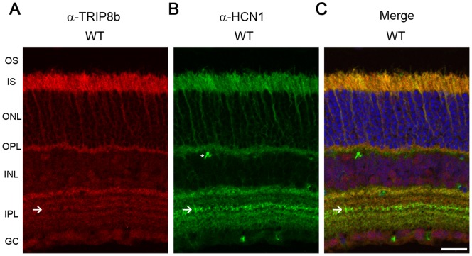
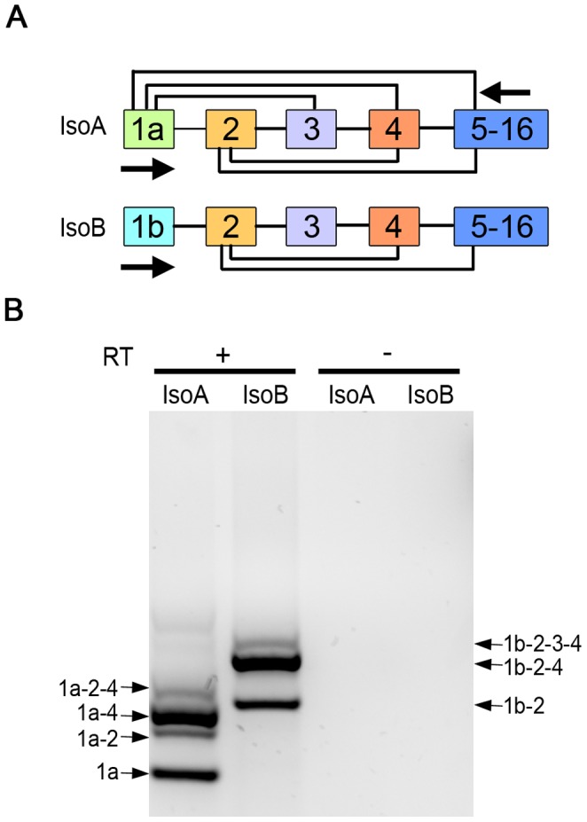
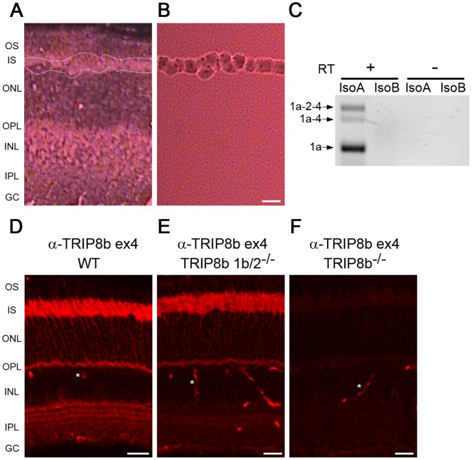
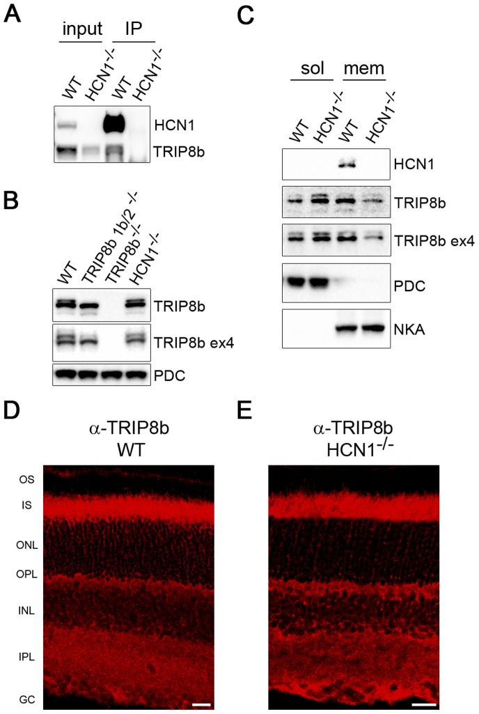
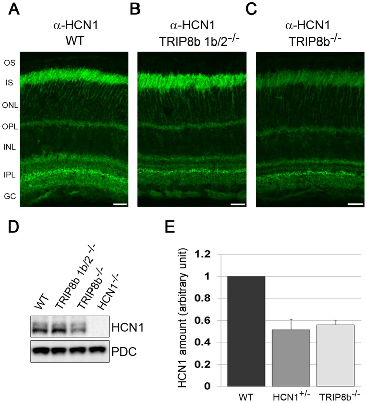
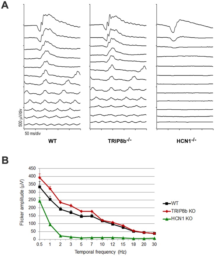
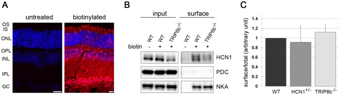
Hyperpolarization-activated cyclic nucleotide-gated (HCN) channels are cation-selective channels present in retina, brain and heart. The activity of HCN channels contributes to signal integration, cell excitability and pacemaker activity. HCN1 channels expressed in photoreceptors participate in keeping light responses transient and are required for normal mesopic vision. The subcellular localization of HCN1 varies among cell types. In photoreceptors HCN1 is concentrated in the inner segments while in other retinal neurons, HCN1 is evenly distributed though the cell. This is in contrast to hippocampal neurons where HCN1 is concentrated in a subset of dendrites. A key regulator of HCN1 trafficking and activity is tetratricopeptide repeat-containing Rab8b interacting protein (TRIP8b). Multiple splice isoforms of TRIP8b are expressed throughout the brain and can differentially regulate the surface expression and activity of HCN1. The purpose of the present study was to determine which isoforms of TRIP8b are expressed in the retina and to test if loss of TRIP8b alters HCN1 expression or trafficking. We found that TRIP8b colocalizes with HCN1 in multiple retina neurons and all major splice isoforms of TRIP8b are expressed in the retina. Photoreceptors express three different isoforms. In TRIP8b knockout mice, the ability of HCN1 to traffic to the surface of retinal neurons is unaffected. However, there is a large decrease in the total amount of HCN1. We conclude that TRIP8b in the retina is needed to achieve maximal expression of HCN1.


Conflict of interest statement

Competing Interests: The authors have declared that no competing interests exist.

Figures

Figure 1
Figure 1. TRIP8b co-localizes with HCN1 in retina.
Mouse retina immunostained with antibodies against A) TRIP8b (red), B) HCN1 (green), C) merged image demonstrating co-localization of these two proteins in the IS and OPL and partial co-localization in the IPL. Asterisks indicate non-specific labeling of blood vessels; arrows indicate IPL sublamina strongly labeled for HCN and containing TRIP8b. The nuclei are counterstained with Hoechst (blue). Abbreviations: OS, outer segment; IS, inner segment; ONL, outer nuclear layer; OPL, outer plexiform layer; INL, inner nuclear layer; IPL, inner plexiform layer; GC, ganglion cell layer. Scale bar is 20 µm.
Figure 2
Figure 2. Multiple TRIP8b splice variants are expressed in retina.
A) Illustration of the alternative splicing that can generate 6, exon 1a containing TRIP8b isoforms (IsoA) or 3, exon 1b TRIP8b isoforms (IsoB). Arrows indicate positions of primers used for RT-PCR. B) RT-PCR products obtained using RNA isolated from mouse retina and primer pairs to amplify TRIP8b IsoA (lane 1) or IsoB splice variants (lane 2). Reverse transcriptase was omitted as a negative control (lanes 3, 4).
Figure 3
Figure 3. Multiple TRIP8b splice variants are expressed in photoreceptors.
A) Mouse retina section before laser capture micro-dissection, dotted white lines indicate region subsequently collected and shown in B. C) RT-PCR products obtained using RNA isolated from tissue collected as in B with the isoform specific primers depicted in Figure 2A. Reverse transcriptase was omitted as a negative control. Immunostaining with an antibody recognizing exon 4 in TRIP8b labels photoreceptors of D) wild type, E) TRIP8b 1b/2−/−, but not F) TRIP8b−/− mouse retina. Asterisks indicate non-specific labeling of blood vessels. Abbreviations as in Figure 1; Scale bars are 20 µm.
Figure 4
Figure 4. HCN1 is required to fully recruit TRIP8b to the membrane.
A) Anti-HCN1 antibodies were used to co-immunoprecipitate TRIP8b from retinal membranes. Membranes prepared from HCN1−/− retinas were used as the negative control. B) Western blot comparing the amount of TRIP8b present in total retina lysates from wild type, both TRIP8b knockout lines, and HCN1−/− mice. Phosducin (PDC) is the loading control. C) Retina lysates from wild type and HCN1−/− mice separated into cytosolic and membrane fractions probed with anti-TRIP8b and anti-HCN1 antibodies. PDC and sodium/potassium ATPase (NKA) are loading controls for each fraction. Immunostaining of TRIP8b in wild type (D) and HCN1−/− retina (E) is indistinguishable. Abbreviations as in Figure 1; Scale bars are 20 µm.
Figure 5
Figure 5. HCN1 protein levels are reduced in the absence of TRIP8b.
Anti-HCN1 antibodies were used to immunostain retina from A) wild type B) TRIP8b 1b/2−/−, or C) TRIP8b−/− mice. Abbreviations as in Figure 1; Scale bars are 20 µm. D) Western blots of total retina lysates probed with anti-HCN1, with PDC used as a loading control. E) Relative amount of HCN1 expressed in the retina of wild type, HCN1 heterozygous, and TRIP8b−/− mice as measured using semi-quantitative Western blots.
Figure 6
Figure 6. Flicker frequency responses are normal in the absence of TRIP8b.
A) Representative waveforms from flicker ERGs of WT, TRIP8b−/−, and HCN1−/− (n = 3 mice for each genotype). Dark-adapted mice were stimulated with a flash intensity of 3.1 cd.s/m2 at frequencies of 0.5, 1, 2, 3, 5, 7, 10, 12, 15, 18, 20, and 30 Hz. B) ERG amplitudes plotted as a function of flicker frequency.
Figure 7
Figure 7. The surface expression of HCN1 is maintained in TRIP8b−/−.
A) Streptavidin staining of control (left) and biotinylated retina (right). B) After biotinylation, surface proteins from either control wild type (WT) or TRIP8b−/− mice were pulled down using NeutrAvidin beads. The level of HCN1 in the total (input) and surface (eluted from NeutrAvidin beads) pools was detected by Western blotting. PDC and NKA were used as negative and positive controls, respectively. C) Densitometry of Western blots represented in (B) was used to calculate the surface to total ratio of HCN1 after normalization to the loading control NKA.

Similar articles

Cited by

References

    1. Fain GL, Quandt FN, Bastian BL, Gerschenfeld HM (1978) Contribution of a caesium-sensitive conductance increase to the rod photoresponse. Nature 272: 466–469. - PubMed
    1. Baylor DA, Matthews G, Nunn BJ (1984) Location and function of voltage-sensitive conductances in retinal rods of the salamander, Ambystoma tigrinum. J Physiol 354: 203–223. - PMC - PubMed
    1. Hestrin S (1987) The properties and function of inward rectification in rod photoreceptors of the tiger salamander. J Physiol 390: 319–333. - PMC - PubMed
    1. Knop GC, Seeliger MW, Thiel F, Mataruga A, Kaupp UB, et al. (2008) Light responses in the mouse retina are prolonged upon targeted deletion of the HCN1 channel gene. Eur J Neurosci 28: 2221–2230. - PubMed
    1. Seeliger MW, Brombas A, Weiler R, Humphries P, Knop G, et al. (2011) Modulation of rod photoreceptor output by HCN1 channels is essential for regular mesopic cone vision. Nat Commun 2: 532. - PubMed

Publication types

LinkOut - more resources