. 2013 Nov 11;4(12):2781-94.
doi: 10.1364/BOE.4.002781. eCollection 2013.

Fiber bundle endocytoscopy

Affiliations

Fiber bundle endocytoscopy

Michael Hughes et al. Biomed Opt Express. .

Abstract

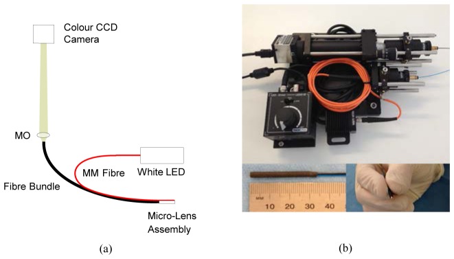
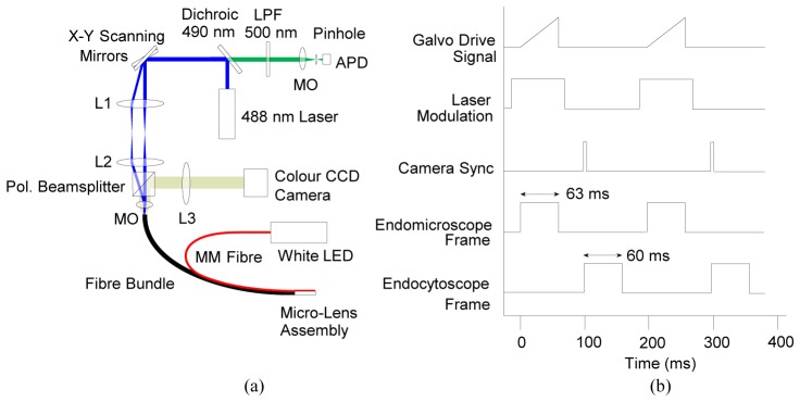
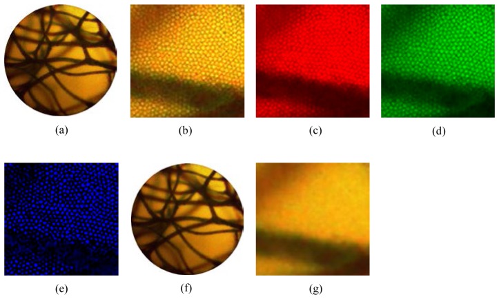
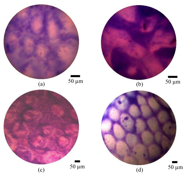
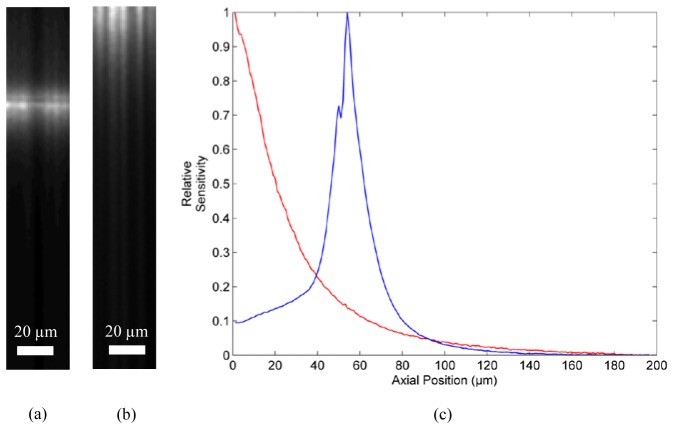
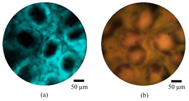
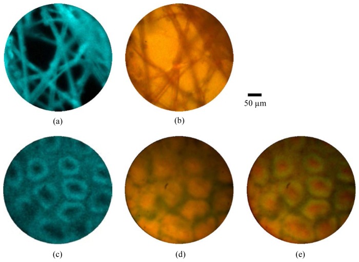
Endocytoscopy is an optical biopsy technique which uses a miniaturized camera to capture white light microscopy images through an endoscope. We have developed an alternative design that instead relays images to an external camera via a coherent fiber bundle. In this paper we characterize the device and demonstrate microscopy of porcine tissue ex vivo. One advantage of our approach is the ease with which other bundle-compatible imaging modalities can be deployed simultaneously. We show this by acquiring quasi-simultaneous endocytoscopy and fluorescence confocal endomicroscopy images through a single fiber bundle. This opens up possibilities for multi-modal endomicroscopy, combining white light and fluorescence imaging.

Keywords: (110.0180) Microscopy; (170.2150) Endoscopic imaging.


Figures

Fig. 1
Fig. 1
Stand-alone fiber-bundle endomicroscopy system. (a) Schematic of optics; (b) photograph of system and close-up view of high resolution probe. Abbreviations: MO: 4X microscope objective, LED: light emitting diode (warm white), MM: multimode.
Fig. 2
Fig. 2
Hybrid endocytoscopy and endomicroscopy system. (a) Schematic of optical system; (b) timing diagram showing synchronization of galvanometer voltage (slow axis scan), laser modulation, camera trigger TTL and acquisition of endomicroscope and endocytoscope image frames. Note that that pulse lengths are illustrative and not precisely to scale, and that the pulse heights are arbitrary. Abbreviations: MM: multimode, Pol.: polarizing, LED: light emitting diode, MO: microscope objective, APD: avalanche photodiode, LPF: long pass wavelength filter, L1 & L3: achromatic doublet, f = 50 mm, L2: achromatic doublet, f = 75 mm.
Fig. 3
Fig. 3
Lateral resolution and field-of-view of (a) high resolution and (b) low resolution probes, evaluated by imaging a USAF resolution target. The three color channels (red, green and blue) of the camera are shown individually and combined (white).
Fig. 4
Fig. 4
Non uniformity of (a) low resolution and (b) high resolution probes. The scale shows the relative image intensity averaged across 400 frames when imaging porcine large bowel mucosa. The non-uniformity for the low resolution probe arises because of the location of the illumination fiber.
Fig. 5
Fig. 5
Removal of fiber bundle core pattern. (a) Raw image of sponge stained with toluidine blue 0.25%, using the high resolution probe; (b) zoom on raw image showing honeycomb pattern of cores; (c,d,e) red, green and blue channels of image in (b), showing that cores can be resolved in each; (f) processed image following application of Gaussian filter; (g) zoom on processed image showing core pattern is no longer visible. Note that the brightness of the color channel images was adjusted for easier viewing. For presentation, the zoomed images were interpolated from 100x100 to 300x300 pixels by cubic interpolation.
Fig. 6
Fig. 6
Ex vivo porcine tissue imaging results showing using high resolution probe (a,b) and low resolution probe (c,d). showing (a,c) large bowel mucosa, and (b,d) stomach mucosa. All samples were stained with toluidine blue 0.25% for approximately 2 minutes and washed before imaging. A selection of videos from the two tissue types are shown in Media 1. Scale bars are 50 µm.
Fig. 7
Fig. 7
Axial sensitivity profile measurements for confocal channel. (a) Cross-sectional image of mirror using high resolution probe; (b) cross-sectional image of mirror using low resolution probe; (c) axial sensitivity profiles for high (blue) and low (red) resolution probes.
Fig. 8
Fig. 8
Ex vivo porcine large bowel mucosa imaged using the combined endocytoscopy and fluorescence confocal endomicroscopy system, using the high resolution probe. The tissue was first stained with acriflavine hydrochloride 0.05% and imaged with the fluorescence channel (a) and subsequently stained with toluidine blue 0.25% and imaged with the endocytoscopy channel (b). Scale bars are 50 µm.
Fig. 9
Fig. 9
Simultaneous fluorescence confocal endomicroscopy (a,c) and endocytoscopy (b,d). (a,b) Lens tissue paper stained with acriflavine hydrochloride 0.05%, imaged with high resolution probe; (b,d) porcine large bowel mucosa ex vivo stained with acriflavine hydrochloride 0.05% and toluidine blue 0.1%, imaged with high resolution probe; (e) merged fluorescence endomicroscopy and endocytoscopy images of large bowel mucosa with fluorescence shown in green. Scale bar is 50 µm

References

    1. Riedl O., Fitzal F., Mader N., Dubsky P., Rudas M., Mittlboeck M., Gnant M., Jakesz R., “Intraoperative frozen section analysis for breast-conserving therapy in 1016 patients with breast cancer,” Eur. J. Surg. Oncol. 35(3), 264–270 (2009).10.1016/j.ejso.2008.05.007 - DOI - PubMed
    1. Shukla R., Abidi W. M., Richards-Kortum R., Anandasabapathy S., “Endoscopic imaging: How far are we from real-time histology?” World J Gastrointest Endosc 3(10), 183–194 (2011).10.4253/wjge.v3.i10.183 - DOI - PMC - PubMed
    1. Liu J. T. C., Loewke N. O., Mandella M. J., Levenson R. M., Group C., “Review article: Modern trends in imaging ii point-of-care pathology with miniature microscopes,” Pathology 34, 81–98 (2011). - PMC - PubMed
    1. Newton R. C., Kemp S. V., Shah P. L., Elson D., Darzi A., Shibuya K., Mulgrew S., Yang G.-Z., “Progress toward optical biopsy: Bringing the microscope to the patient,” Lung 189(2), 111–119 (2011).10.1007/s00408-011-9282-7 - DOI - PubMed
    1. Goualher G. L., Perchant A., Genet M., Cav C., Viellerobe B., Abrat B., Ayache N., “Towards optical biopsies with an integrated fibered confocal fluorescence microscope,” MICCAI 2004, 761–768 (2004).

LinkOut - more resources